Review
. 2020 Jun;17(6):587-599.
doi: 10.1038/s41423-020-0465-0. Epub 2020 May 20.

Antigen presentation by dendritic cells and their instruction of CD4+ T helper cell responses

Affiliations
Review

Antigen presentation by dendritic cells and their instruction of CD4+ T helper cell responses

Kerry L Hilligan et al. Cell Mol Immunol. 2020 Jun.

Abstract

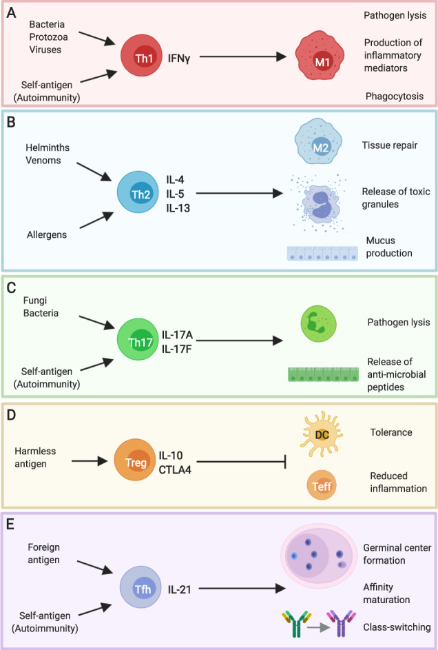
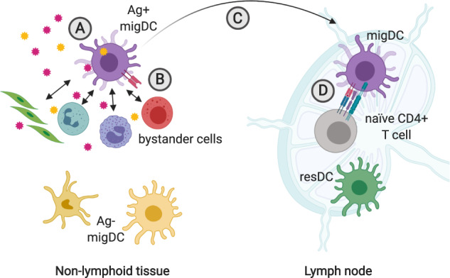
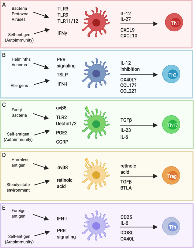
Dendritic cells are powerful antigen-presenting cells that are essential for the priming of T cell responses. In addition to providing T-cell-receptor ligands and co-stimulatory molecules for naive T cell activation and expansion, dendritic cells are thought to also provide signals for the differentiation of CD4+ T cells into effector T cell populations. The mechanisms by which dendritic cells are able to adapt and respond to the great variety of infectious stimuli they are confronted with, and prime an appropriate CD4+ T cell response, are only partly understood. It is known that in the steady-state dendritic cells are highly heterogenous both in phenotype and transcriptional profile, and that this variability is dependent on developmental lineage, maturation stage, and the tissue environment in which dendritic cells are located. Exposure to infectious agents interfaces with this pre-existing heterogeneity by providing ligands for pattern-recognition and toll-like receptors that are variably expressed on different dendritic cell subsets, and elicit production of cytokines and chemokines to support innate cell activation and drive T cell differentiation. Here we review current information on dendritic cell biology, their heterogeneity, and the properties of different dendritic cell subsets. We then consider the signals required for the development of different types of Th immune responses, and the cellular and molecular evidence implicating different subsets of dendritic cells in providing such signals. We outline how dendritic cell subsets tailor their response according to the infectious agent, and how such transcriptional plasticity enables them to drive different types of immune responses.

Keywords: CD4+ T cells; Th effector responses; dendritic cells.


Conflict of interest statement

The authors declare no competing interests.

Figures

Fig. 1
Fig. 1
Distinct effector T helper cell phenotypes are associated with different types of pathogens and immune-driven pathologies. a IFNγ+ Th1 cells are associated with bacterial, protozoan, and viral infections, and promote microbicidal activity in macrophages. Similar pathways are invoked in some autoimmune diseases, where Th1 cells are directed against self-antigens. b Helminths and venoms drive IL-4/IL-5/IL-13+ Th2 cells that induce the release of toxic mediators from mast cells and eosinophils as well as promoting epithelial contractility and mucus production. Macrophages adopt a “M2” phenotype under these conditions to support tissue repair. Aberrant Th2 responses are strongly associated with allergic disease. c Extracellular bacterial and fungal infections drive IL-17+ Th17 cells that promote neutrophil activity and the release of anti-microbial peptides from the epithelium. Pathogenic Th17 cells are also associated with autoimmune diseases, such as multiple sclerosis and psoriasis. d Harmless antigens derived from self or foreign sources, such as the food proteins, are associated with the induction of IL-10+ Treg that act on dendritic cells and effector Th cells to regulate their function. e IL-21+ Tfh cells support antibody responses by promoting germinal center formation, affinity maturation, and antibody class switching. Tfh can be specific for foreign antigen, in the context of infection, or self-antigen, in the context of autoimmune disease. This figure was created using BioRender.com
Fig. 2
Fig. 2
Migratory dendritic cells integrate signals from antigen and bystander cells to inform CD4+ T cell differentiation in local lymph nodes. a Migratory DC (migDC) sample antigen in non-lymphoid tissues. The nature of the antigen (foreign vs. self, immunogenic vs. tolerogenic) determines migDC activation state and expression of CD4+ T cell polarizing factors. b DC communicate with epithelial, stromal, and immune cells in the vicinity. These interactions can inform migDC expression of CD4+ T cell differentiation factors. c Ag+ migDC alter expression of integrins and chemokine receptors to facilitate their migration from the tissue to local lymph nodes. d Ag+ migDC present peptide on MHCII to naive CD4+ T cells expressing a cognate TCR in local lymph nodes. During this interaction, DC provide co-stimulatory signals and instruct CD4+ T cell differentiation through expression of polarizing molecules. This figure was created using BioRender.com
Fig. 3
Fig. 3
Transcriptional plasticity of dendritic cells determines CD4+ T cell differentiation in vivo. Signals from pathogens, immunogens, and antigens, as well as signals from the local stromal, epithelial, and immune cells influence DC expression of CD4+ T cell polarizing factors in a context-specific manner. This ensures activation of the appropriate effector Th cell phenotype. a IFNγ from bystander cells and signaling through pattern-recognition receptors (PRR), such as TLR3, 9, 11, and 12, induces Il12b upregulation by DC. IFNγ also promotes transcription of the CXCR3 ligands Cxcl9 and Cxcl10. Together with IL-27, DC production of these factors facilitates Th1 differentiation. b Helminth products condition DC to promote Th2 differentiation in vivo, potentially via PRR signaling, and DC integration of IFN-I and TSLP signals produced by bystander cells. The DC-derived factors that specifically promote Th2 differentiation are unclear but may include limiting IL-12 production in addition to a “positive” Th2 signal such as OX40L, CCL17, or CCL22. c Engagement of PRR, such as TLR2 and Dectin-1, promote DC production of IL-6 and IL-23. Fibroblast-derived prostaglandin E2 and neuron-derived calcitonin gene-related peptide also promote IL-23 production by DC. Together with αvβ8-mediated activation of TGFβ, DC production of these factors promotes Th17 differentiation. d At steady state, DC are conditioned into a tolerogenic state by their local tissue environment. Retinoic acid may regulate this state in some tissues/settings. Tolerogenic DC express BTLA and the TGFβ activating integrin αvβ8, and produce retinoic acid. In the absence of immunostimulatory signals, the expression of these factors helps support the generation of Treg. e PRR and IFN-I signaling support IL-6 production by DC. Along with co-stimulatory signaling, IL-6 supports Tfh differentiation. DC also express CD25 to limit IL-2 availability to further support Tfh priming. This figure was created using BioRender.com

References

    1. O’Shea JJ, Paul WE. Mechanisms underlying lineage commitment and plasticity of helper CD4+ T cells. Science. 2010;327:1098–1102. - PMC - PubMed
    1. Merad M, Sathe P, Helft J, Miller J, Mortha A. The dendritic cell lineage: ontogeny and function of dendritic cells and their subsets in the steady state and the inflamed setting. Annu. Rev. Immunol. 2013;31:563–604. - PMC - PubMed
    1. Patente TA, Pelgrom LR, Everts B. Dendritic cells are what they eat: how their metabolism shapes T helper cell polarization. Curr. Opin. Immunol. 2019;58:16–23. - PubMed
    1. Blecher-Gonen R, et al. Single-cell analysis of diverse pathogen responses defines a molecular roadmap for generating antigen-specific immunity. Cell Syst. 2019;8:109–121 e106. - PubMed
    1. Meredith MM, et al. Expression of the zinc finger transcription factor zDC (Zbtb46, Btbd4) defines the classical dendritic cell lineage. J. Exp. Med. 2012;209:1153–1165. - PMC - PubMed

Publication types