. 2020 May 20;15(5):e0233502.
doi: 10.1371/journal.pone.0233502. eCollection 2020.

ERp29 as a regulator of Insulin biosynthesis

Affiliations

ERp29 as a regulator of Insulin biosynthesis

Jeffrey Viviano et al. PLoS One. .

Erratum in

Abstract

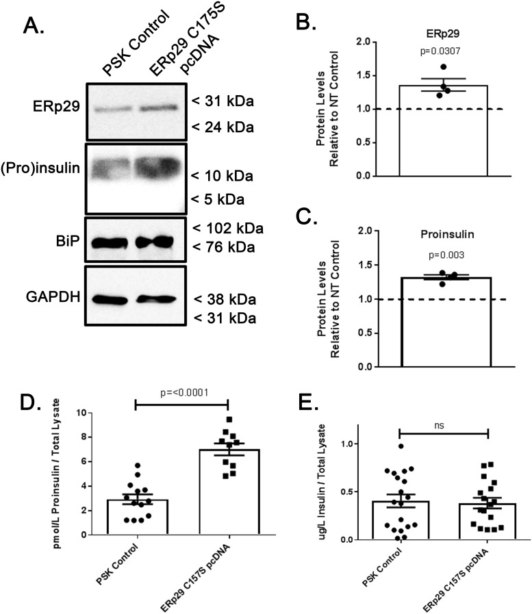
The environment within the Endoplasmic Reticulum (ER) influences Insulin biogenesis. In particular, ER stress may contribute to the development of Type 2 Diabetes (T2D) and Cystic Fibrosis Related Diabetes (CFRD), where evidence of impaired Insulin processing, including elevated secreted Proinsulin/Insulin ratios, are observed. Our group has established the role of a novel ER chaperone ERp29 (ER protein of 29 kDa) in the biogenesis of the Epithelial Sodium Channel, ENaC. The biogenesis of Insulin and ENaC share may key features, including their potential association with COP II machinery, their cleavage into a more active form in the Golgi or later compartments, and their ability to bypass such cleavage and remain in a less active form. Given these similarities we hypothesized that ERp29 is a critical factor in promoting the efficient conversion of Proinsulin to Insulin. Here, we confirmed that Proinsulin associates with the COP II vesicle cargo recognition component, Sec24D. When Sec24D expression was decreased, we observed a corresponding decrease in whole cell Proinsulin levels. In addition, we found that Sec24D associates with ERp29 in co-precipitation experiments and that ERp29 associates with Proinsulin in co-precipitation experiments. When ERp29 was overexpressed, a corresponding increase in whole cell Proinsulin levels was observed, while depletion of ERp29 decreased whole cell Proinsulin levels. Together, these data suggest a potential role for ERp29 in regulating Insulin biosynthesis, perhaps in promoting the exit of Proinsulin from the ER via Sec24D/COPII vesicles.


Conflict of interest statement

The authors have declared that no competing interests exist.

Figures

Fig 1
Fig 1. Proinsulin interacts with the COP II cargo recognition component Sec24D.
(A) Purified recombinant human Insulin, purified human Proinsulin, and whole cell lysate of Min-6 mouse insulinoma cells were resolved by SDS-PAGE and analyzed by immunoblot. (B) Min-6 cells were lysed under non-denaturing conditions and 500 μg of lysate was subject to immunoprecipitation with anti-Insulin/Proinsulin. Precipitated proteins were resolved by SDS-PAGE and immunoblots were probed for Sec24D or Proinsulin as indicated. The Min-6 lysate lane was loaded with 50 μg (10%) of the input lysate. (C) Min-6 cells were lysed under non-denaturing conditions and 50 μg of lysate was subject to immunoprecipitation with a no antibody control (n = 12), anti-V5 (non-specific antibody control, n = 14), or anti-Sec24D antibody (n = 12). Precipitated Proinsulin was quantified with ELISA as described in the materials and methods. Precipitated Proinsulin in anti-V5 Control vs anti-Sec24D, p = <0.0001.
Fig 2
Fig 2. Sec24D regulates intracellular Proinsulin abundance.
Min-6 cells were transiently transfected with non-targeted (control) or Sec24D-targeted siRNA. (A) Whole cell lysates were resolved by SDS-PAGE, followed by analysis by immunoblot. These data are representative of n = 5 independent experiments. (B) Densitometric quantification of the relative abundance of Sec24D in transiently transfected Min-6 cells indicate a decrease in Sec24D levels of ~50% when transfected with Sec24D-targeted siRNA vs. controls (n = 5, p = 0.0117). (C) Densitometric quantification of the relative abundance of Proinsulin indicate a corresponding decrease in whole cell lysate levels of ~60% when transfected with Sec24D-targeted siRNA vs. controls (n = 5, p = 0.0083).
Fig 3
Fig 3. ERp29 interacts with Sec24D and Proinsulin.
Min-6 cells were lysed under non-denaturing conditions, and 500 μg of total lysate protein was subject to immunoprecipitation with either anti-ERp29 (A, D), anti-Sec24D (B), or anti-(Pro)insulin (C). Precipitated proteins were resolved by SDS-PAGE and immunoblots were probed for Sec24D, ERp29, or (Pro)insulin as indicated. The Min-6 lysate lanes were loaded with 50 μg (10%) of the input lysate. These data are representative of n = 4 independent experiments. The co-precipitated Sec24D band is denoted with asterisk in panel (A). (E) Fifty μg of Min-6 whole cell lysate prepared under non-denaturing conditions was subject to immunoprecipitation under the following conditions: No antibody control (n = 12), anti-V5 (non-specific control, n = 11), anti-ERp29 (n = 14), anti-C-peptide from Novus (n = 14), anti-(Pro)Insulin from Sigma (n = 12), and anti-(Pro)Insulin from Cell Signaling Technologies (CST, n = 11). Precipitated Proinsulin was quantified by a rat/mouse Proinsulin ELISA from Mercodia as specified in the materials and methods. Anti-V5 control vs anti-ERp29, p = 0.0262; anti-V5 control vs anti-C-peptide, p = 0.0048; anti-V5 control vs Sigma antibody, p = <0.0001; anti-V5 control vs CST antibody, p = <0.0001.
Fig 4
Fig 4. Depletion of ERp29 decreases cellular Proinsulin.
Min-6 cells were transfected with ERp29-targeting siRNA or with a non-targeting control siRNA. (A) The resulting depletion of ERp29 and its effect on Proinsulin abundance were then assayed by immunoblot. GAPDH was used as a loading control, and BiP was used to monitor ER stress. These data are representative of n = 4 independent experiments. (B) Densitometric quantification of the relative abundance of ERp29 in transiently transfected Min-6 cells indicate a decrease in ERp29 levels of ~70% when transfected with ERp29-targeting siRNA vs. controls (n = 4, p = 0.004). (C) Densitometric quantification of the relative abundance of Proinsulin in transiently transfected Min-6 cells indicate a corresponding decrease in whole cell lysate levels of ~60% when transfected with ERp29-targeting siRNA vs. controls (n = 4, p = 0.0073). (D, E) Transiently transfected Min-6 cells were lysed and analyzed by ELISA. Proinsulin levels decreased when transfected with ERp29-targeting siRNA as compared to controls (D, p = 0.0309), but Insulin content did not significantly change (E, p = ns).
Fig 5
Fig 5. Overexpression of ERp29 increases cellular Proinsulin and Insulin.
Min-6 cells were transiently transfected with a pcDNA4 plasmid expressing WT ERp29 or with a non-expressing PSK control plasmid, and whole cell lysates were prepared. (A) ERp29 and Proinsulin abundance was assayed in whole cell lysates by immunoblot. Immunoreactivity of GAPDH was used as a loading control, and immunoreactivity of BiP was used to monitor for ER stress. These data are representative of n = 5 independent experiments. Densitometry of ERp29 (B, p = 0.020) and Proinsulin (C, p = 0.0149) in immunoblots. (D, E) Lysates of transfected control or WT ERp29 transfected Min-6 cells were analyzed by ELISA, and data demonstrate an increase in both cellular Proinsulin (D, p = 0.0025) and Insulin (E, p = 0.0069) when compared to controls.
Fig 6
Fig 6. Overexpression of ERp29 C157S increases cellular Proinsulin, but not cellular Insulin.
Min-6 cells were transiently transfected with ERp29 C157S pcDNA4 plasmid or mock transfected with non-expressing PSK plasmid as a control. Whole cell lysates were prepared and (A) ERp29 and Proinsulin protein abundance was assayed in whole cell lysates with immunoblot. Immunoreactivity of GAPDH was used as a loading control, and immunoreactivity of BiP was used to monitor for ER stress. These data are representative of n = 4 independent experiments. (B) Densitometry of ERp29 abundance as assessed by immunoblot reveals an ~40% increase in protein levels in ERp29 C157S transfected lysates, relative to controls (p = 0.0307). (C) Densitometry of Proinsulin abundance in immunoblots demonstrates a corresponding increase of ~40% with ERp29 C157S overexpression (p = 0.003). (D, E) Lysates of transfected control or ERp29 C157S transfected Min-6 cells were analyzed by ELISA for cellular content of Proinsulin (D, p = <0.0001) and Insulin (E, p = ns).
Fig 7
Fig 7. Hypothesized role for ERp29 in Insulin synthesis.
Proinsulin (Red) is transported from the ER to the cis-Golgi via COP II vesicles; however, it is currently unclear how Proinsulin is recruited for inclusion in COP II vesicles. The Sec23/24 heterodimer is an essential component of the COP II vesicle pre-budding complex, which also includes Sar1 and the Sec13/31 heterodimer. Our data demonstrate that the Sec24 isoform, Sec24D, interacts with both ERp29 and Proinsulin in pancreatic β-cells, likely as part of a complex. We hypothesize that ERp29 interacts directly with Proinsulin at its putative ERp29 binding domain (24F-F-Y26 in Proinsulin) and promotes inclusion of the Proinsulin/ERp29 complex in COP II vesicles. In this model, ERp29 also associates with a currently unknown integral membrane protein that intermediates the interaction of Proinsulin/ERp29 with Sec24D. One such integral membrane protein may be the KDEL receptor (KDEL-R), which cycles between the ER and cis-Golgi via COP I and II vesicles. Our group previously demonstrated that KDEL-R facilitates the transport of other COP II cargo, specifically the epithelial sodium channel, ENaC, from ER to Golgi, in concert with ERp29 and Sec24D. Future studies will test the hypothesis that KDEL-R intermediates the interaction of ERp29/Proinsulin with Sec24D. Our data also suggest that ERp29 may stabilize Proinsulin, perhaps as it transverses the Golgi cisternae to the trans-Golgi, where it is cleaved to mature Insulin and C-peptide and packaged into granules for secretion.

References

    1. Roden M. and Shulman G.I., The integrative biology of type 2 diabetes. Nature, 2019. 576(7785): p. 51–60. 10.1038/s41586-019-1797-8 - DOI - PubMed
    1. Prentki M. and Nolan C.J., Islet beta cell failure in type 2 diabetes. J Clin Invest, 2006. 116(7): p. 1802–12. 10.1172/JCI29103 - DOI - PMC - PubMed
    1. Sundar Rajan S., et al., Endoplasmic reticulum (ER) stress & diabetes. Indian J Med Res, 2007. 125(3): p. 411–24. - PubMed
    1. Ozcan U., et al., Endoplasmic reticulum stress links obesity, insulin action, and type 2 diabetes. Science, 2004. 306(5695): p. 457–61. 10.1126/science.1103160 - DOI - PubMed
    1. Harding H.P., et al., Regulated translation initiation controls stress-induced gene expression in mammalian cells. Mol Cell, 2000. 6(5): p. 1099–108. 10.1016/s1097-2765(00)00108-8 - DOI - PubMed

Publication types

MeSH terms