. 2020 May 19;31(7):107634.
doi: 10.1016/j.celrep.2020.107634.

β-Glucan Induces Protective Trained Immunity against Mycobacterium tuberculosis Infection: A Key Role for IL-1

Affiliations

β-Glucan Induces Protective Trained Immunity against Mycobacterium tuberculosis Infection: A Key Role for IL-1

Simone J C F M Moorlag et al. Cell Rep. .

Abstract

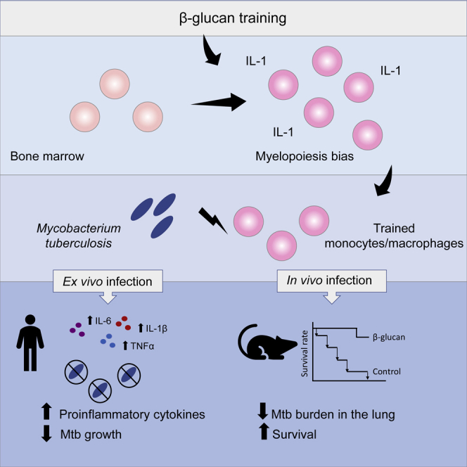
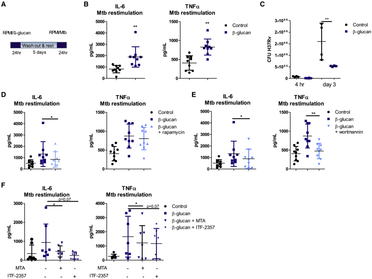
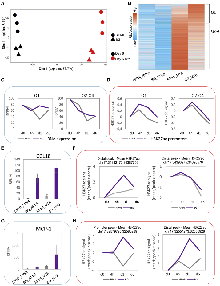
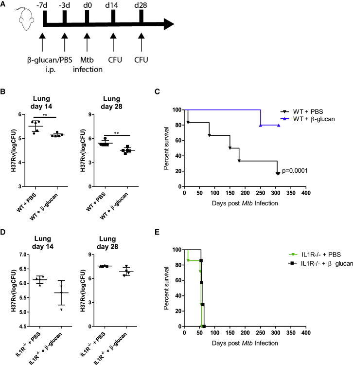
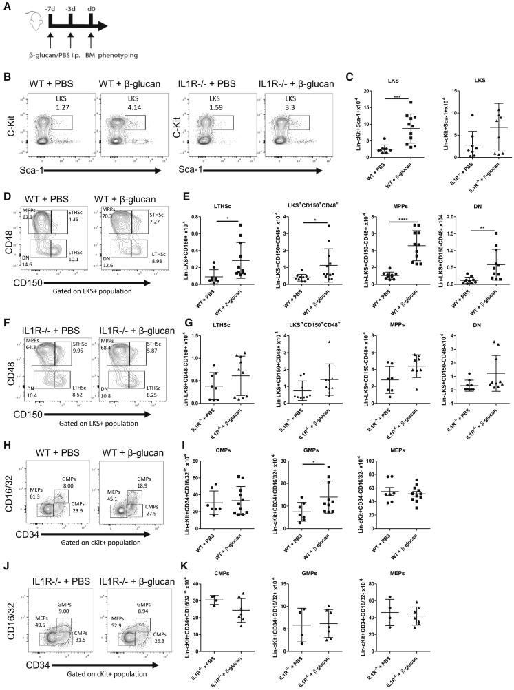
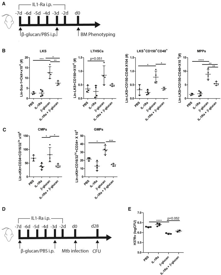
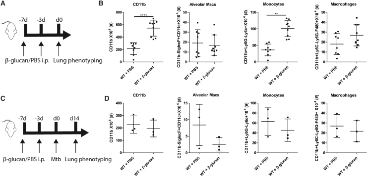
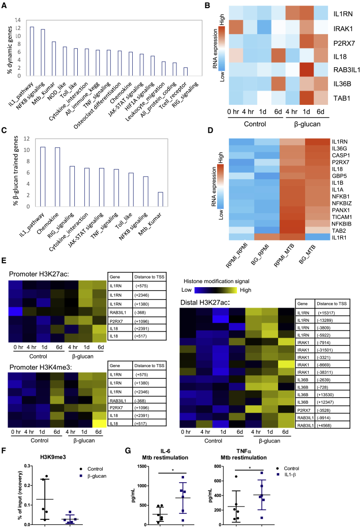
β-glucan is a potent inducer of epigenetic and functional reprogramming of innate immune cells, a process called "trained immunity," resulting in an enhanced host response against secondary infections. We investigate whether β-glucan exposure confers protection against pulmonary Mycobacterium tuberculosis (Mtb) infection. β-glucan induces trained immunity via histone modifications at gene promoters in human monocytes, which is accompanied by the enhanced production of proinflammatory cytokines upon secondary Mtb challenge and inhibition of Mtb growth. Mice treated with β-glucan are significantly protected against pulmonary Mtb infection, which is associated with the expansion of hematopoietic stem and progenitor cells in the bone marrow and increased myelopoiesis. The protective signature of β-glucan is mediated via IL-1 signaling, as β-glucan shows no protection in mice lacking a functional IL-1 receptor (IL1R-/-). The administration of β-glucan may be used as a novel strategy in the treatment of mycobacterial infections and possibly as an adjuvant to improve anti-tuberculosis vaccines.

Keywords: IL-1; Mycobacterium tuberculosis; epigenetics; innate immune memory; monocytes; trained immunity; tuberculosis; β-glucan.


Conflict of interest statement

Declaration of Interests The authors declare no competing interests.

Figures

None
Graphical abstract
Figure 1
Figure 1
β-Glucan Training Increases Antimicrobial Activity of Human Monocytes against M. tuberculosis (A) Schematic representation of the in vitro training model. (B) Human monocytes were trained with β-glucan for 24 h and re-stimulated with heat-killed M. tuberculosis at day 6. IL-6 and TNF-α production was measured in the supernatants (means ± SDs, n = 9, ∗∗p < 0.01, Wilcoxon signed-rank test). See also Figure S1. (C) Monocytes were trained with β-glucan and infected with virulent M. tuberculosis H37Rv at MOI 1 for 4 h. Mtb CFUs were quantified at 4 h and 3 days post-infection (means ± SDs, n = 4, ∗∗p < 0.01, 2-way ANOVA). (D and E) Monocytes were incubated for 24 h with culture medium or β-glucan, with or without an inhibitor of mTOR-glycolysis (rapamycin) (D) or a PI3K inhibitor (wortmannin) (E). After re-stimulation with M. tuberculosis, differences in IL-6 and TNF-α production were assessed between β-glucan-trained monocytes and monocytes that were trained with β-glucan in the presence of rapamycin orwortmannin (means ± SDs, n = 9, p < 0.05, Wilcoxon signed-rank test). (F) Monocytes were stimulated for 24 h with culture medium or β-glucan in the presence or absence of the histone methyltransferase inhibitor MTA or the histone deacetylase inhibitor ITF-2357. On day 6, cells were re-stimulated with M. tuberculosis for 24 h and the difference in cytokine production was assessed between monocytes that were trained in the presence or absence of epigenetic inhibitors (means ± SDs, n = 7, p < 0.05, Wilcoxon signed-rank test).
Figure 2
Figure 2
β-Glucan Training Alters the Transcriptomic Profile of Human Monocytes Exposed to Mtb (A) PCA of RNA-seq data of human monocytes that were trained with β-glucan and exposed to heat-killed M. tuberculosis for 4 h on day 6 (p < 0.05, FC > 1.5, RPKM > 5, n = 3). (B) Heatmap of 2,105 genes that are differentially expressed between β-glucan-treated cells and controls upon exposure to Mtb. The first quartile (Q1) indicates 526 genes that are upregulated (trained) after Mtb exposure in β-glucan-trained monocytes compared to the control. Non-trained genes (1,579 genes) are indicated by Q2–Q4. (C) Average gene expression of genes in Q1 and average expression of non-trained genes (Q2–Q4). (D) Average H3K27ac signal at promoter sites of genes that are present in Q1 and average signal at non-trained genes (Q2–Q4). (E–H) RNA expression of CCL18 (E) and MCP-1 (G) in control versus β-glucan-treated cells upon RPMI/Mtb exposure. H3K27ac signal at promoter/distal regions of CCL18 (F) and MCP-1 (H) upon β-glucan exposure. See also Figure S2 and Table S1.
Figure 3
Figure 3
Training with β-Glucan Enhances Resistance of Mice to Mtb Infection via an IL-1-Dependent Mechanism (A) WT and IL1R−/− mice were treated with β-glucan or PBS at day 0 and day 3, followed by aerosolized infection with Mtb (∼50 H37Rv) at day 7. (B–E) Bacterial burden in the lungs after 14 and 28 days of aerosol infection (means ± SDs, n = 4–5 mice per group, unpaired t test) in WT (B) and IL1R−/− mice (D). Survival of WT (C) and IL1R−/− mice (E) (n = 6–8 mice per group, log-rank test).
Figure 4
Figure 4
Administration of β-Glucan Induces Expansion of HSPCs and Promotes Myelopoiesis in an IL-1-Dependent Manner (A) WT and IL1R−/− mice were treated with β-glucan or PBS at day 0 and day 3. The expansion of cellular subpopulations was assessed in bone marrow 7 days post-β-glucan immunization. (B and C) Representative fluorescence-activated cell sorting (FACS) plots (B) and total cell counts of LKS (C). (D–G) Representative FACS plots (D and F) and total cell counts (E and G) of LTHSc, LKS+CD150+CD48+ cells, MPPs, and DN in WT (D and E) and IL1R−/− mice (F and G). (H–K) Expansion of CMPs, GMPs, and MEPs was assessed in the bone marrow at day 7 post-β-glucan immunization. Representative FACS plots (H and J) for the frequencies of myeloid progenitors and total cell counts of CMPs, GMPs, and MEPs (I and K) in WT (H and I) and IL1R−/− mice (J and K) are shown (means ± SDs, n = 4–7 mice per group, p < 0.05, ∗∗∗p < 0.001 and ∗∗∗∗p < 0.0001 unpaired t test). See Figure S3 for gating strategy.
Figure 5
Figure 5
β-Glucan Fails to Induce Myelopoiesis in the Presence of an IL-1R Antagonist (A) WT mice were treated with β-glucan or PBS at day 0 and day 3 along with IL-1Ra antagonist administered intraperitoneally (i.p.) at day 0 and consecutively until day 6. (B and C) Expansion of cellular subpopulations was assessed in bone marrow 7 days post-β-glucan immunization. Total cell counts of LKS, LT-HSC, LKS+CD150+CD48+ cells, MPPs (B), and CMPs and GMPs (C) are shown (means ± SDs, n = 4 mice per group, p < 0.05, ∗∗p < 0.01, ∗∗∗p < 0.001, and ∗∗∗∗p < 0.00001, 1-way ANOVA). (D) WT mice were treated with β-glucan or PBS at day 0 and day 3 along with IL-1Ra antagonist administered i.p. at day 0 and consecutively until day 6, followed by aerosolized infection with Mtb (∼50 H37Rv) at day 7. (E) Bacterial burden in the lungs after 28 days of aerosol infection (means ± SDs, n = 4–5 mice per group, ∗∗∗∗p < 0.00001, 1-way ANOVA).
Figure 6
Figure 6
β-Glucan Induces an Increase in Myeloid Cell Subsets in the Lungs of Mice (A) WT mice were treated with β-glucan or PBS at day 0 and day 3. Cellular subpopulations were assessed in the lungs 7 days post-β-glucan immunization. (B) Frequency of CD11b, alveolar macrophages, monocytes, and interstitial macrophages 7 days post-β-glucan immunization (means ± SDs, n = 7–8 mice per group). (C) WT mice were treated with β-glucan or PBS at day 0 and day 3, followed by aerosolized infection with Mtb (∼50 H37Rv) at day 7. Cellular subpopulations were assessed in the lungs 14 days post-Mtb infection. (D) Frequency of CD11b, alveolar macrophages, monocytes, and interstitial macrophages 14 days post-Mtb infection (means ± SDs, n = 3 mice per group, ∗∗p < 0.01 and ∗∗∗∗p < 0.0001 unpaired t test). See Figure S4 for gating strategy.
Figure 7
Figure 7
β-Glucan Alters the Transcriptional and Epigenetic Profile of IL-1 Family Genes (A) Percentage of genes in a pathway that are upregulated in human monocytes 24 h after β-glucan exposure (total of 663 genes). (B) Heatmap showing RNA expression of 7 IL-1 family genes induced by exposure to β-glucan compared to the control. Monocytes were incubated with either culture medium or β-glucan. mRNA expression was assessed before incubation, 4 h, 24 h, and 6 days after incubation. See also Figure S5. (C) Enrichment of pathways in genes upregulated after exposure to heat-killed M. tuberculosis in β-glucan-trained cells. (D) Heatmap showing RNA expression of IL-1 family genes upon Mtb exposure on day 6 in β-glucan-trained cells compared to the control. (E) Heatmap of the changes in H3K27ac and H3K4me3 in β-glucan-trained monocytes and control at 4 h, 1 day, and 6 days after the start of incubation near IL-1 family genes shown in (B). (F) Human monocytes were stimulated for 24 h with culture medium or β-glucan. Chromatin was fixated on day 6 and ChIP-qPCR was performed. H3K9me3 was determined at the promoter of IL1B (means ± SDs, n = 5). See Table S2 for primers used. (G) Human monocytes were incubated with culture medium or IL-1β for 24 h. On day 6, cells were re-stimulated with heat-killed M. tuberculosis and levels of IL-6 and TNF-α were assessed (means ± SDs, n = 6, p < 0.05, Wilcoxon signed-rank test).

References

    1. Arts R.J.W., Moorlag S.J.C.F.M., Novakovic B., Li Y., Wang S.Y., Oosting M., Kumar V., Xavier R.J., Wijmenga C., Joosten L.A.B. BCG Vaccination Protects against Experimental Viral Infection in Humans through the Induction of Cytokines Associated with Trained Immunity. Cell Host Microbe. 2018;23:89–100.e5. - PubMed
    1. Barreiro L.B., Neyrolles O., Babb C.L., Tailleux L., Quach H., McElreavey K., Helden P.D., Hoal E.G., Gicquel B., Quintana-Murci L. Promoter variation in the DC-SIGN-encoding gene CD209 is associated with tuberculosis. PLoS Med. 2006;3:e20. - PMC - PubMed
    1. Barreiro L.B., Tailleux L., Pai A.A., Gicquel B., Marioni J.C., Gilad Y. Deciphering the genetic architecture of variation in the immune response to Mycobacterium tuberculosis infection. Proc. Natl. Acad. Sci. 2012;109:1204–1209. - PMC - PubMed
    1. Behar S.M., Divangahi M., Remold H.G. Evasion of innate immunity by Mycobacterium tuberculosis: is death an exit strategy? Nat. Rev. Microbiol. 2010;8:668–674. - PMC - PubMed
    1. Bekkering S., Blok B.A., Joosten L.A., Riksen N.P., van Crevel R., Netea M.G. In Vitro Experimental Model of Trained Innate Immunity in Human Primary Monocytes. Clin. Vaccine Immunol. 2016;23:926–933. - PMC - PubMed

Publication types