. 2020 Jul;40(7):1635-1650.
doi: 10.1161/ATVBAHA.120.314180. Epub 2020 May 21.

Platelets Endocytose Viral Particles and Are Activated via TLR (Toll-Like Receptor) Signaling

Affiliations

Platelets Endocytose Viral Particles and Are Activated via TLR (Toll-Like Receptor) Signaling

Meenakshi Banerjee et al. Arterioscler Thromb Vasc Biol. 2020 Jul.

Abstract

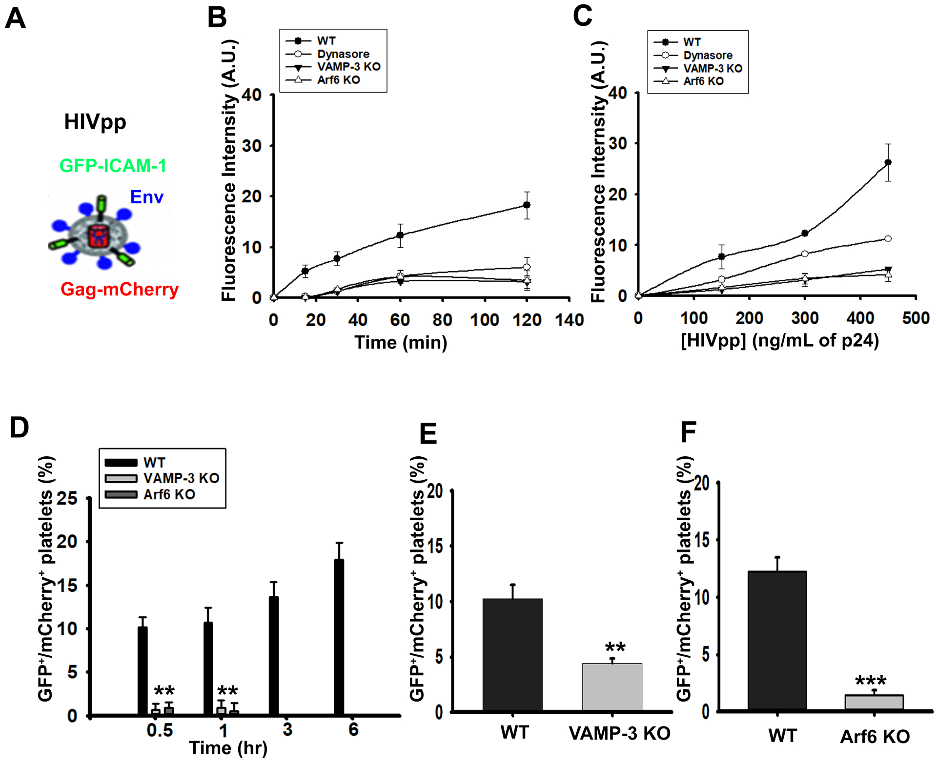
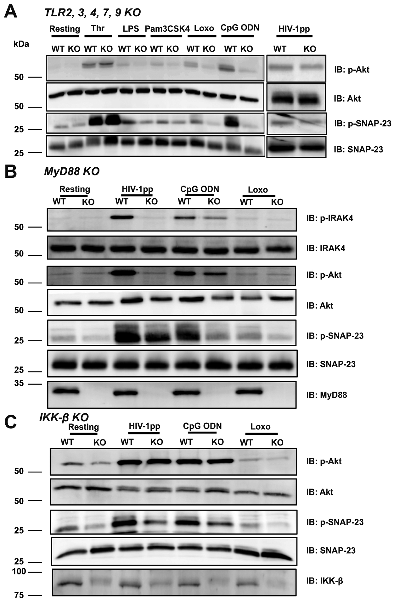
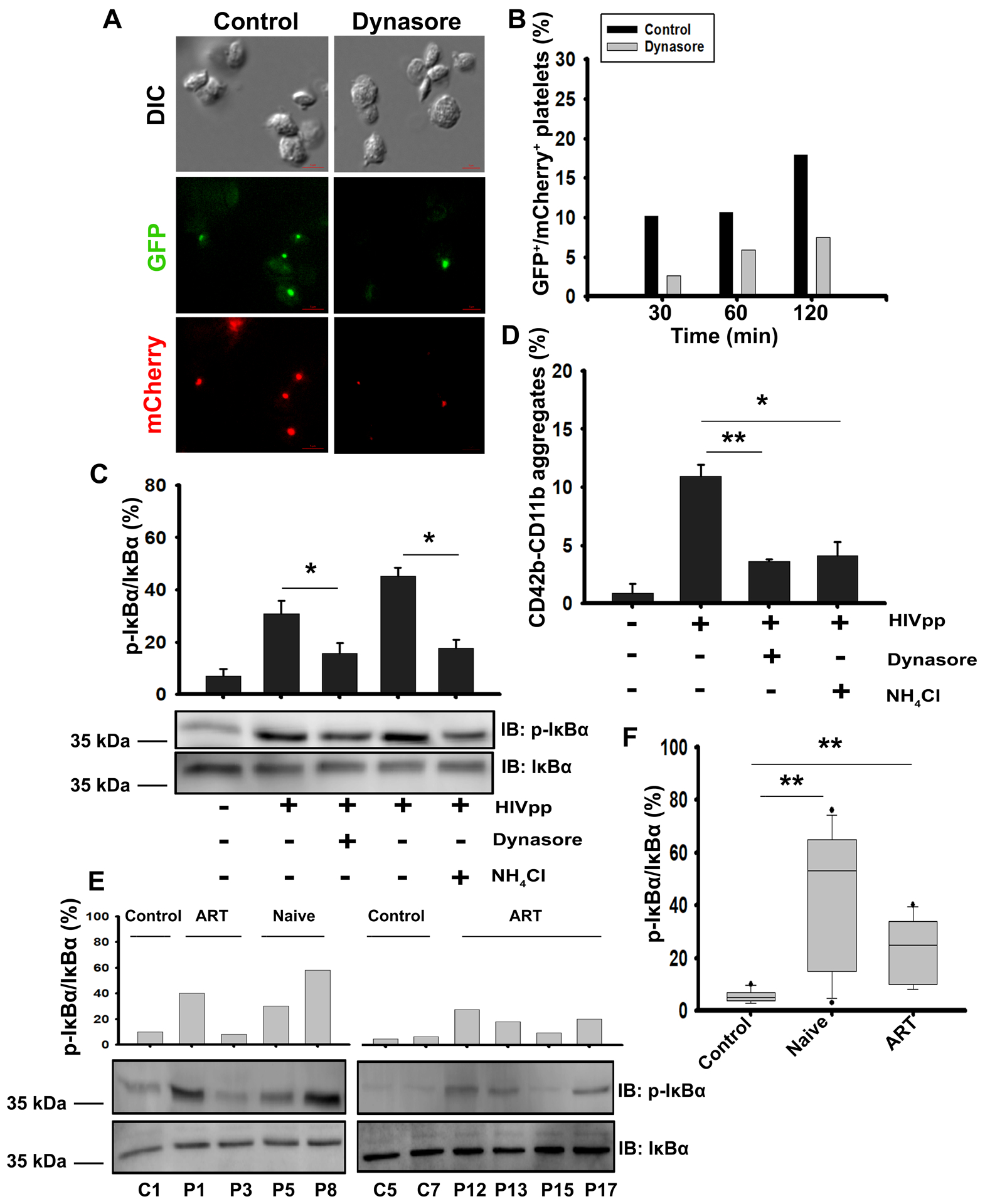
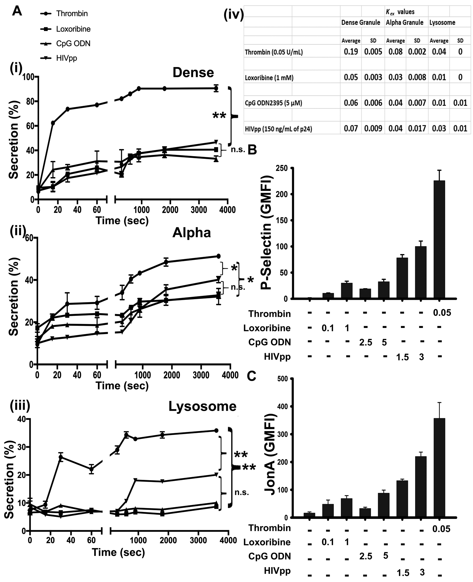
Objective: Thrombocytopenia is associated with many viral infections suggesting virions interact with and affect platelets. Consistently, viral particles are seen inside platelets, and platelet activation markers are detected in viremic patients. In this article, we sought mechanistic insights into these virion/platelet interactions by examining how platelets endocytose, traffic, and are activated by a model virion. Approach and Results: Using fluorescently tagged HIV-1 pseudovirions, 3-dimensional structured illumination microscopy, and transgenic mouse models, we probed the interactions between platelets and virions. Mouse platelets used known endocytic machinery, that is, dynamin, VAMP (vesicle-associated membrane protein)-3, and Arf6 (ADP-ribosylation factor 6), to take up and traffic HIV-1 pseudovirions. Endocytosed HIV-1 pseudovirions trafficked through early (Rab4+) and late endosomes (Rab7+), and then to an LC3+ (microtubule-associated protein 1A/1B-light chain 3) compartment. Incubation with virions induced IRAK4 (interleukin 1 receptor-associated kinase 4), Akt (protein kinase B), and IKK (IκB kinase) activation, granule secretion, and platelet-leukocyte aggregate formation. This activation required TLRs (Toll-like receptors) and MyD88 (myeloid differentiation primary response protein 88) but was less extensive and slower than activation with thrombin. In vivo, HIV-1 pseudovirions injection led to virion uptake and platelet activation, as measured by IKK activation, platelet-leukocyte aggregate formation, and mild thrombocytopenia. All were decreased in VAMP-3-/- and, megakaryocyte/platelet-specific, Arf6-/- mice. Similar platelet activation profiles (increased platelet-leukocyte aggregates, plasma platelet factor 4, and phospho-IκBα) were detected in newly diagnosed and antiretroviral therapy-controlled HIV-1+ patients.

Conclusions: Collectively, our data provide mechanistic insights into the cell biology of how platelets endocytose and process virions. We propose a mechanism by which platelets sample the circulation and respond to potential pathogens that they take up.

Keywords: HIV; Toll-like receptors; blood platelets; cardiovascular diseases; endocytosis; inflammation; viremia.


Conflict of interest statement

DISCLOSURES

The authors declare no competing financial interests.

Figures

Figure 1:
Figure 1:. HIV-1 pseudovirions (HIVpps) trafficked from early to late endosomes and into LC3+ compartments.
WT mouse platelets (1 × 10⁹/mL) were incubated with Gag-mCherry HIVpps at 37°C for 5 to 120 min and prepared for immunofluorescence as described in the Methods. Platelets were stained with (A-B) anti-Rab4 antibody, (C-D) anti-Rab7 antibody, and then with Alexa 680-conjugated secondary antibodies, after permeabilization (see Methods). The Alexa 680-Rab signal was faux-colored blue to improve contrast. Images were taken using the 3D-SIM module of Nikon Ti-E N-STORM/N-SIM super-resolution microscope. Scale bar is 1 μm. Pearson’s correlation coefficient was calculated using the NIS-Elements v3.2 N-SIM/STORM suite software to show overlap between: (B) Alexa 680-Rab4 and Gag-mCherry HIVpps, (D) Alexa 680-Rab7 and Gag-mCherry HIVpps, at the indicated time points. Data are representative of 2 independent experiments. (E-F) Platelets (1 × 10⁹/mL) from GFP-LC3 transgenic mice were incubated with Gag-mCherry HIVpps at 37°C for 5 to 120 min and imaged and analyzed as above. Data are representative of 3 independent experiments.
Figure 2:
Figure 2:. Endocytic machinery required for HIV-1 pseudovirion (HIVpps) uptake in platelets.
(A) Schematic representation of HIV-1 pseudovirions (HIVpps) showing envelope protein (Env) in blue, GFP-tagged-transmembrane domain (TM) of the Intercellular Adhesion Molecule-1 (ICAM-1) membrane protein in green and mCherry-tagged Gag protein in red. WT, Dynasore-pretreated (80 μM for 30 min at 37°C), VAMP-3−/− (KO) and Arf6−/− (KO) platelets (5 × 10⁷/mL) were incubated with GFP/Gag-mCherry HIVpps (150 ng/mL of p24) for 0–120 min (B) or with 0–450 ng/mL of p24 of GFP/Gag-mCherry HIVpps for 60 min (C) at 37°C. Reactions were stopped and fluorescence intensities were measured after addition of Trypan Blue. (D) WT, VAMP-3−/− (KO) and Arf6−/− (KO) platelets (8 × 10⁸/mL) were incubated with GFP/Gag-mCherry HIVpps (300 ng/mL of p24) at 37°C, for 0 to 6 hr, fixed with paraformaldehyde (PFA), treated with Trypan Blue, imaged, and counted. The percent of GFP/Gag-mCherry virion-containing platelets (counted as red and green puncta) was calculated. Age-matched VAMP-3−/− (KO) (n=7) and Arf6−/− (KO) mice (n=6), with their corresponding background-matched littermate (n=7 for VAMP-3 WT and n=6 for Arf6 WT) controls, were injected with HIVpps (i.v.) and platelets were harvested 24 hr post-injection and imaged. Quantification of the percent of GFP/Gag-mCherry virion-containing platelets in WT and VAMP-3−/− (KO) shown in (E) and between WT and Arf6−/− (KO) platelets shown in (F) and is shown. Statistical analyses were done by Student’s t-test where *** represents p≤0.001, ** represents p≤0.01 using the SigmaPlot software (v13.0). Data are representative of at least 2 independent experiments.
Figure 3:
Figure 3:. Endocytosis of HIV-1 pseudovirions (HIVpps) activated platelets in vivo and ex vivo.
Mice (20 weeks old; both male and female) WT (n=7), VAMP-3−/− (KO) (n=7) and Arf6−/− (KO) (n=6)) were either injected (i.v.) with HIVpps or saline (Sal). (A) Blood was drawn 24 hr post-injection to determine platelets counts. (Aii) PLAs in blood were measured by FACS using FITC-conjugated anti-CD41/61 and APC-anti-Ly6G antibodies and analyzed using FlowJo software (v7.6.5). (B) Platelets were isolated (ex vivo) from age-matched WT, VAMP-3−/− and Arf6−/− mice and stimulated with HIVpps for 30 min at 37°C, platelet lysates were prepared, and western blotting was performed using the indicated antibodies. (C) Volumetric quantification of (i) phospho-IRAK4, (ii) phospho-Akt, and (iii) phospho-SNAP-23 as a percent of total protein were calculated. (E) Platelets from WT, VAMP-3−/− (KO), and Arf6−/− (KO) mice were either kept resting or stimulated with HIVpps for 30 min. Samples were then incubated with FITC-anti-CD62 antibody to measure P-Selectin exposure by FACS. Data shown are representative of 3 independent experiments (mean ± SEM). Statistical analyses were done by paired Student’s t-test where ** represents p≤0.01, and * represents p≤0.05 using the SigmaPlot software (v13.0).
Figure 4:
Figure 4:. Responses to Loxoribine, CpG ODN2395, and HIV-1 pseudovirions (HIVpps) required TLR, MyD88, and IKKβ.
Platelets from WT and (A) TLR-2,3,4,7,9 quintuple KO mice were either kept resting or stimulated with thrombin (0.1 U/mL), LPS (100 ng/mL), Pam3CSK4 (10 μg/mL), Loxoribine (1 mM), CpG ODN2395 (5 μM), HIVpps (150 ng/mL of p24) for 60 min at 37°C. Platelets from WT or (B) MyD88−/− (KO) mice or (C) IKKβ−/− (KO) were either kept resting or stimulated with Loxoribine, CpG ODN2395, HIVpps for 60 min at 37°C and lysates were prepared. Western blotting was performed using the indicated antibodies. Data are representative of at least 2 independent experiments for (A) and (B) and 3 experiments for (C).
Figure 5:
Figure 5:. Endocytosis of HIV-1 pseudovirions (HIVpps) by human platelets is Dynasore-sensitive and mediates platelet activation similar to that seen in HIV-1+ patients.
(A) Images of human platelets, either vehicle-treated (control) or Dynasore-treated (80 μM) then incubated with GFP/Gag-mCherry HIVpps, for 60 min. Scale bars are 2 μm. (B) Quantification of time-dependent uptake of GFP/Gag-mCherry HIVpps (counted as red and green puncta) into platelets. (C) Human platelets were incubated with HIVpps for 60 min either in the presence or absence of Dynasore (80 μM) or NH4Cl (20 mM) and lysates were probed by western blotting with anti-phospho-IκBα and anti-IκBα antibodies. (D) Blood was pre-incubated with either Dynasore (80 μM) or NH4Cl (20 mM), stimulated with HIVpps for 30 min, and platelet-leukocyte aggregates were detected by FACS with anti-CD42b and anti-CD11b fluorescent antibodies. Data were analyzed using FlowJo (v.7.6.5). (E) Representative western blots of platelet lysates from healthy donors (C1, C5), newly-diagnosed (HIV-1+) patients (naive, P5, P8) or from patients on anti-retroviral therapy drugs (ART-treated; P1, P3, P12, P13, P15, P17) were probed by western blot using anti-phospho-IκBα (Ser32), anti-IκBα antibodies. (F) The percent of phospho-IκBα over total IκBα were determined for platelet lysates from 10 healthy control samples, 11 newly diagnosed naive (HIV-1+) patients (naive-untreated), and 11 patients on anti-retroviral therapeutics (ART-treated). Statistical significance * represented by p ≤0.05, ** represented by p ≤0.01 was determined by Mann-Whitney U test using SigmaPlot software (v13.0). Data are representative of 3 independent experiments (mean ± SEM).
Figure 6:
Figure 6:. Platelets responded differently to “hemostatic activation” vs. “immuno-activation”.
(A) WT Platelets (2.5 × 10⁸/mL) were stimulated with Loxoribine (1 mM), CpG ODN2395 (5 μM), HIVpps (150 ng/mL of p24) or thrombin (0.05 U/mL) for the indicated times and percent secretion was measured (i) [3H]-Serotonin from dense granules, (ii) PF4 from α-granules and (iii) β-hexosaminidase from lysosomes. (iv) Release rates (Kex) were calculated for each type of secretion process and plotted as mean ± SD. WT platelets were stimulated with Loxoribine (0.1 mM and 1 mM), CpG ODN2395 (2.5 μM and 5 μM), HIVpps (1.5 × 10⁵ pg/mL and 3 × 10⁵ pg/mL) and thrombin (0.05 U/mL) for 30 min and incubated with (B) anti-P-Selectin or (C) anti-JonA antibody and samples were analyzed by FACS. Geometric mean fluorescence intensity (GMFI) was measured for each condition and plotted. Data shown are representative of 3 independent experiments (mean ± SEM). Statistical significance * represented by p ≤0.05, ** represented by p ≤0.01, was calculated by one-way ANOVA using GraphPad Prism 8.0 software.
Figure 7:
Figure 7:. Endocytosis of HIVpps activates platelets.
Schematic depicting how pathogenic ligands such as HIV-1, bacterial products, etc., are endocytosed and trafficked to acidic endosomal compartments where proteolytic degradation occurs to release TLR ligands. This activates downstream signaling molecules that lead to IRAK4, Akt, and SNAP-23 phosphorylation and drives P-Selectin exposure to form platelet-leukocyte aggregates.

Comment in

  • Platelets and Immunity: Going Viral.
    Koupenova M, Freedman JE. Koupenova M, et al. Arterioscler Thromb Vasc Biol. 2020 Jul;40(7):1605-1607. doi: 10.1161/ATVBAHA.120.314620. Epub 2020 Jun 24. Arterioscler Thromb Vasc Biol. 2020. PMID: 32579477 Free PMC article. No abstract available.

References

    1. Flaujac C, Boukour S, Cramer-Borde E. Platelets and viruses: An ambivalent relationship. Cell Mol Life Sci. 2010;67:545–556 - PMC - PubMed
    1. Chaipan C, Soilleux EJ, Simpson P, et al. Dc-sign and clec-2 mediate human immunodeficiency virus type 1 capture by platelets. Journal of virology. 2006;80:8951–8960 - PMC - PubMed
    1. Cloutier N, Allaeys I, Marcoux G, et al. Platelets release pathogenic serotonin and return to circulation after immune complex-mediated sequestration. Proceedings of the National Academy of Sciences of the United States of America. 2018;115:E1550–E1559 - PMC - PubMed
    1. Koupenova M, Vitseva O, MacKay CR, Beaulieu LM, Benjamin EJ, Mick E, Kurt-Jones EA, Ravid K, Freedman JE. Platelet-tlr7 mediates host survival and platelet count during viral infection in the absence of platelet-dependent thrombosis. Blood. 2014;124:791–802 - PMC - PubMed
    1. Zucker-Franklin D, Seremetis S, Zheng ZY. Internalization of human immunodeficiency virus type i and other retroviruses by megakaryocytes and platelets. Blood. 1990;75:1920–1923 - PubMed

Publication types

MeSH terms