Comparative Study
. 2020 Jul;40(7):1738-1747.
doi: 10.1161/ATVBAHA.120.314459. Epub 2020 May 21.

Differential Roles of Endothelial Cell-Derived and Smooth Muscle Cell-Derived Fibronectin Containing Extra Domain A in Early and Late Atherosclerosis

Affiliations
Comparative Study

Differential Roles of Endothelial Cell-Derived and Smooth Muscle Cell-Derived Fibronectin Containing Extra Domain A in Early and Late Atherosclerosis

Prakash Doddapattar et al. Arterioscler Thromb Vasc Biol. 2020 Jul.

Abstract

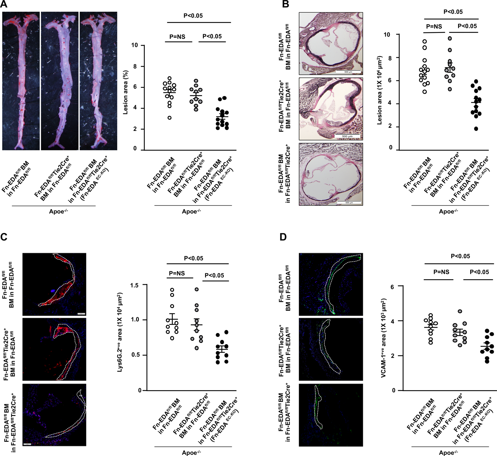
Objective: The extracellular matrix of atherosclerotic arteries contains abundant deposits of cellular Fn-EDA (fibronectin containing extra domain A), suggesting a functional role in the pathophysiology of atherosclerosis. Fn-EDA is synthesized by several cell types, including endothelial cells (ECs) and smooth muscle cells (SMCs), which are known to contribute to different stages of atherosclerosis. Although previous studies using global Fn-EDA-deficient mice have demonstrated that Fn-EDA is proatherogenic, the cell-specific role of EC versus SMC-derived-Fn-EDA in atherosclerosis has not been investigated yet. Approach and Results: To determine the relative contribution of different pools of Fn-EDA in atherosclerosis, we generated mutant strains lacking Fn-EDA in the ECs (Fn-EDAEC-KO) or smooth muscle cells (Fn-EDASMC-KO) on apolipoprotein E-deficient (Apoe-/-) background. The extent of atherosclerotic lesion progression was evaluated in whole aortae, and cross-sections of the aortic sinus in male and female mice fed a high-fat Western diet for either 4 weeks (early atherosclerosis) or 14 weeks (late atherosclerosis). Irrespective of sex, Fn-EDAEC-KO, but not Fn-EDASMC-KO mice, exhibited significantly reduced early atherogenesis concomitant with decrease in inflammatory cells (neutrophil and macrophage) and VCAM-1 (vascular cell adhesion molecule-1) expression levels within the plaques. In late atherosclerosis model, irrespective of sex, Fn-EDASMC-KO mice exhibited significantly reduced atherogenesis, but not Fn-EDAEC-KO mice, that was concomitant with decreased macrophage content within plaques. Lesional SMCs, collagen content, and plasma inflammatory cytokines (TNF-α [tumor necrosis factor-α] and IL-1β [interleukin-1β]), total cholesterol, and triglyceride levels were comparable among groups.

Conclusions: EC-derived Fn-EDA contributes to early atherosclerosis, whereas SMC-derived Fn-EDA contributes to late atherosclerosis.

Keywords: atherosclerosis; cytokines; endothelial cells; fibronectin; macrophages.


Figures

Figure 1.
Figure 1.. Fn-EDAEC-KO mice exhibit reduced early atherosclerosis.
All the mice were females and fed a high-fat Western diet for 4 weeks. A, Representative photomicrographs and quantification of Oil red O -stained en face lesion area in the whole aortae. B, Representative photomicrographs of VerHoeffs/Van Geison-staining and quantification of cross-sectional lesion area in aortic sinuses. C, Representative photomicrographs and quantification of neutrophil-stained (Lys6G.2-positive cells) in aortic sinuses. Scale bar = 100 μm D, Representative photomicrographs and quantification of vascular cell adhesion molecule-stained (VCAM1-positive cells) area in aortic sinuses. Scale bar = 100 μm. Values are expressed as mean ± SEM. Each dot represents a single mouse (n=10–13/group). Statistical analysis: One way ANOVA followed by Tukeys’s multiple comparisons test.
Figure 2.
Figure 2.. Endothelial-derived Fn-EDA does not contribute to late atherosclerosis.
All the mice were females and fed a high-fat Western diet for 14 weeks. A, Representative photomicrographs and quantification of Oil red O -stained en face lesion area in the whole aortae. B, Representative photomicrographs of VerHoeffs/Van Geison-staining and quantification of cross-sectional lesion area in aortic sinuses. Scale bar = 500 μm C, Representative photomicrographs and quantification of macrophage (mac3-positive cells) in aortic sinuses. Scale bar = 500 μm D, Representative photomicrographs and quantification of smooth muscle cell-stained (⍺-SMA-positive) area in aortic sinuses. Scale bar = 100 μm Values are expressed as mean ± SEM. Each dot represents a single mouse (n=10–13/group). Statistical analysis: One way ANOVA followed by Tukeys’s multiple comparisons test.
Figure 3.
Figure 3.. Smooth muscle cell-derived Fn-EDA does not contribute to early atherosclerosis.
All the mice were females and fed a high-fat Western diet for 4 weeks. A, Representative photomicrographs and quantification of Oil red O-stained en face lesion area in the whole aortae. B, Representative photomicrographs of VerHoeffs/Van Geison-staining and quantification of cross-sectional lesion area in aortic sinuses. Scale bar = 500 μm. Lower panels. C, Representative photomicrographs and quantification of neutrophil-stained (Lys6G.2-positive cells) in aortic sinuses. Scale bar = 100 μm. D, Representative photomicrographs and quantification of vascular cell adhesion molecule-stained (VCAM1-positive cells) in aortic sinuses. Scale bar = 100 μm. Each dot represents a single mouse (n=8–14/group). Statistical analysis: unpaired student t-test.
Figure 4.
Figure 4.. Fn-EDASMC-KO mice exhibit reduced late atherosclerosis.
All the mice were females and fed a high-fat Western diet for 14 weeks. A, Representative photomicrographs and quantification of Oil red O-stained en face lesion area in the whole aortae. B, Representative photomicrographs of VerHoeffs/Van Geison-staining and quantification of cross-sectional lesion area in aortic sinuses. Scale bar = 500 μm. C, Representative photomicrographs and quantification of macrophage (mac3-positive cells) area in aortic sinuses. Scale bar = 500 μm. D, Representative photomicrographs and quantification of smooth muscle cell-stained (⍺-SMA-positive) area in aortic sinuses. Scale bar = 100 μm. Each dot represents a single mouse (n=8–14/group). Statistical analysis: unpaired student t-test.

References

    1. Raines EW. The extracellular matrix can regulate vascular cell migration, proliferation, and survival: Relationships to vascular disease. Int J Exp Pathol. 2000;81:173–182 - PMC - PubMed
    1. Shekhonin BV, Domogatsky SP, Idelson GL, Koteliansky VE, Rukosuev VS. Relative distribution of fibronectin and type i, iii, iv, v collagens in normal and atherosclerotic intima of human arteries. Atherosclerosis. 1987;67:9–16 - PubMed
    1. Pedretti M, Rancic Z, Soltermann A, Herzog BA, Schliemann C, Lachat M, Neri D, Kaufmann PA. Comparative immunohistochemical staining of atherosclerotic plaques using f16, f8 and l19: Three clinical-grade fully human antibodies. Atherosclerosis. 2010;208:382–389 - PubMed
    1. Hynes RO. The evolution of metazoan extracellular matrix. The Journal of cell biology. 2012;196:671–679 - PMC - PubMed
    1. Dubin D, Peters JH, Brown LF, Logan B, Kent KC, Berse B, Berven S, Cercek B, Sharifi BG, Pratt RE, et al. Balloon catheterization induced arterial expression of embryonic fibronectins. Arterioscler Thromb Vasc Biol. 1995;15:1958–1967 - PubMed

Publication types

MeSH terms