. 2020 May 20;18(1):208.
doi: 10.1186/s12967-020-02377-x.

Development and optimization of orthotopic liver metastasis xenograft mouse models in uveal melanoma

Affiliations

Development and optimization of orthotopic liver metastasis xenograft mouse models in uveal melanoma

Takahito Sugase et al. J Transl Med. .

Abstract

Background: Patients with metastatic uveal melanoma (MUM) in the liver usually die within 1 year. The development of new treatments for MUM has been limited by the lack of diverse MUM cell lines and appropriate animal models. We previously reported that orthotopic xenograft mouse models established by direct injection of MUM cells into the liver were useful for the analysis associated with tumor microenvironment in the liver. However, considering that patients with UM metastasize to the liver hematogenously, direct liver injection model might not be suitable for investigation on various mechanisms of liver metastasis. Here, we aim to establish new orthotopic xenograft models via hematogenous dissemination of tumor cells to the liver, and to compare their characteristics with the hepatic injection model. We also determine if hepatic tumors could be effectively monitored with non-invasive live imaging.

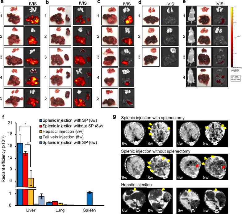
Methods: tdtTomate-labeled, patient-derived MUM cells were injected into the liver, spleen or tail vein of immunodeficient NSG mice. Tumor growth was serially assessed with In Vivo Imaging System (IVIS) images once every week. Established hepatic tumors were evaluated with CT scan and then analyzed histologically.

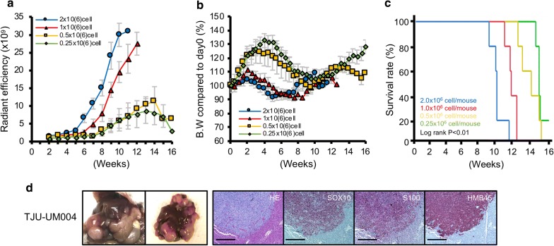
Results: We found that splenic injection could consistently establish hepatic tumors. Non-invasive imaging showed that the splenic injection model had more consistent and stronger fluorescent intensity compared to the hepatic injection model. There were no significant differences in tumor growth between splenic injection with splenectomy and without splenectomy. The splenic injection established hepatic tumors diffusely throughout the liver, while the hepatic injection of tumor cells established a single localized tumor. Long-term monitoring of tumor development showed that tumor growth, tumor distribution in the liver, and overall survival depended on the number of tumor cells injected to the spleen.

Conclusion: We established a new orthotopic hepatic metastatic xenograft mouse model by splenic injection of MUM cells. The growth of orthotopic hepatic tumors could be monitored with non-invasive IVIS imaging. Moreover, we evaluated the therapeutic effect of a MEK inhibitor by using this model. Our findings suggest that our new orthotopic liver metastatic mouse model may be useful for preclinical drug screening experiments and for the analysis of liver metastasis mechanisms.

Keywords: Liver; Liver metastasis; Orthotopic xenograft model; Spleen; Uveal melanoma.


Conflict of interest statement

The authors declare that they have no competing interests.

Figures

Fig. 1
Fig. 1
a TJU-UM001-tdTomato cells express orange-red fluorescence (× 100). NSG mouse (6- to 8-weeks old) were injected with tumor cells (1.0x10⁶ cells/mouse) into the spleen, the liver, or the tail vein. Surgical procedure b splenic injection, c splenectomy, d tail vein injection, and e hepatic injection. f Monitoring schedule
Fig. 2
Fig. 2
Each mouse model was taken IVIS image every week. IVIS images at 2, 6, 7 and 8 weeks after injection were shown: a splenic injection with splenectomy (SP), b splenic injection without SP, c hepatic injection, and d tail vein injection. e Weekly measurement of fluorescent intensity. Data were shown with the mean radiant efficiency ± SEMs of 5 mice in each cohort. f Shift of mouse body weights. Values were shown with the mean ± SEMs
Fig. 3
Fig. 3
Ex vivo image and IVIS image of removed liver, lung, and spleen at 8 weeks after injection: a splenic injection with splenectomy, b splenic injection without splenectomy, c hepatic injection, and d tail vein injection. e Live IVIS images (left) and IVIS images of extracted liver and lung (right) at 6 weeks after splenic injection with splenectomy. f Fluorescent intensity of liver, lung and spleen in each mouse model. Data were the mean radiant efficiency ± SEMs of 5 mice in each mouse model. g CT scan images of tumors in 6 weeks and 8 weeks after injections. Yellow arrows: tumor in the liver. Red arrows: tumor in the spleen
Fig. 4
Fig. 4
Immunohistochemical analysis of hepatic tumor. a Comparison of SOX10, S100 and HMB45 expression in the liver of splenic injection model and hepatic injection model. Liver tissues in each mouse model at 6 and 8 weeks after injection were stained with SOX10 (100x). Liver 6 weeks or 8 weeks after injection of tumor cells; b splenic injection with splenectomy, c splenic injection without splenectomy, d hepatic injection and e tail vein injection (100 × and 200x). f Lung in each model were stained with SOX10 (100x). Scale bar = 500 μm
Fig. 5
Fig. 5
Long term monitoring of hepatic tumor with IVIS image was performed in splenic injection with splenectomy model. NSG mice were injected UM001-tdTomato cells (2.0, 1.0, 0.5, and 0.25x10⁶ cells/mouse, 5 mice of each group) into spleen, then spleen was removed 15 min after injection. IVIS image has been taken every week and mice will be euthanized using C02 exposure followed by cervical dislocation if they show 20% weight loss or severe weakness. a 2.0x10⁶ cells/mouse, b 1.0x10⁶ cells/mouse, c 0.5x10⁶ cells/mouse and d 0.25x10⁶ cells/mouse. The fluorescent intensity between the anterior portion and the posterior portion of the liver in the e 1.0 and f 0.25x10⁶ cells injected mice were compared
Fig. 6
Fig. 6
a Fluorescent intensity in the splenic injection with splenectomy was monitored every week. Data were the mean radiant efficiency ± SEMs of 5 mice in each mouse model. b Mice body weights were measured twice per week. Values shown represent the mean ± SEMs. c Survival curve based on the number of injected cells (5 mice in each mouse model). Survivals were compared using the log rank test. d NSG mouse was injected with TJU-UM004 cells (1.0x10⁶ cells/mouse)into the spleen and sacrificed 9 weeks after injection. HE staining and immunohistochemistry of SOX10, S100, and HMB45 were performed (100x). Scale bar = 500 μm
Fig. 7
Fig. 7
6 weeks after UM001tdTomato cell injection into the spleen, mice were treated with control (dilute solution) or MEK inhibitor, trametinib, (1.0 mg/kg) intraperitoneally once a day for 3 weeks. a IVIS images in each mouse were shown. b The measurement of fluorescent intensity. Data were shown with the mean radiant efficiency ± SEM in each cohort. c IVIS image of removed liver at 21 days after treatment. d Fluorescent intensity of liver in each cohort. Data were the mean radiant efficiency ± SEMs in each cohort. e Mice body weight were measured twice per week in each cohort. Values were shown with the mean ± SEMs

References

    1. Amaro A, Gangemi R, Piaggio F, Angelini G, Barisione G, Ferrini S, Pfeffer U. The biology of uveal melanoma. Cancer Metastasis Rev. 2017;36:109–140. - PMC - PubMed
    1. Chattopadhyay C, Kim DW, Gombos DS, Oba J, Qin Y, Williams MD, Esmaeli B, Grimm EA, Wargo JA, Woodman SE, Patel SP. Uveal melanoma: from diagnosis to treatment and the science in between. Cancer. 2016;122:2299–2312. - PMC - PubMed
    1. Krantz BA, Dave N, Komatsubara KM, Marr BP, Carvajal RD. Uveal melanoma: epidemiology, etiology, and treatment of primary disease. Clin Ophthalmol. 2017;11:279–289. - PMC - PubMed
    1. Gragoudas ES, Egan KM, Seddon JM, Glynn RJ, Walsh SM, Finn SM, Munzenrider JE, Spar MD. Survival of patients with metastases from uveal melanoma. Ophthalmology. 1991;98:383–389. - PubMed
    1. Singh AD, Borden EC. Metastatic uveal melanoma. Ophthalmol Clin North Am. 2005;18:143–150. - PubMed

Publication types