Review
. 2020 Jul;43(7):493-504.
doi: 10.1016/j.tins.2020.04.007. Epub 2020 May 17.

Resurrecting the Mysteries of Big Tau

Affiliations
Review

Resurrecting the Mysteries of Big Tau

Itzhak Fischer et al. Trends Neurosci. 2020 Jul.

Abstract

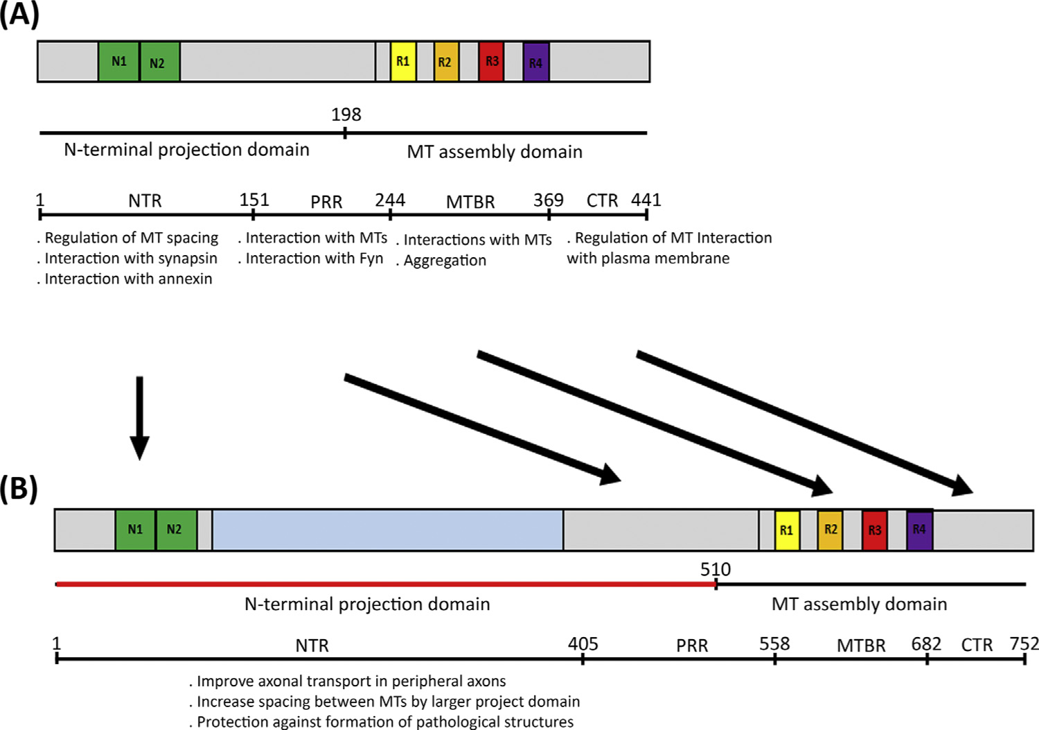
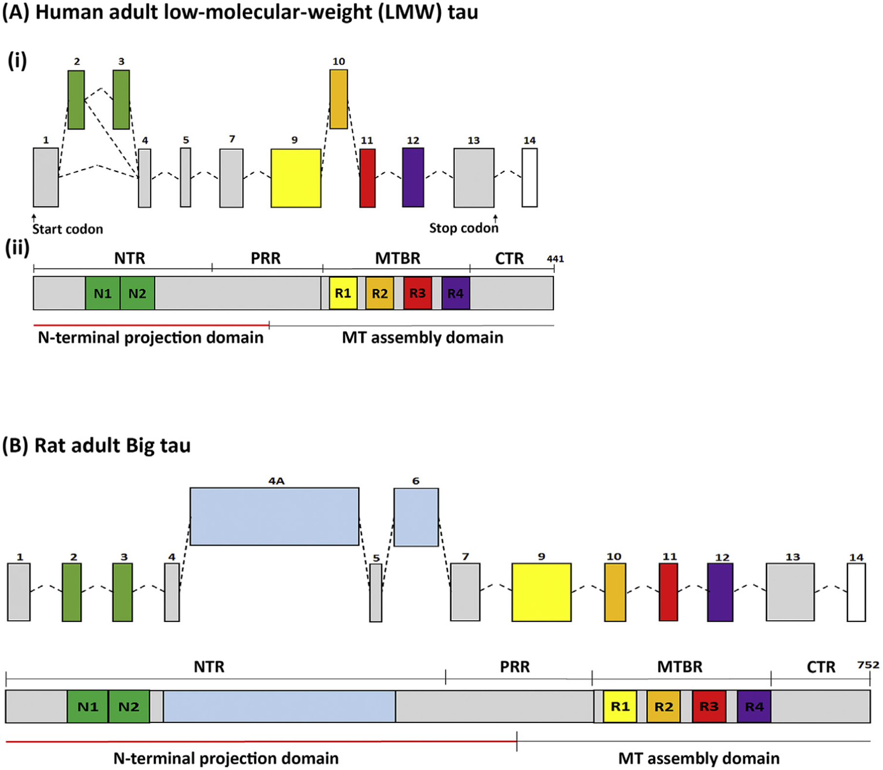
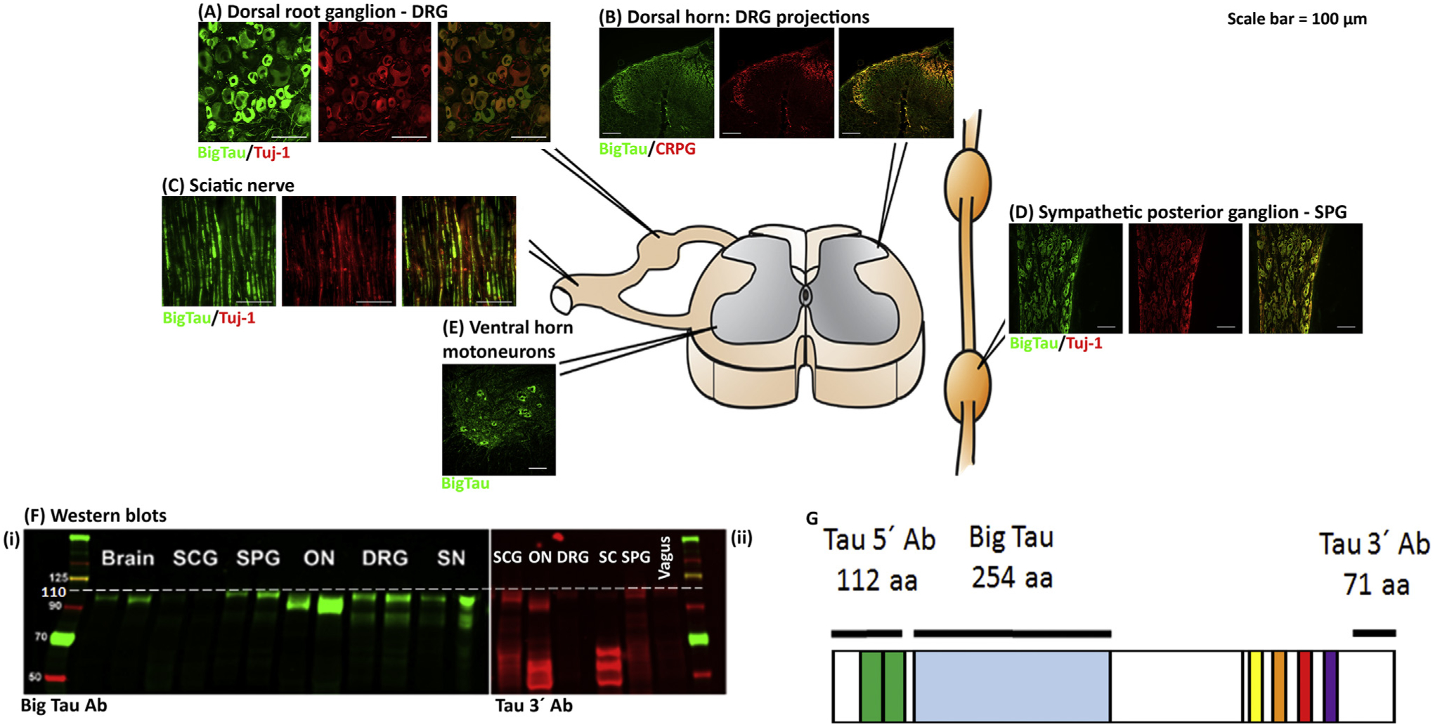
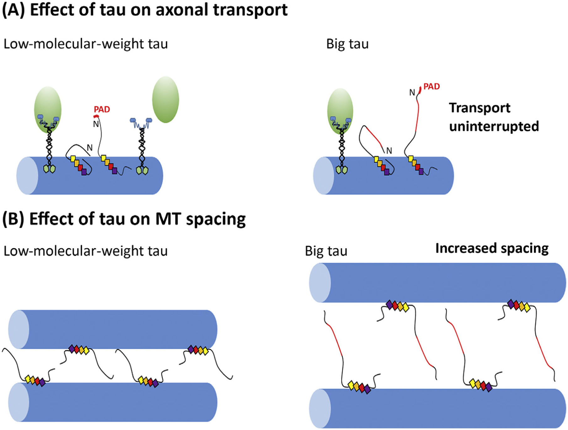
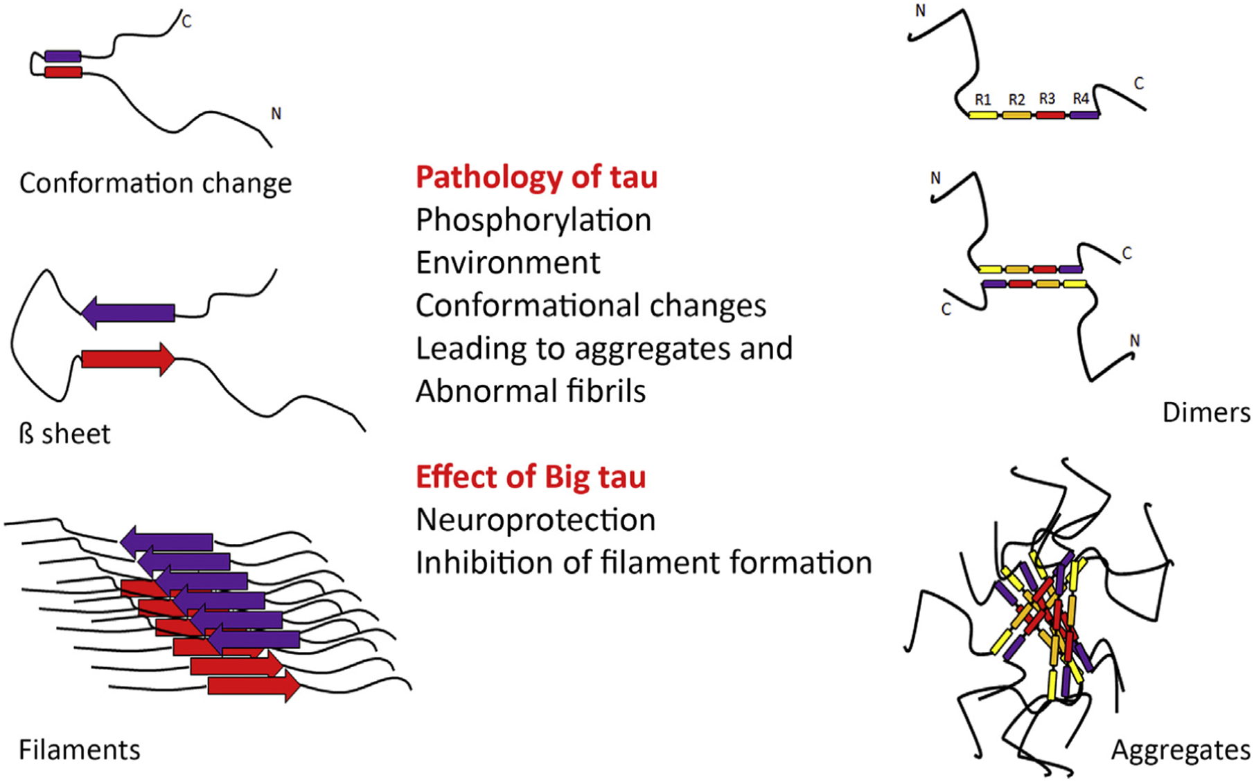
Tau, a microtubule-associated protein that modifies the dynamic properties and organization of microtubules in neurons and affects axonal transport, shows remarkable heterogeneity, with multiple isoforms (45-65 kDa) generated by alternative splicing. A high-molecular-weight (HMW) isoform (110 kDa) that contains an additional large exon termed 4a was discovered more than 25 years ago. This isoform, called Big tau, is expressed mainly in the adult peripheral nervous system (PNS), but also in adult neurons of the central nervous system (CNS) that extend processes into the periphery. Surprisingly little has been learned about Big tau since its initial characterization, leaving a significant gap in knowledge about how the dramatic switch to Big tau affects the properties of neurons in the context of development, disease, or injury. Here we review what was learned about the structure and distribution of Big tau in those earlier studies, and add contemporary insights to resurrect interest in the mysteries of Big tau and thereby set a path for future studies.

Keywords: Alzheimer's disease; Big tau; axon; microtubule; neuron; peripheral nervous system; tau; tauopathy.


Figures

Figure 3.
Figure 3.
The figure shows a partial list of known functions ascribed to different domains of the tau molecule: N-terminal region (NTR), proline-rich region (PRR), microtubule-binding region (MTBR), and C-terminal region (CTR). These same domains are present in low-molecular-weight (LMW) tau (A) and Big tau (B). The knowledge of these various other domains underscores the unknown function of the large 4a insert of the Big tau molecule. Speculated in (B) are potential functional modifications in the molecule due to the unique presence in Big tau of the large 4a insert in the projection domain. Abbreviation: MT, microtubule.
Figure 1.
Figure 1.. Structure of (A) Low-Molecular-Weight (LMW) Tau and (B) Big Tau.
(A) (i) The transcriptional organization of LMW human tau with exons 2/3 (N1 and N2 at the N-terminal) and exon 10 (R2 at the microtubule binding domain) as alternative splicing exons, which are developmentally regulated. LMW tau lacks exons 6 and 8. These transcriptional events give rise to 6 LMW isoforms. (ii) The largest 4R isoforms, with both N1/N2 comprising 441 amino acids, show on Western blots with apparent MW of 48–67 kDa (the true MW being 37–46 kDa). The tau protein comprises the microtubule (MT)-assembly domain, which includes the C-terminal region and the four microtubule-binding repeats (R1–4) and the N-terminal projection domain, including the N-terminal N1 and N2 exons and part of the proline-rich region. (B) Big tau (structure shown for rat) is an isoform that includes exons 4a and alternatively spliced exon 6, expressing proteins of apparent MW of 110 kDa. Big tau has a similar microtubule-assembly domain to LMW tau, but has an N-terminal projection domain that is 510 aa, which is more than double the size relative to the projection domain of LMW tau. Abbreviations: CTR, C-terminal region; MTBR, MT-binding region; NTR, N-terminal region; PRR, proline-rich region.
Figure 2.
Figure 2.. Distribution of Big Tau in the Adult Nervous System.
The images show the distribution of Big tau in the adult rat nervous system, consistent with published data [15,16], assessed using antibodies specific to Big tau. Staining of dorsal root ganglia (DRGs) (A) with the Big tau antibody compared with Tuj-1 (neuronal marker) shows expression of Big tau in small and medium-sized neurons, but not the large ones. (B) and (D) show the expression of Big tau in the spinal cord, with intense staining of motor neurons and their dendrites (D) as well as the dorsal horn (B), where the calcitonin gene-related peptide (CGRP)-positive central processes of DRG axons project and terminate. (C) Big tau expression in sciatic nerve colocalized with Tuj. Analysis of the sympathetic chain of ganglion cells (E) shows an example of the superior posterior ganglion (SPG), where all neurons appear to express Big tau. Not shown but discussed in the main text is the expression in brain, where all cranial nerves express Big tau with the exception of olfactory, vestibular, and spiral ganglia [15]. (F) Western blot results of different tissues from peripheral and central nervous systems (PNS and CNS) using an antibody specific to Big tau (i) as well as tau 3′ antibody (ii). (G) Specificity of Big tau antibodies [prepared against the entire 4a exon of 254 amino acids (aa) of rat tau], which confirms the expression of Big tau using Western blot (Fi) in SPG, optic nerve (ON), DRGs, and sciatic nerve (SN). The pattern of Big tau expression was verified with the tau 3′ antibodies (prepared against 71 amino acids at the C terminus), which recognize all tau isoforms (Fii). Images courtesy of Ying Jin and Theresa Connors.
Figure 4.
Figure 4.. Putative Models of Tau Interactions with Microtubules (MT).
(A) A putative model of tau structural changes that affect axonal transport, in this case the interaction of kinesin with the hidden and exposed phosphatase-activating domain (PAD) of tau [40]. (B) A putative model of the effects of tau on microtubule spacing and potential differences in the distances in the case of Big tau compared with low-molecular-weight (LMW) tau.
Figure 5.
Figure 5.. Aspects of the Pathology of Low-Molecular-Weight (LMW) Tau that May be Partially or Completely Ameliorated by the Presence of the 4a Insert in Big Tau.
The figure depicts putative models of abnormal fibrils and aggregates generated by conformational changes and misfolding of tau. On the left is shown the conformational transition to β sheets and filaments, and on the right the transition to dimers and aggregates, which are known to be affected by the phosphorylation of tau and environmental factors. The schematic introduces the idea that Big tau may not misfold into pathological filaments or toxic aggregates because of the large insert that appears to be a ‘phospho-dead island’ in a phospho-rich molecule.

References

    1. Tapia-Rojas C et al. (2019) It’s all about tau. Prog. Neurobiol 175, 54–76 - PMC - PubMed
    1. Kellogg EH et al. (2018) Near-atomic model of microtubule-tau interactions. Science 360, 1242–1246 - PMC - PubMed
    1. Baas PW and Qiang L (2019) Tau: it’s not what you think. Trends Cell Biol 29, 452–461 - PMC - PubMed
    1. Kadavath H et al. (2015) Tau stabilizes microtubules by binding at the interface between tubulin heterodimers. Proc. Natl. Acad. Sci. U. S. A 112, 7501–7506 - PMC - PubMed
    1. Montejo de Garcini E et al. (1994) Overexpression of tau protein in COS-1 cells results in the stabilization of centrosome-independent microtubules and extension of cytoplasmic processes. Mol. Cell. Biochem 130, 187–196 - PubMed

Publication types