. 2020 Sep;26(9):1247-1256.
doi: 10.1261/rna.076042.120. Epub 2020 May 20.

Pseudouridine-mediated stop codon readthrough in S. cerevisiae is sequence context-independent

Affiliations

Pseudouridine-mediated stop codon readthrough in S. cerevisiae is sequence context-independent

Hironori Adachi et al. RNA. 2020 Sep.

Abstract

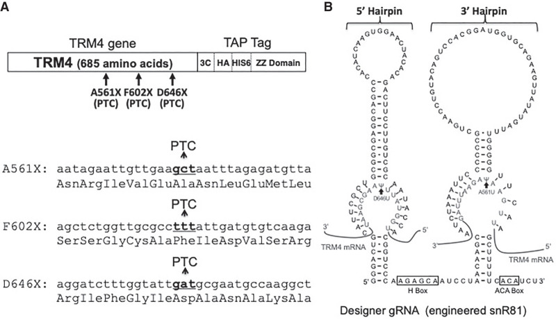
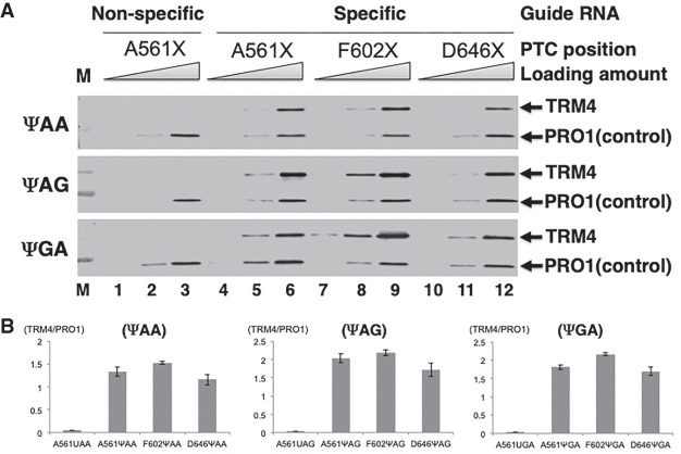
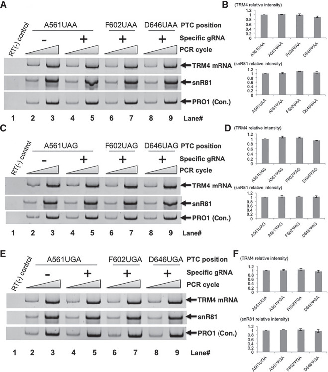
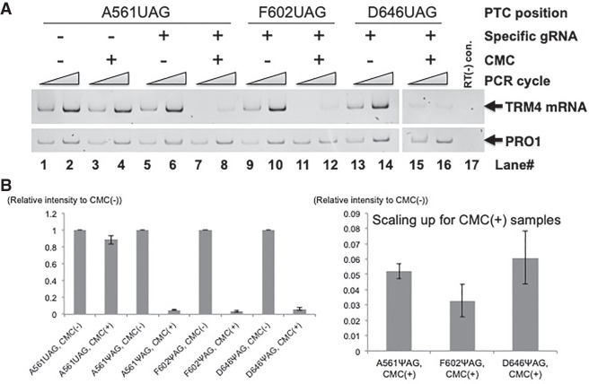
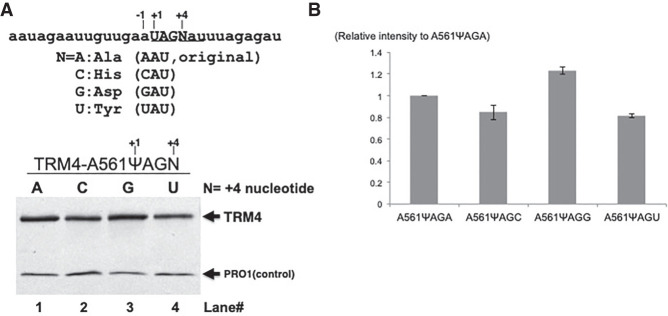
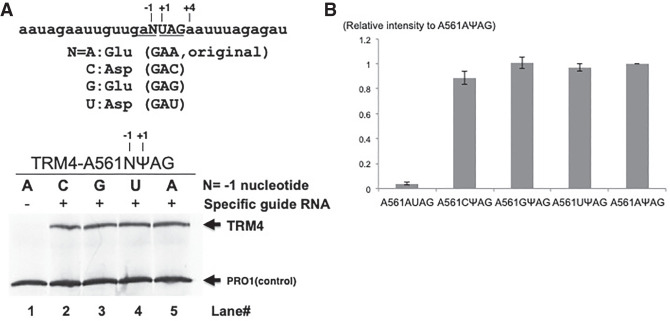
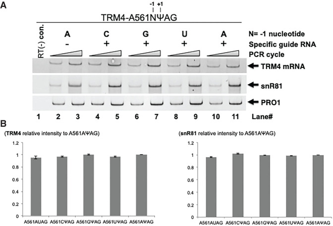
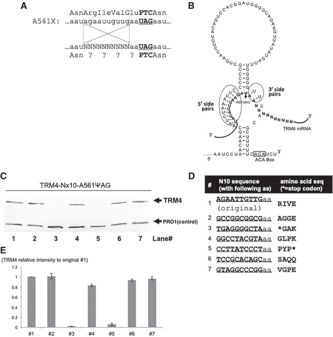
We have previously shown that when the uridine of a stop codon (UAA, UAG, or UGA) is pseudouridylated, the ribosome reads through the modified stop codon. However, it is not clear as to whether or not the pseudouridine (Ψ)-mediated readthrough is dependent on the sequence context of mRNA. Here, we use several different approaches and the yeast system to address this question. We show that when a stop codon (premature termination codon, PTC) is introduced into the coding region of a reporter mRNA at several different positions (with different sequence contexts) and pseudouridylated, we detect similar levels of readthrough. Using mutational and selection/screen analyses, we also show that the upstream sequence (relative to PTC) as well as the nucleotides surrounding the PTC (upstream and downstream) play a minimal role (if at all) in Ψ-mediated ribosome readthrough. Interestingly, we detect no suppression of NMD (nonsense-mediated mRNA decay) by targeted PTC pseudouridylation in the yeast system. Our results indicate that Ψ-mediated nonsense suppression occurs at the translational level, and that the suppression is sequence context-independent, unlike some previously characterized rare stop codon readthrough events.

Keywords: PTC; nonsense suppression; pseudouridylation; sequence context; stop codon.


Figures

FIGURE 1.
FIGURE 1.
Sequence and structure of TRM4 reporter gene and snR81-derived box H/ACA gRNA. (A) TRM4 reporter gene. The carboxy-terminally TAP tagged Trm4 reporter gene is diagrammed (top). Three sites (codons 561, 602 and 646) that were targeted for nonsense mutation (mutated to PTC) are indicated. The sequences surrounding the PTC sites are also shown (bottom). The DNA sequences are depicted in lower case letters. The target codons (that were changed to PTC) are in bold letters. The amino acid sequences are shown below each DNA sequence. (B) Sequence and structure of a box H/ACA RNA. Shown is the designer box H/ACA gRNA derived from snR81 (a naturally occurring yeast box H/ACA RNA). In the designer gRNA, the 5′ pseudouridylation pocket was designed to target the uridine of PTC646 (UAA/UGA), and the 3′ pocket was targeting the uridine of PTC561 (UAG). The two hairpins (5′ and 3′ hairpins) as well as box H and box ACA are indicated. Base-pairing interactions between the guide sequences and the substrate sequences are also depicted. The two arrows indicate the target uridines. The 5′ and 3′ pockets work independently, and they direct U-to-Ψ conversion with essentially the same efficiency.
FIGURE 2.
FIGURE 2.
Ψ-mediated nonsense suppression using TRM4 reporter containing a PTC at different sites. (A) Western blot analysis. Total proteins were isolated from cells cotransformed with a TRM4 reporter construct containing a PTC (either UAA, top panel; UAG, middle panel; and UGA, bottom panel) at codon 561 (lanes 16), codon 602 (lanes 79), or codon 646 (lanes 1012) and the PTC-specific designer guide RNA (lanes 412) or a nonspecific guide RNA (lanes 13). After immunoprecipitation with antibody against protein A (part of the TAP tag at the carboxyl termini of Trm4 and Pro1), the proteins were resolved on SDS-PAGE, blotted, and probed with anti-Protein A antibody. The Trm4 PTC readthrough protein and the control protein (Pro1) are indicated. Loading amount was titrated in each lane (indicated). Modified PTCs (ΨAA, ΨAG, and ΨGA) are indicated on the left (but for the control, lanes 13, the PTCs remain unpseudouridylated). (B) Quantification of the western blot shown in A. The Trm4 signal was normalized against the signal of Pro1 (control) in each lane, and the quantification results are shown in three panels. (Left panel) UAA readthroughs (UAA at three different sites, 561, 602, and 646); (middle panel) UAG readthroughs (UAG at positions 561, 602, and 646); (right panel) UGA readthroughs (UGA at positions 561, 602, and 646).
FIGURE 3.
FIGURE 3.
Quantitation of RNA levels. (A) Total RNA was isolated from cells cotransformed with a TRM4 reporter construct containing a UAA at codon 561 (lanes 25), codon 602 (lanes 6 and 7), or codon 646 (lanes 8,9) and a PTC-specific designer guide RNA (lanes 49) or a nonspecific guide RNA (lanes 2,3). RT-PCR was then carried out (lanes 29). A control, PCR without RT, was also shown (lane 1). The bands of TRM4, designer box H/ACA gRNA (derived from snR81) and PRO1 are indicated. (B) Quantification of the RT-PCR experiments shown in A. (Top panel) TRM4 signal was normalized against the signal of PRO1 in each lane, and then compared with the control of nonspecific gRNA sample (set as 1) where the uridine of the PTC was not converted to Ψ. (Bottom panel) The designer gRNA level was quantified as in the top panel (TRM4 quantification). (C) As in A, except that the PTC is UAG. (D) Quantitation of the experiments shown in C. Quantification was carried out exactly as in B. (E) As in A, except that the PTC is UGA. (F) Quantitation of the experiments shown in E. Quantification was carried out exactly as in B.
FIGURE 4.
FIGURE 4.
Quantification of mRNA pseudouridylation. (A) Total RNA was extracted from cells cotransformed with the TRM4 reporter construct containing a PTC (UAG) at codon 561 (lanes 18), codon 602 (lanes 912), or codon 646 (lanes 1316) and the PTC-specific designer guide RNA (lanes 516) or a nonspecific guide RNA (lanes 14), and treated with CMC [N-cyclohexyl-N′-b-(4-methylmorpholinium) ethylcarbodiimide] (lanes 3,4,7,8,11,12,15,16) or buffer (lanes 1,2,5,6,9,10,13,14). After hydrolysis with alkaline, the RNA samples were then used for RT with a primer complementary to a sequence at the 5′ end of the TAP tag sequence. PCR was then carried out, using a forward primer corresponding to an upstream sequence (relative to the target uridine) and a reverse primer complementary to a downstream sequence (relative to the target uridine). In each reaction, a different set of PCR primers was also included to measure the level of PRO1 mRNA (as a control). The positions of TRM4 and PRO1 are indicated. Lane 17 is a control (PCR without RT). (B) Quantitation of the experiments shown in A. (Left panel) The TRM4 signal was normalized against the PRO1 signal in each lane. CMC-plus samples were then compared with the CMC-minus samples (which are set as 1). (Right panel) Amplification of the three samples shown in the left panel, A561ΨAG CMC(+), A602ΨAG CMC(+), and A646ΨAG CMC(+). The signal intensity (Y-axis) inversely correlates to pseudouridylation level. We estimate that the level of pseudouridylation at every target site (in the presence of site-specific gRNA) is ∼10%–15%.
FIGURE 5.
FIGURE 5.
Measurement of the effect of +4 nt on Ψ-mediated nonsense suppression. (A) Western blot analysis. Total proteins were isolated from cells cotransformed with two constructs: (1) A TRM4 reporter construct, where a PTC (TAG, or UAG in RNA) was placed at codon 561 and the +4 nt (relative to the uridine of PTC) was A (lane 1; original sequence), or changed to C (lane 2), G (lane 3), or T (or U in RNA) (lane 4), and (2) a PTC-specific designer guide RNA (lanes 14). After immunoprecipitation with antibody against protein A (part of the TAP tag at the C-termini of Trm4 and Pro1), the proteins were resolved on SDS-PAGE, blotted, and probed with anti-Protein A antibody. The Trm4 PTC readthrough protein and the control protein (Pro1) are indicated. The RNA sequence surrounding the PTC (UAG at codon 561) is also shown (top). Capital letters indicate the PTC stop codon and N represents the +4 nt, which can be either A, C, G or U. +1 and +4 nt are indicated. Below the RNA sequence are the four possible codons when +4 nucleotide (N) is changed to A, C, G, or U, and the amino acids they code for. (B) Quantitation of the experiments shown in A. The intensity of Trm4 signal is normalized against Pro1 signal in the same lane and compared with the signal (set as 1) of the original construct where +4 nt is A (lane 1 in A).
FIGURE 6.
FIGURE 6.
Measurement of the effect of −1 nt on Ψ-mediated nonsense suppression. (A) Western blot analysis. Total proteins were isolated from cells cotransformed with two constructs: (1) A TRM4 reporter construct, where a PTC (TAG, or UAG in RNA) was placed at codon 561 and the −1 nt (relative to the uridine of PTC) was A (lanes 1,5; original sequence), or changed to C (lane 2), G (lane 3) or T (or U in RNA) (lane 4), and (2) a PTC-specific designer guide RNA (lanes 25) or a nonspecific designer guide RNA (lane 1). After immunoprecipitation with antibody against protein A (part of the TAP tag at the carboxyl termini of Trm4 and Pro1), the proteins were resolved on SDS-PAGE, blotted, and probed with anti-Protein A antibody. The Trm4 PTC readthrough protein and the control protein (Pro1) are indicated. The RNA sequence surrounding the PTC (UAG at codon 561) is also shown (top). Capital letters indicate the PTC stop codon and N represents the −1 nt, which can be either A, C, G, or U. −1, +1, and +4 nt are indicated. Below the RNA sequence are the four possible codons when the −1 nt (N) is changed to A, C, G, or U, and the amino acids they code for. (B) Quantitation of the experiments shown in A. The intensity of Trm4 signal is normalized against Pro1 signal in the same lane, and compared with the signal (set as 1) of the original construct (the −1 nt is A) cotransformed with the PTC-specific guide RNA (Sample A561AΨAG, lane 5 in A).
FIGURE 7.
FIGURE 7.
Quantitation of RNA levels. (A) Total RNA was isolated from cells cotransformed with two constructs: (1) A TRM4 reporter construct, where a PTC (TAG, or UAG in RNA) was placed at codon 561 and the −1 nt (relative to the uridine of PTC) was changed to either A (lanes 2,3,10,11), C (lane 4,5), G (lane 6,7), or T (or U in RNA) (lane 8,9), and (2) a PTC-specific designer guide RNA (lanes 411) or a nonspecific designer guide RNA (lanes 2,3). RT-PCR was then carried out (lanes 211). A control, PCR without RT, was also shown (lane 1). The bands of TRM4, designer box H/ACA gRNA (derived from snR81) and PRO1 are indicated. (B) Quantification of the RT-PCR experiments shown in A. (Left panel) The TRM4 signal was normalized against the signal of PRO1 in the same lane, and then compared with the signal of the control—the original construct (the −1 nt is A) cotransformed with the PTC-specific guide RNA (Sample A561AΨAG, lanes 10,11) (set as 1). (Right panel) The designer gRNA level was quantified as in the left panel (TRM4 quantification).
FIGURE 8.
FIGURE 8.
The effect of an upstream sequence on Ψ-mediated nonsense suppression. (A) A short sequence upstream of the PTC (UAG) at codon 561 (A561X) is shown. Above the A561X nucleotide sequence is the amino acid sequence. The bold letters indicate the stop codon 561 (or PTC561). Below the A561X nucleotide sequence is a mutant A561X sequence where the 10 nt from −12 to −3 (relative to the uridine [+1] of the PTC) are randomized (Ns), and this mutant sequence was used for screen experiments. Below the mutant A561X sequence is the amino acid sequence with question marks representing unknown amino acids (due to the 10 randomized nucleotides within the A561X mRNA). (B) The 3′ hairpin of designer box H/ACA gRNA (targeting the uridine of the A561X stop codon) is shown. Also shown are the base-pairing interactions between the guide sequence and the substrate sequence (bold letters). While there are 10 bp on the 5′ side of pseudouridylation pocket (dotted circle), there are only 2 bp on the 3′ side (dotted circle). The 10 randomized nucleotides (Ns) are also shown. (C) Western blotting analysis of some representative samples (after screen experiments, see text). Yeast cells were cotransformed with the 10 nt randomized A561X construct (described in A) and the PTC-specific designer gRNA (shown in B). Several colonies were selected and total proteins were isolated from these individual colonies. Western blotting was carried out exactly as in Fig. 2A. Trm4 and Pro1 were indicated. (D) Sequences of the mutant A561X analyzed in C. The seven samples tested in C were sequenced, and their nucleotide sequences (left) as well as amino acid sequences (right) are shown. Two of the seven sequences, #3 and #5 corresponding to lanes 3 and 5 in C, have a stop codon (asterisks). (E) Quantitation of the experiments shown in C. The Trm4 signal was normalized against the signal of Pro1 (control) in each lane and compared with the readthrough signal generated from the original (wild-type) sequence (#1) (set as 1).

Similar articles

Cited by

References

    1. Adachi H, De Zoysa MD, Yu YT. 2019. Detection and quantification of pseudouridine in RNA. Methods Mol Biol 1870: 219–235. 10.1007/978-1-4939-8808-2_17 - DOI - PubMed
    1. Bakin A, Ofengand J. 1993. Four newly located pseudouridylate residues in Escherichia coli 23S ribosomal RNA are all at the peptidyltransferase center: analysis by the application of a new sequencing technique. Biochemistry 32: 9754–9762. 10.1021/bi00088a030 - DOI - PubMed
    1. Bonetti B, Fu L, Moon J, Bedwell DM. 1995. The efficiency of translation termination is determined by a synergistic interplay between upstream and downstream sequences in Saccharomyces cerevisiae. J Mol Biol 251: 334–345. 10.1006/jmbi.1995.0438 - DOI - PubMed
    1. Cassan M, Rousset JP. 2001. UAG readthrough in mammalian cells: effect of upstream and downstream stop codon contexts reveal different signals. BMC Mol Biol 2: 3 10.1186/1471-2199-2-3 - DOI - PMC - PubMed
    1. Cridge AG, Crowe-McAuliffe C, Mathew SF, Tate WP. 2018. Eukaryotic translational termination efficiency is influenced by the 3′ nucleotides within the ribosomal mRNA channel. Nucleic Acids Res 46: 1927–1944. 10.1093/nar/gkx1315 - DOI - PMC - PubMed

Publication types

MeSH terms

LinkOut - more resources