. 2020 Jul 16;94(15):e00957-20.
doi: 10.1128/JVI.00957-20. Print 2020 Jul 16.

Replication of Severe Acute Respiratory Syndrome Coronavirus 2 in Human Respiratory Epithelium

Affiliations

Replication of Severe Acute Respiratory Syndrome Coronavirus 2 in Human Respiratory Epithelium

Aleksandra Milewska et al. J Virol. .

Abstract

Currently, there are four seasonal coronaviruses associated with relatively mild respiratory tract disease in humans. However, there is also a plethora of animal coronaviruses which have the potential to cross the species border. This regularly results in the emergence of new viruses in humans. In 2002, severe acute respiratory syndrome coronavirus (SARS-CoV) emerged and rapidly disappeared in May 2003. In 2012, Middle East respiratory syndrome coronavirus (MERS-CoV) was identified as a possible threat to humans, but its pandemic potential so far is minimal, as human-to-human transmission is ineffective. The end of 2019 brought us information about severe acute respiratory syndrome coronavirus 2 (SARS-CoV-2) emergence, and the virus rapidly spread in 2020, causing an unprecedented pandemic. At present, studies on the virus are carried out using a surrogate system based on the immortalized simian Vero E6 cell line. This model is convenient for diagnostics, but it has serious limitations and does not allow for understanding of the biology and evolution of the virus. Here, we show that fully differentiated human airway epithelium cultures constitute an excellent model to study infection with the novel human coronavirus SARS-CoV-2. We observed efficient replication of the virus in the tissue, with maximal replication at 2 days postinfection. The virus replicated in ciliated cells and was released apically.IMPORTANCE Severe acute respiratory syndrome coronavirus 2 (SARS-CoV-2) emerged by the end of 2019 and rapidly spread in 2020. At present, it is of utmost importance to understand the biology of the virus, rapidly assess the treatment potential of existing drugs, and develop new active compounds. While some animal models for such studies are under development, most of the research is carried out in Vero E6 cells. Here, we propose fully differentiated human airway epithelium cultures as a model for studies on SARS-CoV-2.

Keywords: COVID-19; Coronaviridae; FISH; HAE; NCoV-2019; SARS; SARS-CoV-2; coronavirus; human airway epithelium; model.


Figures

FIG 1
FIG 1
SARS-CoV-2 replicates in HAE cultures. (A) Replication of SARS-CoV-2 in HAE and Vero E6 cultures was evaluated using RT-qPCR, and the data are presented as RNA copy numbers per ml. The experiment was carried out twice, each time in triplicate, and average values with standard deviations are presented. (B) Plaque assay of SARS-CoV-2 stock collected from HAE and Vero E6 cells. The assay was performed using A549 cells overexpressing the ACE2 protein. (C) Inhibition of SARS-CoV-2 replication by patient serum evaluated using RT-qPCR. The data are presented as log reduction values (LRVs) compared to results for the untreated control. The experiment was carried out in triplicate, and average values with standard deviations are presented.
FIG 2
FIG 2
sgmRNAs of SARS-CoV-2 in HAE cultures. The presence of the N sgmRNAs 4 days p.i. in the HAE cultures infected with SARS-CoV-2 was evaluated using RT-PCR. NC, negative control; PC, positive control. (A) N sgmRNA in HAEs obtained after the first PCR. (B) N sgmRNA obtained after the second PCR. An additional band of ∼600 bp represents sgmRNA8. The experiment was performed three times, each time using cells obtained from different donors.
FIG 3
FIG 3
SARS-CoV-2 infects ciliated cells of the human airway epithelium. Three-dimensional immuno-RNA FISH demonstrating localization of SARS-CoV-2 subgenomic RNA in ciliated HAE cultures. Three-dimensionally reconstructed confocal image stacks of mock-inoculated control cells (A) or cells infected with SARS-CoV-2 (B). The bottom lanes of panels A and B show the xz plane in orthogonal views. SARS-CoV-2 RNA was visualized by FISH using a set of probes against viral nucleocapsid RNA and is shown in red. Cilia were visualized by an anti-β5 tubulin antibody and are shown in green. Nuclei were stained with DAPI (4′,6-diamidino-2-phenylindole) and are shown in blue. Bar = 20 μM.
FIG 4
FIG 4
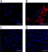
Specificity of the SARS-CoV-2 RNA FISH assay in HAE cultures. Three-dimensionally reconstructed confocal image stacks of mock-infected control cells (A) and cells infected with SARS-CoV-2 (B), NL63 (C), and HKU1 (D) subjected to RNA FISH using a set of probes against SARS-CoV-2 nucleocapsid RNA. SARS-CoV-2 RNA is shown in red. Nuclei were stained with DAPI and are shown in blue. Bar = 20 μM.

References

    1. Cui J, Li F, Shi ZL. 2019. Origin and evolution of pathogenic coronaviruses. Nat Rev Microbiol 17:181–192. doi:10.1038/s41579-018-0118-9. - DOI - PMC - PubMed
    1. Pyrc K, Dijkman R, Deng L, Jebbink MF, Ross HA, Berkhout B, van der Hoek L. 2006. Mosaic structure of human coronavirus NL63, one thousand years of evolution. J Mol Biol 364:964–973. doi:10.1016/j.jmb.2006.09.074. - DOI - PMC - PubMed
    1. van der Hoek L, Pyrc K, Jebbink MF, Vermeulen-Oost W, Berkhout RJ, Wolthers KC, Wertheim-van Dillen PM, Kaandorp J, Spaargaren J, Berkhout B. 2004. Identification of a new human coronavirus. Nat Med 10:368–373. doi:10.1038/nm1024. - DOI - PMC - PubMed
    1. Forni D, Cagliani R, Clerici M, Sironi M. 2017. Molecular evolution of human coronavirus genomes. Trends Microbiol 25:35–48. doi:10.1016/j.tim.2016.09.001. - DOI - PMC - PubMed
    1. Ramadan N, Shaib H. 2019. Middle East respiratory syndrome coronavirus (MERS-CoV): a review. Germs 9:35–42. doi:10.18683/germs.2019.1155. - DOI - PMC - PubMed

Publication types

LinkOut - more resources