. 2020 May 20;12(11):10527-10543.
doi: 10.18632/aging.103277. Epub 2020 May 20.

Human amnion-derived mesenchymal stem cells promote osteogenic differentiation of human bone marrow mesenchymal stem cells via H19/miR-675/APC axis

Affiliations

Human amnion-derived mesenchymal stem cells promote osteogenic differentiation of human bone marrow mesenchymal stem cells via H19/miR-675/APC axis

Xiaojie Ma et al. Aging (Albany NY). .

Abstract

Bone volume inadequacy is an emerging clinical problem impairing the feasibility and longevity of dental implants. Human bone marrow mesenchymal stem cells (HBMSCs) have been widely used in bone remodeling and regeneration. This study examined the effect of long noncoding RNAs (lncRNAs)-H19 on the human amnion-derived mesenchymal stem cells (HAMSCs)-droved osteogenesis in HBMSCs. HAMSCs and HBMSCs were isolated from abandoned amniotic membrane samples and bone marrow. The coculture system was conducted using transwells, and H19 level was measured by quantitative real-time reverse transcription-polymerase chain reaction (RT-PCR). The mechanism was further verified. We here discovered that osteogenesis of HBMSCs was induced by HAMSCs, while H19 level in HAMSCs was increased during coculturing. H19 had no significant effect on the proliferative behaviors of HBMSCs, while its overexpression of H19 in HAMSCs led to the upregulated osteogenesis of HBMSCs in vivo and in vitro; whereas its knockdown reversed these effects. Mechanistically, H19 promoted miR-675 expression and contributed to the competitively bounding of miR-675 and Adenomatous polyposis coli (APC), thus significantly activating the Wnt/β-catenin pathway. The results suggested that HAMSCs promote osteogenic differentiation of HBMSCs via H19/miR-675/APC pathway, and supply a potential target for the therapeutic treatment of bone-destructive diseases.

Keywords: adenomatous polyposis coli; human amnion-derived mesenchymal stem cells; long noncoding RNA H19; miR-675; osteogenic differentiation.


Conflict of interest statement

CONFLICTS OF INTEREST: The authors declare no conflicts of interest.

Figures

Figure 1
Figure 1
Osteogenic differentiation of HBMSCs cocultured with HAMSCs, lncRNA-H19 expression in HAMSCs and effects of lncRNA-H19 in HAMSCs on the proliferation of HBMSCs. (A) Relative mRNA expressions of ALP, RUNX2 and OCN in HBMSCs cocultured with HAMSCs were measured by RT-PCR analysis. (B) LncRNA-H19 expression in HAMSCs during coculturing was measured by RT-PCR analysis. (C) HBMSCs proliferation was demonstrated by flow cytometry. Data are shown as mean ± SD. *P < 0.05 and **P < 0.01.
Figure 2
Figure 2
LncRNA-H19 in HAMSCs promotes osteogenic differentiation of HBMSCs. (A) Protein levels of ALP, RUNX2, OCN, and OSX were assessed by western blot assay in HBMSCs, NC, H19, shNC and shH19 groups. (B) Relative mRNA expressions of ALP, RUNX2, OCN and OSX were measured by RT-PCR analysis in HBMSCs, NC, H19, shNC and shH19 groups. (C) ALP staining and activity in HBMSCs, NC, H19, shNC, and shH19 groups. Scale bar, 100 μm. (D) Alizarin red staining and quantification in HBMSCs, NC, H19, shNC, and shH19 groups. Scale bar, 1cm. Data are shown as mean ± SD. *P < 0.05 and **P < 0.01.
Figure 3
Figure 3
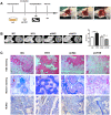
LncRNA-H19 in HAMSCs promotes osteogenesis in vivo. (A) NC, H19, shNC and shH19 groups were transplanted subcutaneously into a rat critical-sized mandibular defect model for 8 weeks. (B) Reconstructed 3D micro-CT images of the tissue-engineered bone and percentages of BV/TV. (C) H&E staining, Masson staining and immunohistochemical staining of RUNX2 in NC, H19, shNC and shH19 groups. b: bone-like tissues, h: HA/TCP scaffold, f: fibrous. Scale bar, 200 μm. Data are shown as mean ± SD. **P < 0.01.
Figure 4
Figure 4
MiR-675 in HAMSCs is activated by lncRNA-H19 and promotes osteogenic differentiation of HBMSCs. (A) Transfection efficacy of miR-675 was detected by RT-PCR. (B) MiR-675 expression in HAMSCs during coculturing was measured by RT-PCR. (C) MiR-675 expression was measured by RT-PCR in NC, H19, shNC, and shH19 groups. (DF) Protein levels of ALP, RUNX2, OCN and OSX were assessed by western blot assay in NC, mimics, iNC and inhibitor groups. (G, H) Relative mRNA expressions of ALP, RUNX2, OCN, and OSX were measured by RT-PCR analysis in NC, mimics, iNC and inhibitor groups. (I) ALP staining and activity in NC, mimics, iNC and inhibitor groups. Scale bar, 100 μm. (J) Alizarin red staining and quantification in NC, mimics, iNC and inhibitor groups. Scale bar, 1 cm. Data are shown as mean ± SD. *P < 0.05 and **P < 0.01.
Figure 5
Figure 5
MiR-675 downregulates APC expression in HAMSCs. (A) The potential binding sites between APC and miR-675 predicted by biological software. (B) Protein level of APC was assessed by western blot assay in NC, mimics, iNC and inhibitor groups. (C) Relative mRNA expression of APC was measured by RT-PCR analysis in NC, mimics, iNC and inhibitor groups. (D) Luciferase reporter assay was used to validate the target in 293T cells. Relative Renilla luciferase activity was normalized to that of firefly luciferase. Data are shown as mean ± SD. **P < 0.01.
Figure 6
Figure 6
APC inhibits Wnt/β-catenin pathway and HAMSCs-droved osteogenesis. (A, B) Protein levels of β-catenin, Cyclin D1 and c-Myc were assessed by western blot assay in NC and APC groups. (C) Relative mRNA expression of β-catenin was measured by RT-PCR analysis in NC and APC groups. (D) Immunofluorescence staining showed the β-catenin location in NC and APC groups. Scale bar, 20 μm. (E, F) Protein levels of ALP, RUNX2, OCN and OSX were assessed by western blot assay in NC and APC groups. (G) Relative mRNA expressions of ALP, RUNX2, OCN, and OSX were measured by RT-PCR analysis in NC and APC groups. Data are shown as mean ± SD. **P < 0.01.
Figure 7
Figure 7
MiR-675 mimic could rescue the shH19 mediated inhibitory effects. (A) Relative mRNA expression of APC was measured by RT-PCR analysis. (B, C) Protein levels of ALP, RUNX2, OCN, OSX, and APC were assessed by western blot assay. (D) Relative mRNA expressions of ALP, RUNX2, OCN, and OSX were measured by RT-PCR analysis. (E) Alizarin red staining analysis. Scale bar, 1 cm. Data are shown as mean ± SD. *P < 0.05 and **P < 0.01.
Figure 8
Figure 8
MiR-675 mimic could rescue the APC overexpression mediated inhibitory effects. (A) Relative mRNA expressions of ALP, RUNX2, OCN, and OSX were measured by RT-PCR analysis. (B) Alizarin red staining analysis. Scale bar, 1 cm. Data are shown as mean ± SD. **P < 0.01.
Figure 9
Figure 9
The schematic diagram for lncRNA-H19/miR-675/APC/Wnt/β-catenin axis in this study.

References

    1. Kim HJ, Yi SW, Oh HJ, Lee JS, Park JS, Park KH. Transfection of gene regulation nanoparticles complexed with pDNA and shRNA controls multilineage differentiation of hMSCs. Biomaterials. 2018; 177:1–13. 10.1016/j.biomaterials.2018.05.035 - DOI - PubMed
    1. Augello A, De Bari C. The regulation of differentiation in mesenchymal stem cells. Hum Gene Ther. 2010; 21:1226–38. 10.1089/hum.2010.173 - DOI - PubMed
    1. Hayashi M, Nakashima T, Yoshimura N, Okamoto K, Tanaka S, Takayanagi H. Autoregulation of osteocyte Sema3A orchestrates estrogen action and counteracts bone aging. Cell Metab. 2019; 29:627–637.e5 10.1016/j.cmet.2018.12.021 - DOI - PubMed
    1. Ling L, Wei T, He L, Wang Y, Wang Y, Feng X, Zhang W, Xiong Z. Low-intensity pulsed ultrasound activates ERK1/2 and PI3K-akt signalling pathways and promotes the proliferation of human amnion-derived mesenchymal stem cells. Cell Prolif. 2017; 50:e12383. 10.1111/cpr.12383 - DOI - PMC - PubMed
    1. Soncini M, Vertua E, Gibelli L, Zorzi F, Denegri M, Albertini A, Wengler GS, Parolini O. Isolation and characterization of mesenchymal cells from human fetal membranes. J Tissue Eng Regen Med. 2007; 1:296–305. 10.1002/term.40 - DOI - PubMed

Publication types

MeSH terms

LinkOut - more resources