. 2020 May 16;9(1):1-14.
doi: 10.1302/2046-3758.91.BJR-2019-0043.R2. eCollection 2020 Jan.

Mechanotransduction in osteogenesis

Affiliations

Mechanotransduction in osteogenesis

Sarah Stewart et al. Bone Joint Res. .

Abstract

Bone is one of the most highly adaptive tissues in the body, possessing the capability to alter its morphology and function in response to stimuli in its surrounding environment. The ability of bone to sense and convert external mechanical stimuli into a biochemical response, which ultimately alters the phenotype and function of the cell, is described as mechanotransduction. This review aims to describe the fundamental physiology and biomechanisms that occur to induce osteogenic adaptation of a cell following application of a physical stimulus. Considerable developments have been made in recent years in our understanding of how cells orchestrate this complex interplay of processes, and have become the focus of research in osteogenesis. We will discuss current areas of preclinical and clinical research exploring the harnessing of mechanotransductive properties of cells and applying them therapeutically, both in the context of fracture healing and de novo bone formation in situations such as nonunion. Cite this article: Bone Joint Res 2019;9(1):1-14.

Keywords: Bone; Mechanoreceptor; Mechanotransduction.


Conflict of interest statement

Conflict of interest statement: S. Masouros reports an institutional grant (paid to the Centre for Blast Injury Studies) from the Royal British Legion, related to this study. C. Higgins reports an institutional grant (paid to the Centre for Blast Injury Studies) from the Medical Research Council, related to this study.

Figures
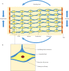
Fig. 1
Fig. 1
Bone cellular architecture. a) Mechanical loading of bone causes tension and compression forces across the bone’s lacuno-canalicular (LC) network. b) The tension/compression forces cause interstitial fluid shift within the LC network in an oscillatory manner across the cell membrane. BLC, bone lining cell; ECM, extracellular matrix. Adapted with permission from Duncan RL, Turner CH. Mechanotransduction and the functional response of bone to mechanical strain. Calcif Tissue Int. 1995;57(5):344-358.
Fig. 2
Fig. 2
Mechanocoupling. a) Axial section of osteocyte process with surrounding interstitial fluid in canaliculus. b) Longitudinal section of osteocyte process. Actin filaments span the process and are attached to the canalicular wall by tethering elements. Applied loading produces interstitial fluid movement producing a drag force on the tethering elements. c) Force balance on tethering elements: as the tethering elements deform, they pull the cell membrane outwards. d) Force balance on osteocyte skeleton: deformation of the cell membrane produces amplified strain on the actin cytoskeleton. The small vertical arrows indicate the direction of loading throughout the fibrin filaments. Adapted with permission from You L, Cowin SC, Schaffler MB, Weinbaum S. A model for strain amplification in the actin cytoskeleton of osteocytes due to fluid drag on pericellular matrix. J Biomech. 2001;34(11):1375-1386.
Fig. 3
Fig. 3
Biochemical coupling. Mechanosensors: a) Transmembrane proteins termed ‘integrins’ form focal adhesions with linker proteins, stimulating kinase pathways. b) Voltage-sensitive calcium (Ca2+) channels stimulate influx of calcium into the cell. c) Hexagonal connexins allow for efflux of newly synthesized prostaglandin 2 (PGE2), and their presence is upregulated by PGE2 via connexin 43 (Cx43). d) Primary cilia regulate bone cell function in response to fluid shear stress through calcium ion influx (activating signal transducer and activators of transcription (STAT) signalling pathways) and prostaglandin release. e) Cadherins span the cell membrane and dissociate with β-catenin in response to fluid shear stresses. Signal transduction pathways: f) Kinase pathways are activated by integrins and intracellular calcium release, resulting in upregulation of osteogenic transcription factors including runt-related transcription factor 2 (Runx2) and Osterix (OSX). g) An influx of calcium ions stimulates PGE2 synthesis via adenosine triphosphate (ATP), and activates kinase pathways. h) The wingless integrated (Wnt) signalling pathway is activated following dissociation of β-catenin from cadherin receptors. Accumulation and translocation of β-catenin to the cell nucleus stimulates upregulation of RUNX2. Akt, protein kinase B; ERK, extracellular signal-related kinase; FAK, focal adhesion kinase; MAPK, mitogen-activated protein kinase; OPG, osteoprotegerin; OPN, osteopontin; PKA, protein kinase A.
Fig. 4
Fig. 4
Transmission of signal. a) Direct transmission: gap junctions form between adjacent connexins on cell membranes, allowing the passage of small molecules such as calcium, adenosine triphosphate (ATP) and cyclic adenosine monophosphate (cAMP). This propagates mechanotransductive signals through the network of osteocytes to the bone lining cells (BLCs). b) Indirect transmission: BLCs release paracrine factors including prostaglandin E2 (PGE2) and insulin-like growth factor 1 (IGF-1), stimulating osteoprogenitor cells to differentiate in preosteoblasts and subsequently osteoblasts. These new osteoblasts attach to the bone surface and produce new bone matrix. Ca2+, calcium; OB, osteoblast; OC, osteoclast. Adapted with permission from Duncan RL, Turner CH. Mechanotransduction and the functional response of bone to mechanical strain. Calcif Tissue Int. 1995;57(5):344-358.
Fig. 5
Fig. 5
Applications of mechanotransduction. a) Shockwaves induce hyperpolarization of the cell membrane with subsequent induction of the osteogenic growth factor, transforming growth factor beta-1 (TGF-β1), as well as synthesizing oxygen free radicals. b) Vibrations and nanovibrations enhance the oscillatory fluid flow in the interstitial fluid of the lacuno-canalicular (LC) network, increasing the fluid shear forces that stimulate integrin receptors. Upregulation of integrin activity stimulates Sonic Hedgehog pathways and Rho A pathways leading to upregulation of osteopontin (OPN), osteocalcin (OCN), osteonectin (ONN), and Osterix (OSX) proteins. c) Electrical stimulation can increase the influx of calcium through voltage-gated ion channels, modulating calmodulin kinase (CAMK) pathways leading to increased expression of the osteogenic gene Runx2 and OSX. d) Ultrasound waves enhance integrin receptor activity, stimulating cyclo-oxygenase-2 (COX-2) pathway synthesis via protein kinase B (AKT), focal adhesion kinase (FAK), and extracellular-signal related kinase (ERK) pathways. This leads to the upregulation of osteocalcin, bone sialoprotein and insulin growth factor. RhoA, Ras homolog family member A.

References

    1. Jaalouk DE, Lammerding J. Mechanotransduction gone awry. Nat Rev Mol Cell Biol. 2009;10(1):63-73. - PMC - PubMed
    1. Duncan RL, Turner CH. Mechanotransduction and the functional response of bone to mechanical strain. Calcif Tissue Int. 1995;57(5):344-358. - PubMed
    1. Wolff J. The Law of Bone Remodelling. Translated by P. Maquet and R. Furlong. New York, NY: Springer-Verlag; 1986.
    1. Teti A. Bone development: overview of bone cells and signaling. Curr Osteoporos Rep. 2011;9(4):264-273. - PubMed
    1. Klein-Nulend J, van der Plas A, Semeins CM, et al. Sensitivity of osteocytes to biomechanical stress in vitro. FASEB J. 1995;9(5):441-445. - PubMed