. 2020 May 18;5(1):41.
doi: 10.1038/s41541-020-0190-9. eCollection 2020.

New GMP manufacturing processes to obtain thermostable HIV-1 gp41 virosomes under solid forms for various mucosal vaccination routes

Affiliations

New GMP manufacturing processes to obtain thermostable HIV-1 gp41 virosomes under solid forms for various mucosal vaccination routes

Mario Amacker et al. NPJ Vaccines. .

Abstract

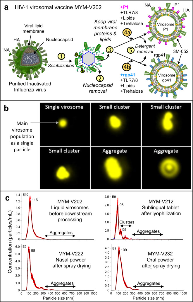
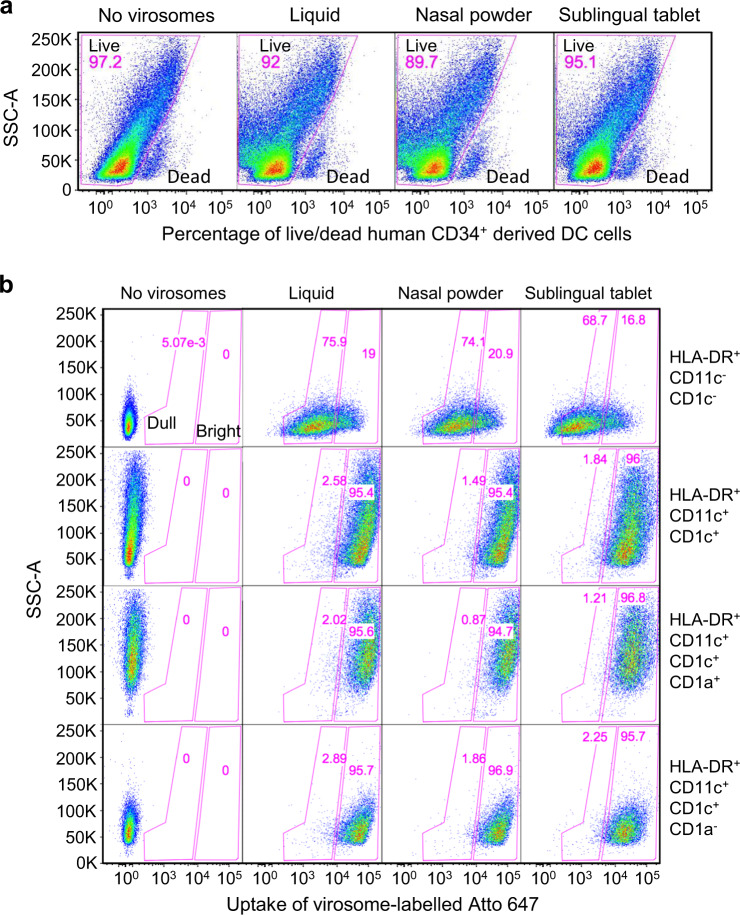
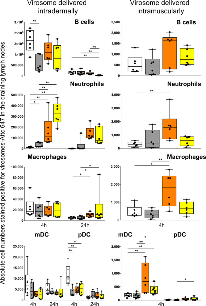
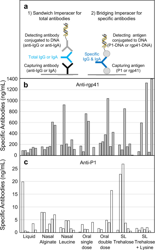
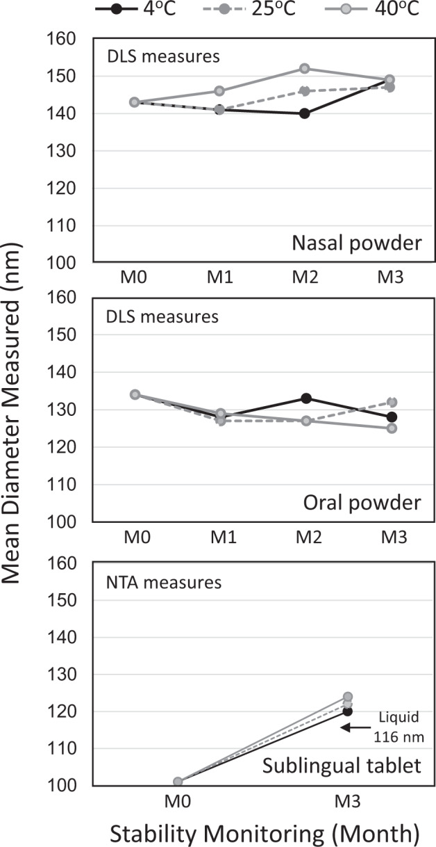
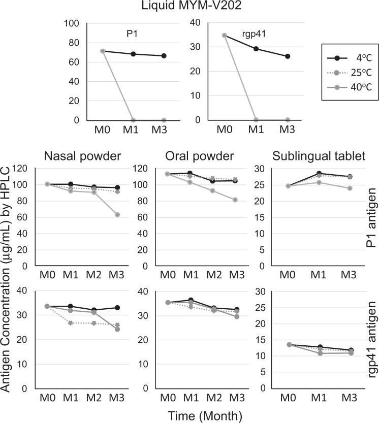
The main objective of the MACIVIVA European consortium was to develop new Good Manufacturing Practice pilot lines for manufacturing thermostable vaccines with stabilized antigens on influenza virosomes as enveloped virus-like particles. The HIV-1 gp41-derived antigens anchored in the virosome membrane, along with the adjuvant 3M-052 (TLR7/8 agonist) on the same particle, served as a candidate vaccine for the proof of concept for establishing manufacturing processes, which can be directly applied or adapted to other virosomal vaccines or lipid-based particles. Heat spray-dried powders suitable for nasal or oral delivery, and freeze-dried sublingual tablets were successfully developed as solid dosage forms for mucosal vaccination. The antigenic properties of vaccinal antigens with key gp41 epitopes were maintained, preserving the original immunogenicity of the starting liquid form, and also when solid forms were exposed to high temperature (40 °C) for up to 3 months, with minimal antigen and adjuvant content variation. Virosomes reconstituted from the powder forms remained as free particles with similar size, virosome uptake by antigen-presenting cells in vitro was comparable to virosomes from the liquid form, and the presence of excipients specific to each solid form did not prevent virosome transport to the draining lymph nodes of immunized mice. Virosome integrity was also preserved during exposure to <-15 °C, mimicking accidental freezing conditions. These "ready to use and all-in-one" thermostable needle-free virosomal HIV-1 mucosal vaccines offer the advantage of simplified logistics with a lower dependence on the cold chain during shipments and distribution.

Keywords: Biotechnology; Immunology; Infectious diseases.


Conflict of interest statement

Competing interestsM. Amacker and S.F. are employees of Mymetics SA, T.S. and F.B. are employees of Mymetics BV. S.F. and T.S. own equity in the mother company Mymetics Corporation. Mymetics Corporation shareholders had no role in study design, data collection and analysis, decision to publish, or preparation of the manuscript. R.J. is an employee of Upperton Ltd. and owns equity in Upperton Ltd. Mymetics Corporation and Catalent U.K. Swindon Zydis Limited have filed a patent application in 2019 (PCT/EP2019/082940—Oral dispersible vaccine comprising virosomes) with inventors D.W., C.S., M. Amacker, S.F., and T.S. that are authors of this manuscript. The remaining authors declare no competing interests.

Figures

Fig. 1
Fig. 1. Influenza virosome-based vaccine manufacturing.
a Production of adjuvanted virosome-P1 (MYM-V111) and virosome-rgp41 (MYM-V112): Step 1, inactivated influenza A/H1N1 are solubilized with detergent; Step 2, nucleocapsides are discarded; Step 3, the viral membrane lipids with the native influenza hemagglutinin (HA) and neuraminidase (NA) are recovered; Steps 4a and 4b, synthetic lipids with 3M-052 adjuvant and antigen P1 or rgp41 are mixed with isolated viral membrane components and trehalose; Step 5, virosomes-P1 (pink rod) and virosome-rgp41 (blue rod) are gradually assembled in vitro during the detergent removal. Each virosome is then diluted and mixed together to generate the HIV-1 liquid vaccine MYM-V202. Universal T help provided by HA/NA. b Amnis® ImageStream on fluorescent Dil dye-labeled virosomes (in yellow) to visualize particles. The liquid virosome population contains mostly single particles (upper left image). Reconstituted powders contain also a major population of single particles, but a minor population of few small virosome clusters and bigger aggregates are also observed. Images were enlarged in Powerpoint because original AMNIS images are only tiny dots. c Mean particle size and population distribution monitored by NTA for the liquid bulk vaccine (MYM-V202, upper left panel), the reconstituted sublingual tablet (MYM-V212, upper right panel), the reconstituted nasal powder (MYM-V222, lower left panel), the reconstituted oral powder (MYM-V232, lower right panel). Black arrows identify the population of small clusters 200–300 nm and aggregates >300 nm, using arbitrary cut-off.
Fig. 2
Fig. 2. In vitro virosome toxicity and uptake by antigen-presenting cells.
a Human CD34+-derived cells in culture were exposed to the various virosome formulations for 1 hour and cells were then stained with LIVE/DEAD dye to determine the percentage of dead cells and cells alive. b Virosomes were labeled with a stable tracer fluorescent lipid labeled with Atto 647 and their uptake after 1 hour by various APC subpopulations defined by markers HLA-DR, CD11c, CD1c, and CD1a were monitored by cytometry, gating on dull and bright virosome signals in pink gates (gating strategy described in Supplementary Methods Fig. 3). Cells that are HLA-DR+CD11c are not differentiated into dendritic cells and they represent the majority of the population. Cells that are HLA-DR+CD11c+CD1c+CD1a+ have a phenotype similar to Langerhans cells, and cells that are HLA-DR+CD11c+CD1c+CD1a are more similar to dermal DC. The stronger is the fluorescent Atto 647 signal, the more virosome uptake took place. The percentage of Atto 647 positive cells is comparable between cells exposed to the starting liquid formulation and the various solid vaccine dosage forms, suggesting that excipients did not interfere with early virosome uptake by APC. Data are from a representative experiment.
Fig. 3
Fig. 3. Impact of excipients on in vivo migration of virosomes from liquid and solid dosage forms.
The absolute number of virosome positive Atto 647 in B cells, neutrophils, macrophages, myeloid DCs (mDCs), and plasmacytoid DCs (pDCs) present in two draining lymph nodes of mice after 4 or 24 hours following intradermal injection (panels on the left column) or after 4 hours following intramuscular injection (panels on the right column). Cell subpopulations were defined (gating strategy described in Supplementary Methods Fig. 4) by the presence or absence of various cell surface antigens (markers): B cells (B220+CD11CLy6C), neutrophils (CD11b+Ly6C+Ly6G+), macrophages (I-Ab+CD11b+F4/80+Ly6CLoLy6GCD11c), myeloid DCs (I-Ab+CD11b+Ly6CLoLy6G+CD11c+), plasmacytoid DCs (I-Ab+B220+Ly6C+CD11c+). Solid dosage forms were dissolved in water prior injection and similar hemagglutinin dose were administered. Liquid virosome (white bars) prior downstream processing, sublingual tablet (gray bars), oral powder (orange bars), and nasal powder (yellow bars). Each vaccine group had six mice, and the corresponding data for each animal is displayed for showing distribution within box-and-whisker plots, with the means and standard deviations for the measured cell subpopulations in the lymph nodes. Statistical analyses used the Mann–Whitney U test and statistical significances between mice groups that received different virosome formulations are indicated: *p < 0.05, **p < 0.01.
Fig. 4
Fig. 4. Vaccine-induced serum antibodies in the presence of various excipients.
Early development research grade lots (not optimized yet) of the virosomal vaccine MYM-V202 under nasal and oral powder, and sublingual (SL) tablets were reconstituted in water and were administered subcutaneously to rats at day 1 and day 28 to determine if excipients of each new solid dosage form were affecting the antibody response. Each final serum at day 42 was analyzed by the Imperacer assays developed during MACIVIVA project, as illustrated in (a). Data of each animal is shown for better appreciating of the inter-individual variation often observed with suboptimal formulations. Approximate net serum (pre-immune deducted) anti-rgp41 antibody concentrations detected at day 42 (b), and anti-P1 antibody concentrations detected at day 42 (c). Data are from a representative Imperacer assay experiment.
Fig. 5
Fig. 5. Stability of the virosome particle size after storage of the vaccine solid forms under various environmental conditions.
A 3 months stability study was performed on the various solid vaccine forms (nasal and oral powders, sublingual tablets) stored under three different temperatures and relative humidity (RH) conditions: 4 °C (black line), 25 °C/65% RH (gray dot line), and 40 °C/75% RH (gray line). At each indicated month time point (M0–M3), samples were reconstituted with water and the mean virosome particle size (nm) was determined. Due to specific excipient interference during particle size analysis and different equipment available at different manufacturing sites, different methods were selected for particle analysis during stability study: DLS for nasal and oral powder and NTA for sublingual tablets. Note that for the sublingual virosomes, the mean particle size (101 nm) is about 10% smaller after lyophilization at M0, respective to the liquid virosomes (116 nm), but it is closer to the starting size after 3 months storage (120 nm at 4 °C and 124 nm at 40 °C). The higher residual moisture content in sublingual tablets (about 4%) and nasal powder (about 3%), respective to the oral powder (about 2.5%) may have contributed to bring back the virosome size closer to the original liquid virosome size over time. Data shown are from representative DLS and NTA measures.
Fig. 6
Fig. 6. Antigen concentrations in the various vaccine forms exposed to different environmental conditions.
The liquid adjuvanted vaccine formulation MYM-V202 containing both P1 and rgp41 antigens served as reference material for comparison to the solid vaccine dosage form for nasal, oral, and sublingual delivery. A 3 months stability study was performed on the various liquid and solid vaccine forms stored under three different temperatures and relative humidity (RH) conditions: 4 °C (black line), 25 °C/65% RH (gray dot line, not done for the liquid form), and 40 °C/75% RH (gray line). P1 and rgp41 antigens were previously shown to be temperature sensitive, which is confirmed again here in the first two upper panels, showing rapid P1 and rgp41 modifications at 40 °C, as compared to the liquid vaccine stored under the recommended temperature at 4 °C. For solid vaccine forms, at each indicated month time point (M0, M1, M2, or M3), samples were reconstituted with water and analyzed by HPLC for the P1 and rgp41 content. Note that chemical modifications such as oxidation or deamidation on antigens are the main reasons to the observed lower antigen concentration, as antigen degradation could not be reported by native immunoblot. Data shown are from representative HPLC measures.
Fig. 7
Fig. 7. Immunogenicity of P1 and rgp41 from liquid and various solid vaccine dosage forms exposed to different temperatures.
The liquid adjuvanted vaccine formulation MYM-V202 containing both P1 and rgp41 antigens were temperature sensitive and served as reference material for comparison with the immunogenicity of the solid vaccine forms with improved thermostability. Black line, vaccines stored 3 months at 2–8 °C; gray dot line, vaccines exposed 1 month at 40 °C; gray line, vaccines exposed 3 months at 40 °C. In each panel, the antibody endpoint titers (specific toward P1 or rgp41 antigen) of the serum pool of 10 rat sera are indicated, data are from a representative experiment. Endpoint titer against each antigen corresponds to the last serum dilution generating an optical density (OD) value >2-fold above the pre-immune background.
Fig. 8
Fig. 8. Key gp41 epitopes on vaccinal antigens are preserved in the new solid vaccine forms.
The vaccine-induced antibody reactivity profile was tested toward six different gp41 biotinylated peptides (S3–S8) that were captured by pre-coated streptavidin ELISA plate. Rat serums from animal immunized with the liquid HIV-1 vaccine MYM-V202 stored at 4 °C serve as reference for determining the starting serum antibody reactivity toward the different gp41 peptides. Then, the liquid MYM-V202 was exposed at 40 °C for 1 and 3 months and used for immunizing animals, and the vaccine-induced serum antibodies were tested for their reactivity profile toward the selected peptides, and compared to the serums from animals immunized with the vaccine stored at 4 °C. Similarly, the nasal, oral, and sublingual solid vaccine forms were also stored at 4 °C or exposed at 40 °C for 3 months for evaluating the impact of heat exposure on antigen immunogenicity. As the antibody repertoire toward the P1 and rgp41 vaccinal antigens corresponds to the sum of various recognized epitopes, detecting the serum reactivity toward a single peptide/epitope is expected to be weak, justifying ELISA assays limited to dilutions from 1/200 to 1/1200. The data shown here correspond to the serum pool diluted 1/600 with pre-immune background removed, it corresponds to the dilution giving sufficient optical density (OD) signals among all peptides and conditions to determine if a given epitope was preserved intact, reduced or lost. OD values measured at 492 nm from representative ELISA tests are reported on the Y-axis.

Similar articles

Cited by

References

    1. Chen D, Kristensen D. Opportunities and challenges of developing thermostable vaccines. Expert Rev. Vaccines. 2009;8:547–557. - PubMed
    1. de Jonge J, et al. Inulin sugar glasses preserve the structural integrity and biological activity of influenza virosomes during freeze-drying and storage. Eur. J. Pharm. Sci. 2007;32:33–44. - PubMed
    1. Karp CL, et al. Evaluating the value proposition for improving vaccine thermostability to increase vaccine impact in low and middle-income countries. Vaccine. 2015;33:3471–3479. - PubMed
    1. Kristensen D, Chen D, Cummings R. Vaccine stabilization: research, commercialization, and potential impact. Vaccine. 2011;29:7122–7124. - PubMed
    1. Lloyd J, Cheyne J. The origins of the vaccine cold chain and a glimpse of the future. Vaccine. 2017;35:2115–2120. - PubMed