. 2020 Apr 8:12:100219.
doi: 10.1016/j.ynstr.2020.100219. eCollection 2020 May.

Dopamine modulates individual differences in avoidance behavior: A pharmacological, immunohistochemical, neurochemical and volumetric investigation

Affiliations

Dopamine modulates individual differences in avoidance behavior: A pharmacological, immunohistochemical, neurochemical and volumetric investigation

Geiza Fernanda Antunes et al. Neurobiol Stress. .

Abstract

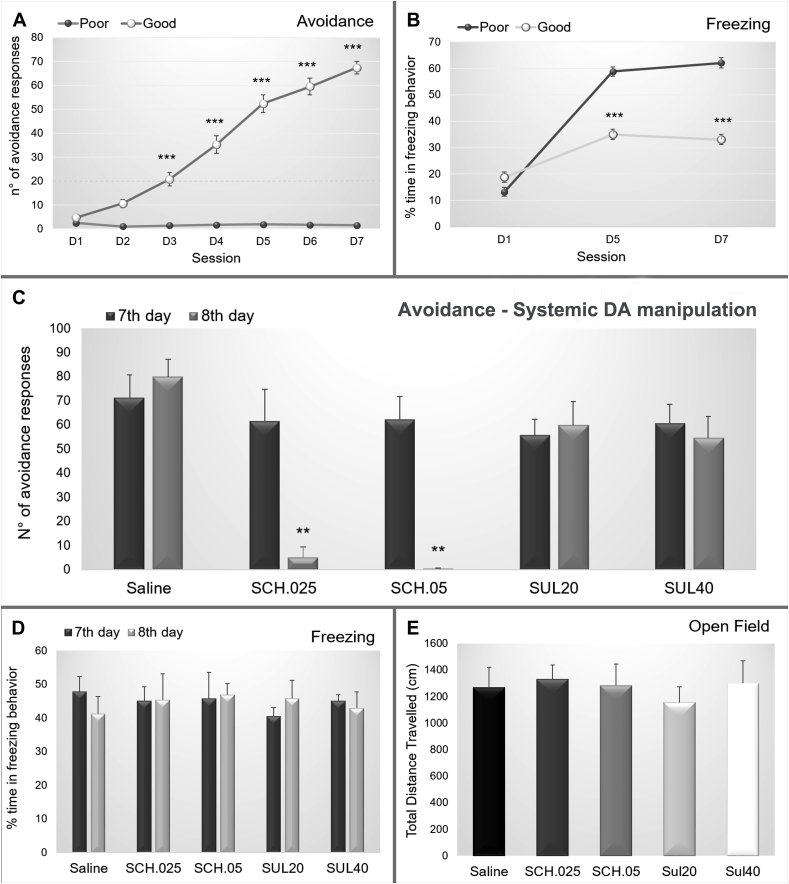
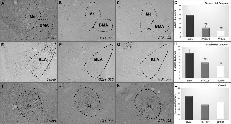
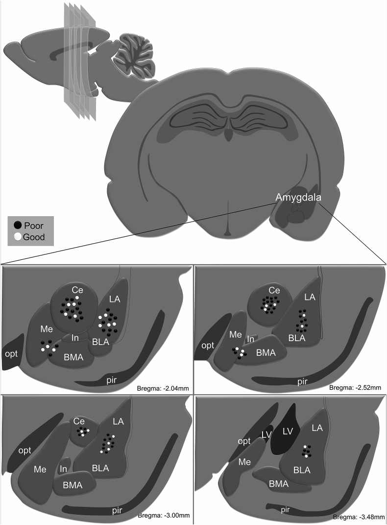
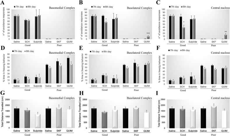
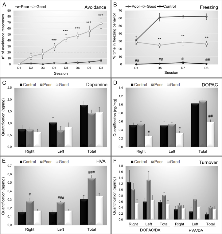
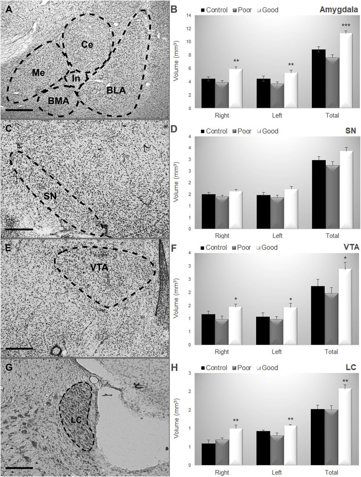
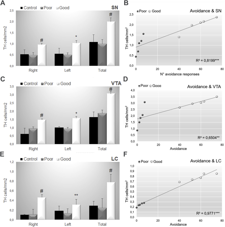
Avoidance behavior is a hallmark in pathological anxiety disorders and results in impairment of daily activities. Individual differences in avoidance responses are critical in determining vulnerability or resistance to anxiety disorders. Dopaminergic activation is implicated in the processing of avoidance responses; however, the mechanisms underlying these responses are unknown. In this sense, we used a preclinical model of avoidance behavior to investigate the possibility of an intrinsic differential dopaminergic pattern between good and poor performers. The specific goal was to assess the participation of dopamine (DA) through pharmacological manipulation, and we further evaluated the effects of systemic injections of the dopaminergic receptor type 1 (D1 antagonist - SCH23390) and dopaminergic receptor type 2 (D2 antagonist - sulpiride) antagonists in the good performers. Additionally, we evaluated the effects of intra-amygdala microinjection of a D1 antagonist (SCH23390) and a D2 antagonist (sulpiride) in good performers as well as intra-amygdala microinjection of a D1 agonist (SKF38393) and D2 agonist (quinpirole) in poor performers. Furthermore, we quantified the contents of dopamine and metabolites (3,4-dihydroxyphenylacetic acid (DOPAC) and homovanillic acid (HVA)) in the amygdala, evaluated the basal levels of tyrosine hydroxylase expression (catecholamine synthesis enzyme) and measured the volume of the substantia nigra, ventral tegmental area and locus coeruleus. Our results showed that it could be possible to convert animals from good to poor performers, and vice versa, by intra-amygdala (basolateral and central nucleus) injections of D1 receptor antagonists in good performers or D2 receptor agonists in poor performers. Additionally, the good performers had lower levels of DOPAC and HVA in the amygdala, an increase in the total volume of the amygdala (AMG), substantia nigra (SN), ventral tegmental area (VTA) and locus coeruleus (LC), and an increase in the number of tyrosine hydroxylase-positive cells in SN, VTA and LC, which positively correlates with the avoidance behavior. Taken together, our data show evidence for a dopaminergic signature of avoidance performers, emphasizing the role of distinct dopaminergic receptors in individual differences in avoidance behavior based on pharmacological, immunohistochemical, neurochemical and volumetric analyses. Our findings provide a better understanding of the role of the dopaminergic system in the execution of avoidance behavior.

Keywords: Amygdala; Anxiety disorders; Aversive learning; Avoidance; Dopamine; Prefrontal cortex.


Conflict of interest statement

The authors report no conflicts of interest.

Figures

Fig. 1
Fig. 1
(A). Experimental design of the study divided in Experiment 1 that consisted of dopaminergic manipulation that was performed systemic and intra-amygdala manipulation in good and poor performers; and Experiment 2 that evaluated intrinsic differences. (B). Flowchart of the animals used in the study illustrating the number of the animals used per experiment in each experimental condition. QUIN: quinpirole. SCH: SCH23390; SKF: SKF38393.
Fig. 2
Fig. 2
(A) Number of avoidance and (B) Percentage of the time spent in freezing exhibited by the poor and good avoiders. (C) Number of avoidance performed and (D) Percentage of time spent in freezing behavior by the good performers on the 7th day of training and on the 8th day of training after the administration of saline (N = 7), SCH 0.025 mg/kg (N = 7), SCH 0.05 mg/kg (N = 6), sulpiride 20 mg/kg (N = 8) or sulpiride 40 mg/kg (N = 8). (E) Total travel distance showed by good performers after the administration saline (N = 7), SCH 0.25 mg/kg (N = 7), SCH 0.5 mg/kg (N = 6), sulpiride 20 mg/kg (N = 8) or sulpiride 40 mg/kg (N = 8). ***: p < 0.001 in comparison with poor performers.
Fig. 3
Fig. 3
Representative photomicrographs of c-Fos staining in the basomedial complex, basolateral complex and central amygdala after the administration of (A, E, I) saline, (B, F, J) SCH 0.25 mg/kg or (C, G, K) SCH 0.5 mg/kg in good performers. Mean density of c-Fos + cells in (D) Basomedial complex, (H) Basolateral complex and (L) Central amygdala for good avoiders after the systemic injection of saline (N = 6), SCH 0.25 mg/kg (N = 6) or SCH 0.5 mg/kg (N = 5). Scale bars represent 400 μm in all photographs. **: p < 0.01 in comparison with the other groups.
Fig. 4
Fig. 4
Representative microinjection sites for basomedial complex, basolateral complex and central nucleus of amygdala along bregma −2.04, −2.52, −3.00 and −3.48 mm. Each dot indicates the site of injection corresponding to poor avoiders (black dots) and good avoiders (white dots). BLA: basolateral nucleus of amygdala, BMA: basomedial nucleus of the amygdala; Ce: central nucleus of amygdala, In: intercalated nucleus of the amygdala, LA: lateral nucleus of amygdala, LV: lateral ventricle, Me: medial nucleus of amygdala; pir: piriform nucleus, opt: optic tract.
Fig. 5
Fig. 5
Number of avoidance performed by good performers on the 7th day of training (basal) and on the 8th day of training after the administration of saline, SCH 0.3 μL, sulpiride 0.2 μL and by poor avoiders on the 7th day of training (basal) and on the 8th day of training after the administration of saline, SKF 0.4 μL, or quinpirole 0.1 μL into the (A) basomedial complex, (B) basolateral complex or (C) central nucleus of amygdala. Percentage of time spent in freezing behavior performed by good performers on the 7th day of training (basal) and on the 8th day of training after the administration of saline, SCH 0.3 μL, sulpiride 0.2 μL and by poor avoiders on the 7th day of training (basal) and on the 8th day of training after the administration of saline, SKF 0.4 μL, or quinpirole 0.1 μL into the (D) basomedial complex, (E) basolateral complex or (F) central nucleus of the amygdala. Total travel distance showed by good performers after the administration of saline, SCH 0.3 μL, sulpiride 0.2 μL and by the poor avoiders on the 7th day of training (basal) and on the 8th day of training after the administration of saline, SKF 0.4 μL, or quinpirole 0.1 μL into the (G) basomedial complex, (H) basolateral complex or (I) central nucleus of amygdala. Group good avoiders: saline (N = 9), SCH 0.3 μL (basomedial N = 6, basolateral N = 9, central N = 9), sulpiride 0.2 μL (basomedial N = 5, basolateral N = 6, central N = 6). Group poor avoiders: saline (N = 9), SKF 0.4 μL (basomedial N = 4, basolateral N = 9, central N = 9) or quinpirole 0.1 μL (basomedial N = 4, basolateral N = 9, central N = 9). *: p < 0.05 in comparison with the corresponding 7th day.
Fig. 6
Fig. 6
Data are reported as the means ± SEM. (A). Number of active avoidance responses in good and poor performers across training sessions. (B). Percentage of time spent in freezing behavior along sessions. (C). Quantification of dopamine (ng/mg of tissue) in the right, left and both hemispheres considering control, poor and good performers. (D). Quantification of 3,4-dihydroxyphenylacetic acid - DOPAC (ng/mg of tissue) in the right, left and both hemispheres considering control, poor and good performers. (E). Quantification of homovanillic acid - HVA (ng/mg of tissue) in the right, left and both hemispheres considering control, poor and good performers. (F). Quantification of dopamine turnover of DOPAC and HVA in the right, left and both hemispheres considering control, poor and good performers. Poor: poor performers, Good: good performers, Control: control group. DOPAC/DA: turnover rate for DOPAC; HVA/DA: turnover rate for HVA, right: right hemisphere, left: left hemisphere, total: right and left hemisphere. For the behavioral data: control (N = 5), good (N = 13) and poor (N = 15). For the HPLC data: control (N = 5), good (N = 8) and poor (N = 10) and **: p < 0.01 in comparison with poor performers, ***: p < 0.001 in comparison with poor performers, #: p < 0.05 in comparison with good and poor performers, ##: p < 0.01 in comparison with good and poor performers; ###: p < 0.001 in comparison with good and poor performers.
Fig. 7
Fig. 7
Representative photomicrographs of (A) the Amygdala nuclei, (C) Substantia Nigra, (E) Ventral Tegmental Area, (G) Locus Coeruleus. Data are reported as the means ± SEM. Volumetric estimates in (B) the Amygdala nuclei, (D) Substantia nigra, (F) Ventral Tegmental Area, (H) Locus Coeruleus considering the right and left hemisphere and the total volume considering control, poor and good performers. BLA: basolateral nucleus of amygdala; BMA: basomedial nucleus of amygdala; Ce: central nucleus of amygdala, LC: locus coeruleus; Me: medial nucleus of amygdala; SN: substantia nigra; VTA: ventral tegmental area. Control (N = 3), Good (N = 5), Poor (N = 5). Scale bars represent 400 μm in all photographs. *: p < 0.05 in comparison with poor performers; **: p < 0.01 in comparison with poor performers, ***: p < 0.001 in comparison with poor performers.
Fig. 8
Fig. 8
Data are reported as the means ± SEM. TH positive cells/mm2 in (A) Substantia nigra, (C) Ventral Tegmental Area, (E) Locus Coeruleus considering the right and left hemisphere and the total volume considering control, poor and good performers. Correlation data between the number of avoidance and tyrosine hydroxylase-positive cells/mm2 in the (B) Substantia Nigra, (D) Ventral Tegmental Area and (E) Locus Coeruleus considering poor and good performers. Control (N = 3), Good (N = 5), Poor (N = 5). LC: locus coeruleus; SN: substantia nigra; VTA: ventral tegmental area. Scale bars represent 400 μm in all photographs. *: p < 0.05 in comparison with poor performers; **: p < 0.01 in comparison with poor performers, ***: p < 0.001 in comparison with poor performers, #: P < 0.05 in comparison with all groups.

References

    1. Aguilar M.A., Marí-Sanmillán M.I., Morant-Deusa J.J., Miñarro J. Different inhibition of conditioned avoidance response by clozapine and DA D1 and D2 antagonists in male mice. Behav. Neurosci. 2000 Apr;114(2):389–400. - PubMed
    1. American Psychiatric Association . American Psychiatric Pub; 2013. Diagnostic and Statistical Manual of Mental Disorders (DSM-5R)
    1. Antelman S.M., Caggiula A.R. Norepinephrine-dopamine interactions and behavior. Science. 1977 Feb 18;195(4279):646–653. - PubMed
    1. Ardeshiri M.R., Hosseinmardi N., Akbari E. The effect of orexin 1 and orexin 2 receptors antagonisms in the basolateral amygdala on memory processing in a passive avoidance task. Physiol. Behav. 2017 May 15;174:42–48. doi: 10.1016/j.physbeh.2017.03.004. - DOI - PubMed
    1. Bardeen J.R., Tull M.T., Stevens E.N., Gratz K.L. Exploring the relationship between positive and negative emotional avoidance and anxiety symptom severity: the moderating role of attentional control. J. Behav. Ther. Exp. Psychiatr. 2014 Sep;45(3):415–420. doi: 10.1016/j.jbtep.2014.04.006. - DOI - PubMed

LinkOut - more resources