Case Reports
. 2020 Aug;267(8):2185-2192.
doi: 10.1007/s00415-020-09885-2. Epub 2020 May 20.

Stroke in patients with SARS-CoV-2 infection: case series

Affiliations
Case Reports

Stroke in patients with SARS-CoV-2 infection: case series

Mauro Morassi et al. J Neurol. 2020 Aug.

Abstract

Background: Italy is one of the most affected countries by the coronavirus disease 2019 (COVID-19). The responsible pathogen is named severe acute respiratory syndrome coronavirus (SARS-CoV-2). The clinical spectrum ranges from asymptomatic infection to severe pneumonia, leading to intensive care unit admission. Evidence of cerebrovascular complications associated with SARS-CoV-2 is limited. We herein report six patients who developed acute stroke during COVID-19 infection.

Methods: A retrospective case series of patients diagnosed with COVID-19 using reverse-transcriptase polymerase chain reaction (RT-PCR) on nasopharyngeal swabs, who developed clinical and neuroimaging evidence of acute stroke during SARS-CoV-2 infection.

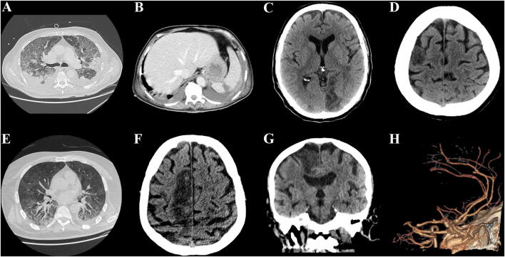
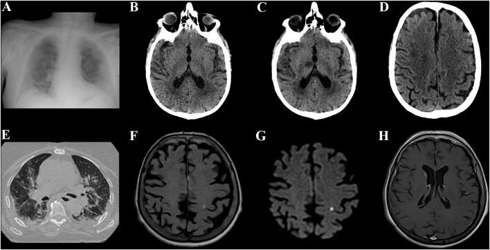
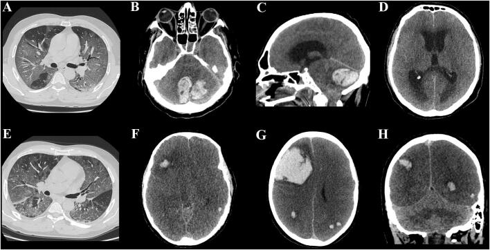
Results: Six patients were identified (5 men); median age was 69 years (range 57-82). Stroke subtypes were ischemic (4, 67%) and hemorrhagic (2, 33%). All patients but one had pre-existing vascular risk factors. One patient developed encephalopathy prior to stroke, characterized by focal seizures and behavioral abnormalities. COVID-19-related pneumonia was severe (i.e., requiring critical care support) in 5/6 cases (83%). Liver enzyme alteration and lactate dehydrogenase (LDH) elevation were registered in all cases. Four patients (67%) manifested acute kidney failure prior to stroke. Four patients (67%) had abnormal coagulation tests. The outcome was poor in the majority of the patients: five died (83%) and the remaining one (17%) remained severely neurologically affected (mRS: 4).

Conclusions: Both ischemic and hemorrhagic stroke can complicate the course of COVI-19 infection. In our series, stroke developed mostly in patients with severe pneumonia and multiorgan failure, liver enzymes and LDH were markedly increased in all cases, and the outcome was poor.

Keywords: Brain hemorrhage; COVID-19; Cerebrovascular disease; Coronavirus; Encephalitis; Neurological complications.


Conflict of interest statement

The authors declare that they have no conflicts of interest.

Figures

Fig. 1
Fig. 1
Total body and brain CT examination of Patient 1 ad showing extensive bilateral consolidations and ground-glass opacities of the lungs (a), a hypodense area in the upper portion of the spleen (b) and two different ischemic lesions involving the left occipital lobe (c) and the right frontal precentral gyrus (d), respectively. Chest and brain CT examination of Patient 2 eh showing bilateral consolidations and ground-glass opacities of the lungs with pleural effusion on the left side (e), an ischemic lesion involving the frontal lobe on the right side (fg), and the occlusion of the right pericallosal artery on a 3D-volume rendering reconstruction of the brain CT angiography examination (h)
Fig. 2
Fig. 2
Chest X-ray and two different brain CT examinations of Patient 3 ad showing multiple opacities involving both lungs and a left lower lobe consolidation (a), the normal aspect of both thalami (b) and the appearance few days later of multiple ischemic lesions involving the right thalamus (c), and the subcortical white matter of the centrum semiovale of the same side (d). Chest CT and brain MRI examinations of patient 4 eh, demonstrating multiple areas of ground-glass opacities and consolidations mainly involving the lower lobes (e), a focal T2-FLAIR hyperintensity lesion in the left precentral gyrus (f) with a bright signal on DWI sequence (g), and mild post-contrast enhancement of the head of the right caudate nucleus (h)
Fig. 3
Fig. 3
Chest and brain CT examination of Patient 5 (ad), showing diffuse bilateral ground-glass opacities involving both lungs (a), a large cerebellar hemorrhage (b) which compresses the brainstem and the fourth ventricle determining a subsequent obstructive hydrocephalus (c, d). Chest and brain CT examination of Patient 6 (eh), demonstrating diffuse bilateral ground-glass opacities with no pleural effusion (e), diffuse cerebral edema with loss of normal gray–white matter differentiation and obliteration of CSF spaces (f), a large right frontal hemorrhage in association with other smaller hemorrhages on the axial and on the coronal multiplanar reconstruction of brain CT (g, h), and a bright spot within the sagittal sinus suspected for dural sinus thrombosis

Comment in

References

    1. COVID-19 Map (2020) In: Johns Hopkins coronavirus resource center. https://coronavirus.jhu.edu/map.html. Accessed 6 Apr 2020
    1. Chen N, Zhou M, Dong X, et al. Epidemiological and clinical characteristics of 99 cases of 2019 novel coronavirus pneumonia in Wuhan, China: a descriptive study. Lancet. 2020;395:507–513. doi: 10.1016/S0140-6736(20)30211-7. - DOI - PMC - PubMed
    1. Guan W-J, Ni Z-Y, Hu Y, et al. Clinical Characteristics of Coronavirus Disease 2019 in China. N Engl J Med. 2020 doi: 10.1056/NEJMoa2002032. - DOI - PMC - PubMed
    1. Wang D, Hu B, Hu C, et al. Clinical characteristics of 138 hospitalized patients with 2019 novel coronavirus-infected pneumonia in Wuhan, China. JAMA. 2020 doi: 10.1001/jama.2020.1585. - DOI - PMC - PubMed
    1. Umapathi T, Kor AC, Venketasubramanian N, et al. Large artery ischaemic stroke in severe acute respiratory syndrome (SARS) J Neurol. 2004;251:1227–1231. doi: 10.1007/s00415-004-0519-8. - DOI - PMC - PubMed

MeSH terms