Case Reports
. 2020 May;7(5):829-838.
doi: 10.1002/acn3.51051. Epub 2020 May 20.

Reversing 21 years of chronic paralysis via non-invasive spinal cord neuromodulation: a case study

Affiliations
Case Reports

Reversing 21 years of chronic paralysis via non-invasive spinal cord neuromodulation: a case study

Monzurul Alam et al. Ann Clin Transl Neurol. 2020 May.

Abstract

Objective: The objective of the current study was to investigate if a non-invasive spinal cord neuromodulation modality could restore sensorimotor functions in a patient with chronic spinal cord injury (SCI).

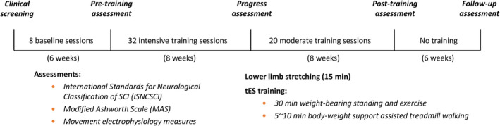
Methods: In this study, transcutaneous electrical stimulation (tES) to the spinal cord was utilized to restore sensorimotor functions in a chronic SCI patient who sustained a traumatic C7 cervical cord injury 21 years ago. At baseline, the patient had very limited volitional movement in her right leg, and her left leg was completely paralyzed. tES parameters were optimized in eight stimulation sessions before the treatment. The therapeutic stimulation involved biphasic tES, applied to T11 and L1 spinal levels during a 1-hour standing and walking training, 2-4 times per week for 16 weeks.

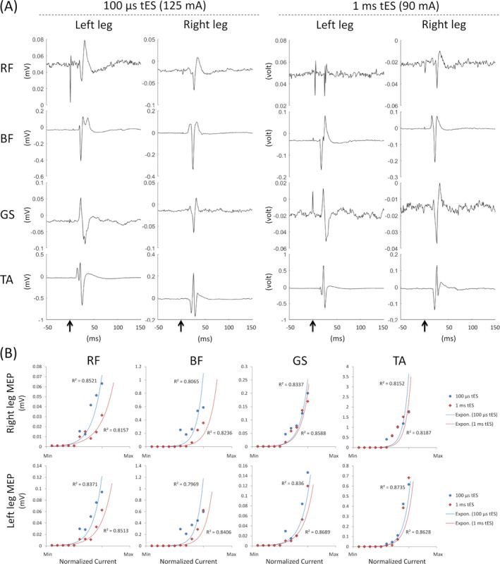
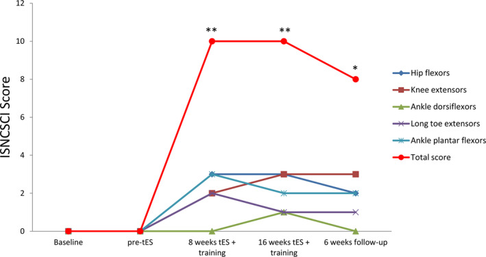
Results: Our pre-treatment tests indicated that a shorter burst duration (100 µsec) was more effective than a longer burst duration of tES in improving functional movements. After 32 training sessions with tES, the patient regained significant left-leg volitional movements (grade 0 to grade 10 according to the ISNCSCI scale). Right-leg motor scores also increased from 17 to 21. The tES treatment also improved her pinprick sensation (from 73 to 79). Upon completion of the treatment (52 sessions), the patient's standing ability noticeably improved. She could stabilize her knee to stand without any assistance. She could also squat while holding onto a walker.

Interpretation: These promising results demonstrate beneficial effects of non-invasive tES in regaining volitional control of plegic lower limbs in patients with chronic paralysis.


Conflict of interest statement

The authors declare no conflict of interest.

Figures

Figure 1
Figure 1
Study procedures. After passing the clinical screening, eight transcutaneous electrical stimulation (tES) sessions were conducted on the study participant to determine the optimum stimulation parameters for training, followed by pre‐training assessments (listed in the figure). tES sessions were then applied 4 times/week in conjunction with physical training. After 32 intensive training sessions, functional reassessments were conducted to determine the participant’s improvement. Upon completion of the 32 intensive training and progress assessments, the participant underwent another 20 moderate training sessions (2–3 times/week) with tES before the post‐training assessment. Furthermore, 6‐week post‐treatment follow up was conducted to assess the participant’s functional ability.
Figure 2
Figure 2
Transcutaneous electrical stimulation (tES) at T11 and L1 spinal level induced motor evoked potential (MEP) in different lower‐limb muscles every 2 sec. (A) MEP induced with 100 µsec at 125 mA and with 1 msec tES at 90 mA stimulation intensities. (B) Motor recruitments were calculated from peak‐to‐peak amplitude of MEP signals at different stimulation intensities for both stimulation configurations (100 µsec and 1 msec tES settings). Each R 2 value indicates the exponential fit of motor recruitments. RF, rectus femoris, BF, biceps femoris; GS, gastrocnemius; TA, tibialis anterior.
Figure 3
Figure 3
Volitional hip and knee flexion and extension movements without stimulation in the supine position after 32 sessions of tES treatment and training. (A and B) Stick diagrams in sagittal view. Motion tracking reflective markers were placed on the upper lateral 1/3 surface of the thigh, lateral femoral epicondyle, medial femoral epicondyle, medial malleolus, and the second metatarsal head. The motions of the knee moving in the cephalic and caudal directions are plotted from yellow‐to‐red, and from blue‐to‐cyan respectively. Each line plotted is separated in time by 200 msec. (C and D) Knee flexion angles calculated from the motion tracking markers, where zero degree marks angle at the initial resting state, positive slope represents knee flexion, and negative slope represents knee extension.
Figure 4
Figure 4
Independent sit‐to‐stand and stand‐to‐sit function with the aid of a walker after 32 treatment sessions. (A) Vertical force acted by the patient onto a floor‐embedded force plate. The shaded areas indicate sit‐to‐stand and stand‐to‐sit transitions, respectively; and (B) changes in the center of mass with (red) and without (black) tES. (C) surface electromyography amplitudes (mV) during the whole sit‐to‐stand and stand‐to‐sit task without stimulation.
Figure 5
Figure 5
Assessments of International Standards for Neurological Classification of SCI (ISNCSCI) of the left leg at the baseline, pre‐tES, after 8‐ and 16‐week tES with training, and at 6‐week follow up without spinal cord stimulation or training. The total score of the five individual movements (hip flexion, knee extension, ankle dorsiflexion, long toe extension, and ankle plantar flexion) exhibited significant improvements (**P < 0.001; one‐way ANOVA, post hoc Tukey’s multiple comparison test) after stimulation as compared to the baseline. The score remains significantly higher than the baseline (*P < 0.05; one‐way ANOVA, post hoc Tukey’s multiple comparison test) even after stimulation and training had been stopped for 6 weeks (follow up).

References

    1. Alam M, He J. Lower‐limb neuroprostheses: restoring walking after spinal cord injury In: Naik G. R. and Guo Y., eds. Emerging theory and practice in neuroprosthetics. pp. 153–180. Hershey, PA: IGI Global, 2014.
    1. Kumar R, Lim J, Mekary RA, et al. Traumatic spinal injury: global epidemiology and worldwide volume. World Neurosurg 2018;113:e345–e363. - PubMed
    1. Angeli CA, Edgerton VR, Gerasimenko YP, Harkema SJ. Altering spinal cord excitability enables voluntary movements after chronic complete paralysis in humans. Brain 2014;137(Pt 5):1394–1409. - PMC - PubMed
    1. Harkema S, Gerasimenko Y, Hodes J, et al. Effect of epidural stimulation of the lumbosacral spinal cord on voluntary movement, standing, and assisted stepping after motor complete paraplegia: a case study. Lancet 2011;377:1938–1947. - PMC - PubMed
    1. Angeli CA, Boakye M, Morton RA, et al. Recovery of over‐ground walking after chronic motor complete spinal cord injury. N Engl J Med 2018;379(13):1244–1250. - PubMed

Publication types