. 2020 Jul 14;16(7):4726-4733.
doi: 10.1021/acs.jctc.0c00338. Epub 2020 Jun 12.

Pseudo-Improper-Dihedral Model for Intrinsically Disordered Proteins

Affiliations

Pseudo-Improper-Dihedral Model for Intrinsically Disordered Proteins

Łukasz Mioduszewski et al. J Chem Theory Comput. .

Abstract

We present a new coarse-grained Cα-based protein model with a nonradial multibody pseudo-improper-dihedral potential that is transferable, time-independent, and suitable for molecular dynamics. It captures the nature of backbone and side-chain interactions between amino acid residues by adapting a simple improper dihedral term for a one-bead-per-residue model. It is parameterized for intrinsically disordered proteins and applicable to simulations of such proteins and their assemblies on millisecond time scales.


Conflict of interest statement

The authors declare no competing financial interest.

Figures

Figure 1
Figure 1
Idea of the PID angles. The interaction between residues i and j involves angles Ψij (defined by i – 1, i, i + 1, and j beads) and Ψji (defined by j – 1, j, j + 1, and i beads).
Figure 2
Figure 2
Distributions of the PID angles in the contacts from the PDB survey that are only of one type (green histograms). Local i, i + 3 and i, i + 4 contacts are excluded. Each contact has two angles. Distribution of the first (ΨPIDij) is on the top panels, and that of the second (ΨPIDji) is on the bottom panels. Subdistributions made from contacts that obey the directional criteria defined in ref (37) are shown as red histograms. The potential resulting from the Boltzmann inversion procedure (blue dots; unit of energy, ε ≈ 1.5 kcal/mol) was fitted to an analytical function (purple line).
Figure 3
Figure 3
Two-dimensional distributions of contacts, where the PID angle ΨPID is on one axis, and the Cα–Cα distance r is on the other axis. The i, i + 3 and i, i + 4 contacts are excluded. The distributions include each contact obtained in the PDB survey where at least one residue in the pair was of the given amino acid type (PRO, GLY, ALA, and GLN). The PID angle ΨPID for each contact is the one associated with this residue (if both residues were the same amino acid, then it corresponds to two counts with two different PID angles).
Figure 4
Figure 4
Comparison between the simulated and measured radius of gyration for 23 IDPs. Panels (a)–(f) show simulation results obtained within six different models. The model names are specified in the panels and defined in the text. The value of the Pearson coefficient is also indicated in each panel. Colors indicate the number of residues constituting a given IDP.
Figure 5
Figure 5
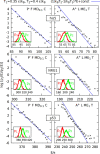
Histogram test for the new model (P F MD0.1 C) and previous model (A+ L ME1 T) based on simulations of proteins his5, NRG1, and p53 at temperatures 0.35 and 0.4 ε/kB. Insets show the energy histograms binned every 1 ε and used to construct the quantity log(p1(E)/p2(E)) shown on the main plots as a function of energy.
Figure 6
Figure 6
Physical time of 1 ns-simulation run on a single core for the new model (P F MD0.1 C, squares) and previous model (A+ L ME1 T, stars) as a function of the system size. The number of protein chains in the simulation is indicated by color. The system density of multichain simulations was set to 1 residue/nm3. Fitted lines have coefficients of 0.003 and 0.0005 s/residue, respectively, with a fit error in the order of 0.0001 s/residue.
Figure 7
Figure 7
Top seven variants of the PID model ranked by their Pearson coefficient.

Similar articles

Cited by

References

    1. Bixon M.; Lifson S. Potential functions and conformations in cycloalkanes. Tetrahedron 1967, 23, 769–784. 10.1016/0040-4020(67)85023-3. - DOI
    1. Lifson S.; Warshel A. Consistent force field for calculations of conformations, vibrational spectra, and enthalpies of cycloalkane and n-alkane molecules. J. Chem. Phys. 1968, 49, 5116–5129. 10.1063/1.1670007. - DOI
    1. Levitt M.; Lifson S. Refinement of protein conformations using a macromolecular energy minimization procedure. J. Mol. Biol. 1969, 46, 269–279. 10.1016/0022-2836(69)90421-5. - DOI - PubMed
    1. McCammon J. A.; Gelin B. R.; Karplus M. Dynamics of folded proteins. Nature 1977, 267, 585.10.1038/267585a0. - DOI - PubMed
    1. Levitt M.; Warshel A. Computer simulation of protein folding. Nature 1975, 253, 694–698. 10.1038/253694a0. - DOI - PubMed

MeSH terms

Substances

LinkOut - more resources