

Observational Study
doi: 10.1148/radiol.2020201933.
Epub 2020 May 21.
Imaging of Neurologic Disease in Hospitalized Patients with COVID-19: An Italian Multicenter Retrospective Observational Study
Affiliations
- PMID: 32437313
- PMCID: PMC7587295
- DOI: 10.1148/radiol.2020201933
Item in Clipboard
Observational Study
Imaging of Neurologic Disease in Hospitalized Patients with COVID-19: An Italian Multicenter Retrospective Observational Study
Radiology.
2020 Nov.
Abstract
Of 725 consecutive hospitalized patients with coronavirus disease 2019, 108 (15%) had acute neurologic symptoms necessitating neurologic imaging.
Figures
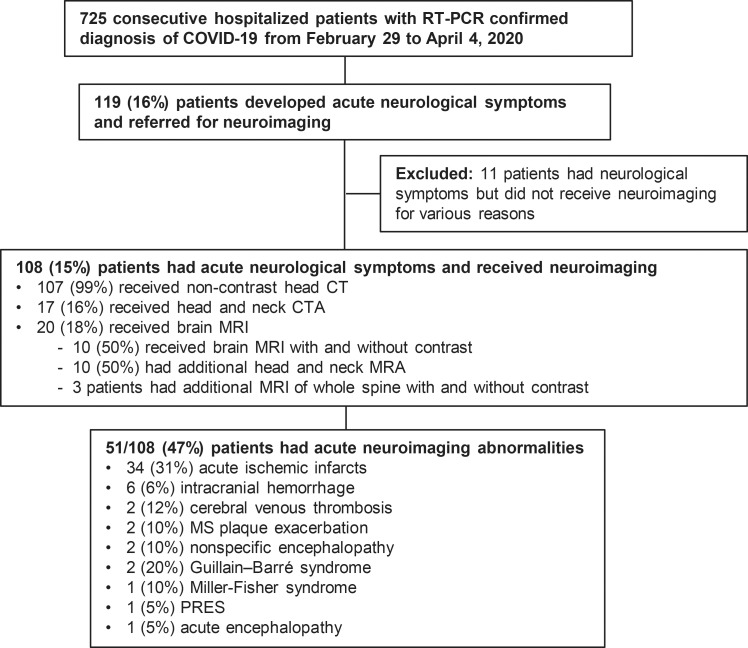
Study flowchart. COVID-19 = coronavirus disease 2019, CTA = CT angiography, MRA = MR angiography, MS = multiple sclerosis, PRES = posterior reversible encephalopathy syndrome, RT-PCR = real-time reverse-transcriptase polymerase chain reaction.

Images of acute encephalopathy in a 60-year-old-man without history of seizures who presented with convulsion. A, B, Fluid-attenuated inversion-recovery images show multifocal areas of hyperintensity in the right cerebellum (arrow in A), left anterior cingular cortex, and superior frontal gyrus (arrows in B). C–E, Diffusion-weighted images show restricted diffusion in the left anterior cingulate cortex and superior frontal gyrus (arrows in C), superior frontal and middle temporal gyrus (arrows in D), and right cerebellum (arrows in E), consistent with cerebellar diaschisis. F, MRI scan obtained with gradient-echo sequence shows no hemosiderin deposits.
Comment in
-
Neurologic Involvement of Patients with Coronavirus Disease 2019: Making the Most of MRI.Radiology. 2020 Oct;297(1):E239. doi: 10.1148/radiol.2020202466. Epub 2020 Jun 9. Radiology. 2020. PMID: 32515675 Free PMC article. No abstract available.
References
-
- Coronavirus disease 2019 (COVID-19) Situation Report – 115. https://www.who.int/docs/default-source/coronaviruse/situation-reports/2.... Published May 14, 2020.
-
- Li Y, Wang M, Zhou Y, et al. Acute Cerebrovascular Disease Following COVID-19: A Single Center, Retrospective, Observational Study. SSRN Electronic Journal 2020 https://www.ncbi.nlm.nih.gov/pmc/articles/PMC7371480/. Published online July 2, 2020. - PMC - PubMed
Publication types
MeSH terms
LinkOut - more resources
Full Text Sources
Medical
