. 2020 Oct;297(1):E223-E227.
doi: 10.1148/radiol.2020202040. Epub 2020 May 21.

COVID-19-associated Diffuse Leukoencephalopathy and Microhemorrhages

Affiliations

COVID-19-associated Diffuse Leukoencephalopathy and Microhemorrhages

Alireza Radmanesh et al. Radiology. 2020 Oct.

Abstract

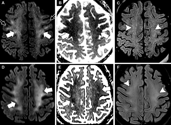
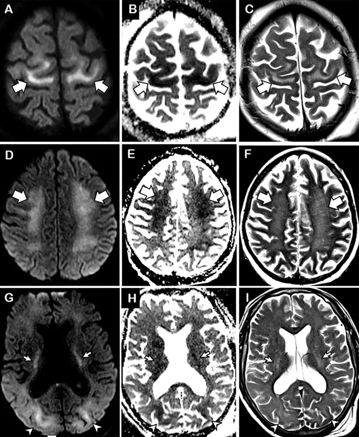
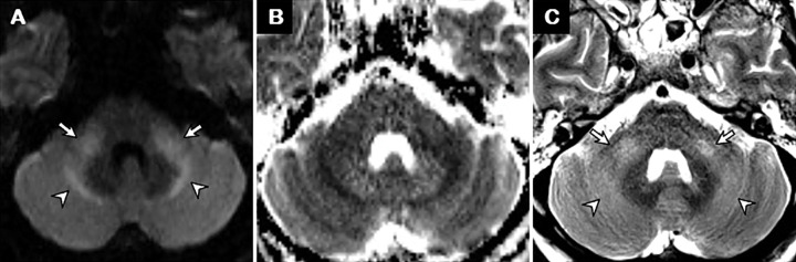
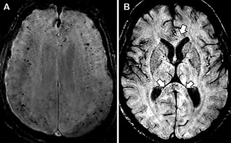
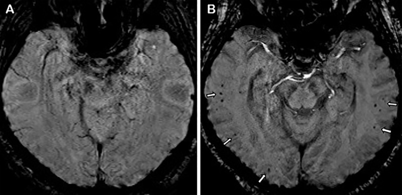
Diffuse leukoencephalopathy and juxtacortical and/or callosal microhemorrhages were brain imaging features in critically ill patients with coronavirus disease 2019. Coronavirus disease 2019 (COVID-19) has been reported in association with a variety of brain imaging findings such as ischemic infarct, hemorrhage, and acute hemorrhagic necrotizing encephalopathy. Herein, the authors report brain imaging features in 11 critically ill patients with COVID-19 with persistently diminished mental status who underwent MRI between April 5 and April 25, 2020. These imaging features include (a) confluent T2 hyperintensity and mild restricted diffusion in bilateral supratentorial deep and subcortical white matter (in 10 of 11 patients) and (b) multiple punctate microhemorrhages in juxtacortical and callosal white matter (in seven of 11 patients). The authors also discuss potential pathogeneses.


Figures

Axial brain MRI scans in two critically ill patients with coronavirus disease 2019 with persistently diminished mental status. A–C, Images in a 56-year-old man. D–F, Images in a 64-year-old man. A, D, Diffusion-weighted images, B, E, apparent diffusion coefficient maps, and, C, F, fluid-attenuated inversion recovery images at the level of centrum semiovale demonstrate symmetric diffuse T2 hyperintensity (arrowheads) and mild restricted diffusion (thick arrows) involving the deep and subcortical white matter with relative sparing of juxtacortical white matter (thin arrows) in both patients. The restricted diffusion is more conspicuous than the T2 hyperintensity.
Figure 1:
Axial brain MRI scans in two critically ill patients with coronavirus disease 2019 with persistently diminished mental status. A–C, Images in a 56-year-old man. D–F, Images in a 64-year-old man. A, D, Diffusion-weighted images, B, E, apparent diffusion coefficient maps, and, C, F, fluid-attenuated inversion recovery images at the level of centrum semiovale demonstrate symmetric diffuse T2 hyperintensity (arrowheads) and mild restricted diffusion (thick arrows) involving the deep and subcortical white matter with relative sparing of juxtacortical white matter (thin arrows) in both patients. The restricted diffusion is more conspicuous than the T2 hyperintensity.
Axial brain MRI scans in a 63-year-old woman who was critically ill due to coronavirus disease 2019. The patient had persistently diminished mental status after 27 days of mechanical ventilation. A, D, G, Diffusion-weighted images, B, E, H, apparent diffusion coefficient maps, and, C, F, I, T2-weighted images at the levels of, A–C, paracentral lobule, D–F, centrum semiovale, and, G–I, corona radiata demonstrate confluent symmetric T2 hyperintensity and restricted diffusion extending from the juxtacortical and subcortical white matter of the precentral gyrus (arrows in A–C) down to the centrum semiovale (arrows in D–F) and down through the posterior limbs of the internal capsules (arrows in G–I) and occipital lobe juxtacortical and subcortical white matter (arrowheads in G–I).
Figure 2:
Axial brain MRI scans in a 63-year-old woman who was critically ill due to coronavirus disease 2019. The patient had persistently diminished mental status after 27 days of mechanical ventilation. A, D, G, Diffusion-weighted images, B, E, H, apparent diffusion coefficient maps, and, C, F, I, T2-weighted images at the levels of, A–C, paracentral lobule, D–F, centrum semiovale, and, G–I, corona radiata demonstrate confluent symmetric T2 hyperintensity and restricted diffusion extending from the juxtacortical and subcortical white matter of the precentral gyrus (arrows in A–C) down to the centrum semiovale (arrows in D–F) and down through the posterior limbs of the internal capsules (arrows in G–I) and occipital lobe juxtacortical and subcortical white matter (arrowheads in G–I).
Axial brain MRI scans in a 64-year-old man who was critically ill due to coronavirus disease 2019. Images were obtained after 28 days of mechanical ventilation. A, Diffusion-weighted image, B, apparent diffusion coefficient map, and, C, T2-weighted image demonstrate patchy faint areas of restricted diffusion and T2 hyperintensity in the middle cerebellar peduncles (arrows) and in the white matter lateral to the deep cerebellar nuclei (arrowheads). The remaining parts of the brainstem and cerebellum are normal.
Figure 3:
Axial brain MRI scans in a 64-year-old man who was critically ill due to coronavirus disease 2019. Images were obtained after 28 days of mechanical ventilation. A, Diffusion-weighted image, B, apparent diffusion coefficient map, and, C, T2-weighted image demonstrate patchy faint areas of restricted diffusion and T2 hyperintensity in the middle cerebellar peduncles (arrows) and in the white matter lateral to the deep cerebellar nuclei (arrowheads). The remaining parts of the brainstem and cerebellum are normal.
Axial susceptibility-weighted images in two critically ill patients with coronavirus disease 2019. A, Image obtained in a 45-year-old man after 23 days of mechanical ventilation. B, Image obtained in a 56-year-old man after 17 days of mechanical ventilation. Images demonstrate numerous punctate microhemorrhagic foci within the juxtacortical white matter, particularly near the depth of the sulci (A), and multiple punctate microhemorrhages within the corpus callosum, particularly in the splenium (arrows in B).
Figure 4:
Axial susceptibility-weighted images in two critically ill patients with coronavirus disease 2019. A, Image obtained in a 45-year-old man after 23 days of mechanical ventilation. B, Image obtained in a 56-year-old man after 17 days of mechanical ventilation. Images demonstrate numerous punctate microhemorrhagic foci within the juxtacortical white matter, particularly near the depth of the sulci (A), and multiple punctate microhemorrhages within the corpus callosum, particularly in the splenium (arrows in B).
Axial susceptibility-weighted images in a 64-year-old man. A, Image obtained 1 week before the current hospital encounter, during work-up for headaches. B, Image obtained after 23 days of mechanical ventilation in the hospital intensive care unit due to coronavirus disease 2019 show multiple juxtacortical punctate microhemorrhages in bilateral temporal and right occipital lobes (arrows) that were not on the previous MRI scan.
Figure 5:
Axial susceptibility-weighted images in a 64-year-old man. A, Image obtained 1 week before the current hospital encounter, during work-up for headaches. B, Image obtained after 23 days of mechanical ventilation in the hospital intensive care unit due to coronavirus disease 2019 show multiple juxtacortical punctate microhemorrhages in bilateral temporal and right occipital lobes (arrows) that were not on the previous MRI scan.

Similar articles

Cited by

References

    1. Mao L, Jin H, Wang M, et al. Neurologic Manifestations of Hospitalized Patients With Coronavirus Disease 2019 in Wuhan, China. JAMA Neurol 2020;77(6):683. - PMC - PubMed
    1. Radmanesh A, Raz E, Zan E, Derman A, Kaminetzky M. Brain Imaging Utilization and Findings in COVID-19: A Single Academic Center Experience in the Epicenter of Disease in the United States. AJNR Am J Neuroradiol 10.3174/ajnr.A6610. Published online May 28, 2020. - DOI - PMC - PubMed
    1. Poyiadji N, Shahin G, Noujaim D, Stone M, Patel S, Griffith B. COVID-19-associated acute hemorrhagic necrotizing encephalopathy: CT and MRI features. Radiology 10.1148/radiol.2020201187. Published online March 31, 2020. Accessed May 2, 2020. - DOI - PMC - PubMed
    1. Levi M, Toh CH, Thachil J, Watson HG. Guidelines for the diagnosis and management of disseminated intravascular coagulation. British Committee for Standards in Haematology. Br J Haematol 2009;145(1):24–33. - PubMed
    1. Li Y, Wang M, Zhou Y, et al. Acute cerebrovascular disease following COVID-19: a single center, retrospective, observational study. Stroke and Vascular Neurology. 10.1136/svn-2020-000431. Published January 2020. Accessed May 2, 2020. - DOI - PMC - PubMed

MeSH terms

LinkOut - more resources