. 2020 Sep;26(9):2005-2015.
doi: 10.3201/eid2609.202095. Epub 2020 May 21.

Pathology and Pathogenesis of SARS-CoV-2 Associated with Fatal Coronavirus Disease, United States

Pathology and Pathogenesis of SARS-CoV-2 Associated with Fatal Coronavirus Disease, United States

Roosecelis B Martines et al. Emerg Infect Dis. 2020 Sep.

Abstract

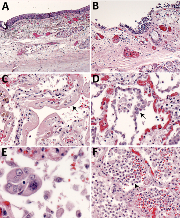
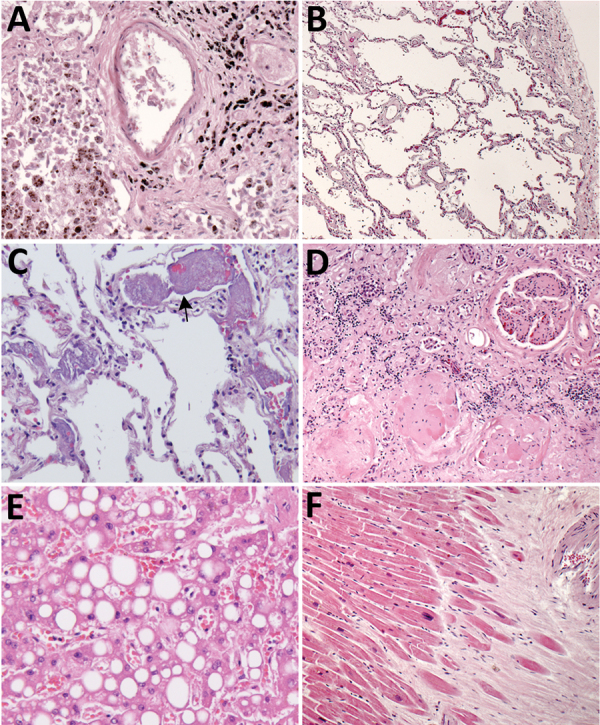
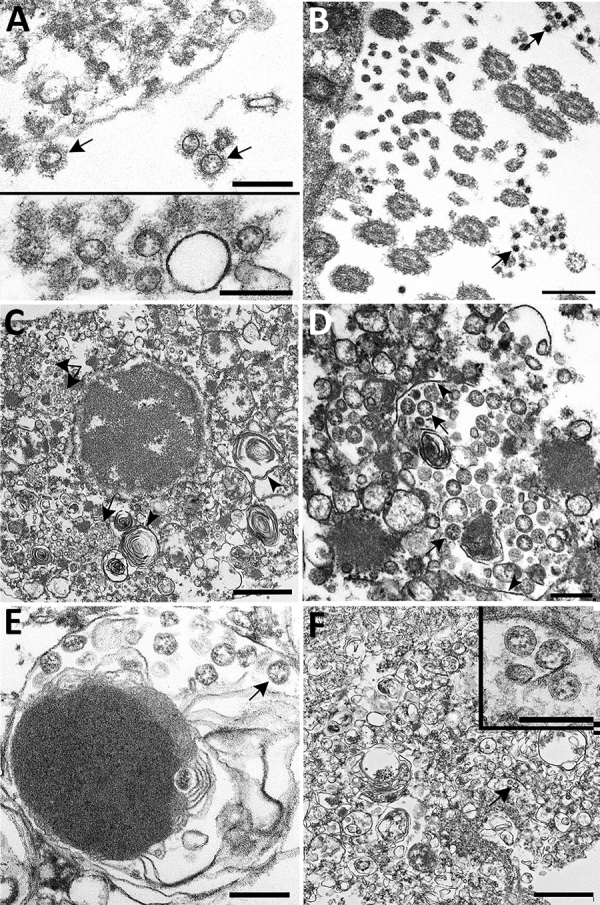
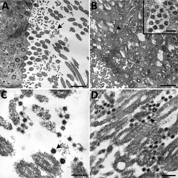
An ongoing pandemic of coronavirus disease (COVID-19) is caused by infection with severe acute respiratory syndrome coronavirus 2 (SARS-CoV-2). Characterization of the histopathology and cellular localization of SARS-CoV-2 in the tissues of patients with fatal COVID-19 is critical to further understand its pathogenesis and transmission and for public health prevention measures. We report clinicopathologic, immunohistochemical, and electron microscopic findings in tissues from 8 fatal laboratory-confirmed cases of SARS-CoV-2 infection in the United States. All cases except 1 were in residents of long-term care facilities. In these patients, SARS-CoV-2 infected epithelium of the upper and lower airways with diffuse alveolar damage as the predominant pulmonary pathology. SARS-CoV-2 was detectable by immunohistochemistry and electron microscopy in conducting airways, pneumocytes, alveolar macrophages, and a hilar lymph node but was not identified in other extrapulmonary tissues. Respiratory viral co-infections were identified in 3 cases; 3 cases had evidence of bacterial co-infection.

Keywords: 2019 novel coronavirus disease; COVID-19; SARS-CoV-2; coronavirus; coronavirus disease; diffuse alveolar damage; electron microscopy; histopathology; immunohistochemistry; pathology; respiratory infections; severe acute respiratory syndrome coronavirus 2; viruses; zoonoses.


Figures

Figure 1
Figure 1
Pulmonary histopathology in fatal coronavirus disease cases caused by severe acute respiratory syndrome coronavirus 2 infection. A) Patient no. 5: tracheitis characterized by moderate mononuclear inflammation within the submucosa (original magnification ×10). B) Patient no. 3: extensive denudation of tracheal epithelium; submucosal congestion, mild edema, and mononuclear inflammation (original magnification ×10). C) Patient no. 4: exudative phase of diffuse alveolar damage characterized by abundant hyaline membranes lining alveolar spaces (arrow) (original magnification ×20). D) Patient no. 8: proliferative phase of diffuse alveolar damage characterized by proliferation of type II pneumocytes (arrow) (original magnification ×20). E) Patient no. 1: atypical pneumocytes with enlarged and multiple nuclei, and expanded cytoplasm in a case with proliferative DAD (original magnification ×40). F) Patient no. 7: bronchopneumonia with filling of alveolar spaces by neutrophils and patchy hemorrhage (arrow) (original magnification ×10).
Figure 2
Figure 2
Histopathologic findings associated with underlying conditions in fatal coronavirus disease. A) Patient no. 2: lung, hemosiderin-laden macrophages (brown pigment, bottom left), and anthracosis (black pigment, top right) in a patient with congestive heart failure (original magnification ×20). B) Patient no. 3: lung, emphysema in a patient with chronic obstructive pulmonary disease (original magnification ×5). C) Patient no. 7: lung, pulmonary microthrombosis (arrow) (original magnification ×20). D) Patient no. 2: kidney, extensive glomerulosclerosis in a patient with renal disease (original magnification ×10). E) Patient no. 3: liver, steatosis in a patient with morbid obesity (original magnification ×20). F) Patient no. 2: heart, myocardial fibrosis and mild cardiomyocyte hypertrophy in a patient with cardiomegaly (original magnification ×5).
Figure 3
Figure 3
Immunostaining of severe acute respiratory syndrome coronavirus 2 in pulmonary tissues from fatal coronavirus disease cases. A) Patient no. 5: scattered immunostaining of tracheal epithelial cells (original magnification ×40). B) Patient no. 5: higher magnification shows immunostaining of ciliated cells (original magnification ×63). C) Patient no. 8: immunostaining of desquamated type I pneumocyte in an alveolar lumen (original magnification ×63). D) Patient no. 4: colocalization of SARS-CoV-2 viral antigen (red) with type II pneumocyte stained by surfactant (brown; arrow) (original magnification ×63). E) Patient no. 4: colocalization of SARS-CoV-2 viral antigen (red) with macrophages stained by CD163 (brown; arrows); virus immunostaining within type II pneumocytes is also seen (arrowheads) (original magnification ×40). F) Patient no. 4: extensive immunostaining of hyaline membranes in a region of exudative DAD (original magnification ×20). G) Patient no. 3: scattered immunostaining within macrophage in hilar lymph node; anthracosis is also present (original magnification ×63).
Figure 4
Figure 4
Ultrastructural features of severe acute respiratory syndrome coronavirus 2 lung infection in fatal coronavirus disease. A) Top: alveolar space containing extracellular virions (arrows) with prominent surface projections. Bottom: cluster of virions in the alveolar space. Scale bars indicate 200 nm. B) Extracellular virions (arrow) associated with ciliated cells of the upper airway. Scale bar indicates 200 nm. C) Membrane-bound vacuoles (arrows) containing viral particles within the cytoplasm of an infected type II pneumocyte; surfactant (lamellated material) indicted by arrowheads. Scale bar indicates 1 μm. D) Membrane-bound vacuole (double-headed arrow in panel C) containing virus particles (arrows) with the characteristic black dots that are cross-sections through the viral nucleocapsid. Arrowheads indicate vacuolar membrane. Scale bar indicates 200 nm. E) Viral particles (arrow) within a phagosome of an alveolar macrophage. Scale bar: 200 nm. F) Viral particles within a portion of a hyaline membrane. Scale bar indicates 800 nm. Inset: Higher magnification of virus particles indicated by arrow; scale bar indicates 200 nm.
Figure 5
Figure 5
Ultrastructural features of severe acute respiratory syndrome coronavirus 2 infection within the upper airway of a fatal coronavirus disease case from formalin-fixed paraffin-embedded (FFPE) tissue. Viral particles associated with the cilia of ciliated cells (A, C, and D) and the cytoplasm of respiratory epithelial cells (B) in the upper airway are indicted by arrows. Images in panels A and C were obtained from FFPE tissue removed from a paraffin block using a 2-mm biopsy punch. Images in panels B and D were collected from a 3 μm section of FFPE tissue affixed to a glass slide. Viral particles visualized in FFPE samples were smaller than those observed from fresh tissue; extracellular viral particles in fresh tissue samples were 105 nm in diameter and those from FFPE tissues were 75 nm in diameter. Scale bars indicate 1 μm (panel A), 800 nm (panel B), and 200 nm (panels C and D).

References

    1. Zhu N, Zhang D, Wang W, Li X, Yang B, Song J, et al.; China Novel Coronavirus Investigating and Research Team. A novel coronavirus from patients with pneumonia in China, 2019. N Engl J Med. 2020;382:727–33. 10.1056/NEJMoa2001017 - DOI - PMC - PubMed
    1. World Health Organization. Coronavirus disease (COVID-19) pandemic. 2020. May 4 [cited 2020 May 5]. https://www.who.int/emergencies/diseases/novel-coronavirus-2019
    1. Holshue ML, DeBolt C, Lindquist S, Lofy KH, Wiesman J, Bruce H, et al.; Washington State 2019-nCoV Case Investigation Team. First case of 2019 novel coronavirus in the United States. N Engl J Med. 2020;382:929–36. 10.1056/NEJMoa2001191 - DOI - PMC - PubMed
    1. Harcourt J, Tamin A, Lu X, Kamili S, Sakthivel SK, Murray J, et al. Severe acute respiratory syndrome coronavirus 2 from patient with 2019 novel coronavirus disease, United States. Emerg Infect Dis. 2020;26:1266–73. 10.3201/eid2606.200516 - DOI - PMC - PubMed
    1. Zhou P, Yang XL, Wang XG, Hu B, Zhang L, Zhang W, et al. A pneumonia outbreak associated with a new coronavirus of probable bat origin. Nature. 2020;579:270–3. 10.1038/s41586-020-2012-7 - DOI - PMC - PubMed

LinkOut - more resources