Review
. 2020 Aug 1;143(8):2341-2368.
doi: 10.1093/brain/awaa072.

Clinical and experimental insight into pathophysiology, comorbidity and therapy of absence seizures

Affiliations
Review

Clinical and experimental insight into pathophysiology, comorbidity and therapy of absence seizures

Vincenzo Crunelli et al. Brain. .

Abstract

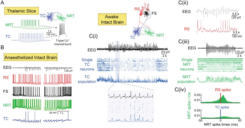
Absence seizures in children and teenagers are generally considered relatively benign because of their non-convulsive nature and the large incidence of remittance in early adulthood. Recent studies, however, show that 30% of children with absence seizures are pharmaco-resistant and 60% are affected by severe neuropsychiatric comorbid conditions, including impairments in attention, cognition, memory and mood. In particular, attention deficits can be detected before the epilepsy diagnosis, may persist even when seizures are pharmacologically controlled and are aggravated by valproic acid monotherapy. New functional MRI-magnetoencephalography and functional MRI-EEG studies provide conclusive evidence that changes in blood oxygenation level-dependent signal amplitude and frequency in children with absence seizures can be detected in specific cortical networks at least 1 min before the start of a seizure, spike-wave discharges are not generalized at seizure onset and abnormal cortical network states remain during interictal periods. From a neurobiological perspective, recent electrical recordings and imaging of large neuronal ensembles with single-cell resolution in non-anaesthetized models show that, in contrast to the predominant opinion, cortical mechanisms, rather than an exclusively thalamic rhythmogenesis, are key in driving seizure ictogenesis and determining spike-wave frequency. Though synchronous ictal firing characterizes cortical and thalamic activity at the population level, individual cortico-thalamic and thalamocortical neurons are sparsely recruited to successive seizures and consecutive paroxysmal cycles within a seizure. New evidence strengthens previous findings on the essential role for basal ganglia networks in absence seizures, in particular the ictal increase in firing of substantia nigra GABAergic neurons. Thus, a key feature of thalamic ictogenesis is the powerful increase in the inhibition of thalamocortical neurons that originates at least from two sources, substantia nigra and thalamic reticular nucleus. This undoubtedly provides a major contribution to the ictal decrease in total firing and the ictal increase of T-type calcium channel-mediated burst firing of thalamocortical neurons, though the latter is not essential for seizure expression. Moreover, in some children and animal models with absence seizures, the ictal increase in thalamic inhibition is enhanced by the loss-of-function of the astrocytic GABA transporter GAT-1 that does not necessarily derive from a mutation in its gene. Together, these novel clinical and experimental findings bring about paradigm-shifting views of our understanding of absence seizures and demand careful choice of initial monotherapy and continuous neuropsychiatric evaluation of affected children. These issues are discussed here to focus future clinical and experimental research and help to identify novel therapeutic targets for treating both absence seizures and their comorbidities.

Keywords: anti-absence drugs; attention deficits; basal ganglia; cortico-thalamo-cortical loop; limbic system.


Figures

Figure 1
Figure 1
Spectrum of SWD features and attention levels in children with absence seizures. (A) Characteristic EEG presentation of SWD in CAE. [B(i)] A long SWD with variable cycle waveform (left trace) was associated with lack of consciousness (indicated by the interruption of counting in the period marked by the two arrows) while a short SWD (right trace) in the same CAE child was not. [B(ii)] SWD with double spikes (enlarged on the right). [B(iii)] A long, hyperventilation-induced SWD from another child with CAE. (C) Different absence seizures, characterized by small and large changes in BOLD signal amplitude and 2.5–4 Hz EEG power, are associated with spared and impaired attention, respectively. DMN = default-mode network; TPN = task-positive network; SMT = sensorimotor-thalamic network. A is modified from Cerminara et al. (2012), and C from Guo et al. (2016).
Figure 2
Figure 2
BOLD signal changes precede absence seizures and persist interictally. (A) Changes in functional MRI BOLD signal amplitude are present in children with absence seizures 14 s before the clinical and electrographic signatures of a seizure are manifested. (B) In individuals with generalized SWD, including a small CAE cohort, an increase in the phase-synchrony of functional MRI BOLD signals is present in a ‘spike-wave cortical network’ from a few seconds before until 20 s after a seizure (top left) (nodes on the far right column). There is also a decreased phase-synchrony in an ‘occipital cortical network’ that starts at least 1 min prior to seizure onset (bottom left). Altered phase-synchrony of BOLD signals in the ‘sensorimotor cortical network’ persists interictally but is not present in healthy controls (middle). Modified from Bai et al. (2010) and Tangwiriyasakul et al. (2018).
Figure 3
Figure 3
Absence seizure ictogenesis in cortico-thalamo-cortical networks. (A) In thalamic slices, glutamatergic thalamocortical (TC) neurons and GABAergic reticular thalamic (NRT) neurons elicit a T-type Ca2+ channel-mediated burst of action potentials at each cycle of the putative paroxysmal activity. (B) In anaesthetized absence seizure models, except TC neurons that fire rarely, all other constituent neurons of cortico-thalamo-cortical networks—cortical regular-spiking excitatory pyramidal neurons (RS), cortical fast-spiking inhibitory neurons (FS) and NRT neurons—show strong (mostly burst) firing at each cycle of a SWD. (C) In sharp contrast, in awake animal models, RS neurons [C(ii)] show a marked ictal decrease of firing (bottom trace is an intracellular recording). The vast majority of TC neurons decrease their firing rate during seizures [raster plot in C(i)] though there is a consistent output at each cycle of the SWD when the firing of these neurons is grouped together [see enlarged population activity in C(i)]. Two groups of NRT neurons can be distinguished on the basis of their ictal firing: one group that increases their firing ictally (mostly consisting of T-type Ca2+ channel-mediated bursts of action potentials) [top three cells in the raster plot in C (iii)] and another group that shows a decreased activity [bottom three cells in the raster plot in C(iii)]. [C(iv)] Histograms of the activity of NRT neurons recorded simultaneously with an RS (top) or and a TC neuron (bottom) in a freely moving absence seizure model. Note how, in contrast to results in thalamic slices (A), the ictal NRT neuron firing (green bars) is mainly driven by the cortical RS neuron spikes rather than the TC neuron spikes. Time zero marks the firing of the RS and TC neurons (red and blue line, respectively). Modified from Bal et al. (1995b); Pinault et al. (1998); Polack et al. (2007); Chipaux et al. (2011); McCafferty et al. (2018b) and Meyer et al. (2018).
Figure 4
Figure 4
Pre-ictal and ictal temporal firing dynamics of absence seizures. (A) Average fluorescence changes indicate a decrease in firing in unidentified cortical layer 5 and 6 (but not layer 4) neurons of the visual cortex of non-anaesthetized, head-restrained stargazer mice that begins more than 2 s before and ends almost 2 s after the electrographic seizure. A similar decrease in total firing rate that start 2 s before seizure onset and terminates at seizure offset also occurs in identified fast-spiking (FS) interneurons of the cortical initiation network (CIN) and in ventrobasal thalamocortical (TC) neurons (blue trace) of freely moving GAERS rats, whereas the total firing GABAergic thalamic reticular neurons (green trace) decreases from 3 s before seizure onset but then show a persistent ictal increase until seizure offset. In contrast, there is an increase in T-channel mediated burst firing in ventrobasal thalamocortical (blue) and thalamic reticular neurons (green) neurons that begins ∼1 s prior to seizure onset and continues until seizure offset. (B) Timing of action potential output with respect to the EEG-spike of different neurons in cortico-thalamo-cortical and basal ganglia pathways are superimposed on a schematic spike-and-wave (light black line). Time zero indicates the peak of the EEG spike and individual brain regions are shown on the right. Data for cortico-thalamic CIN layer 5/6, substantia nigra, ventrobasal thalamocortical and reticular nucleus are from freely moving GAERS rats (Deransart et al., 2003; McCafferty et al., 2018b), whereas data from striatum, subthalamic nucleus, and globus pallidus are from GAERS rats under neurolept anaesthesia (Slaght et al., 2004; Paz et al., 2005, 2007). As the firing time of cortico-thalamic layer 5/6 neurons in the CIN of freely moving GAERS rats occurs about 10 ms earlier than that of the same GAERS neurons recorded under neurolept anaesthesia, the timing of cortico-subthalamic, cortico-striatal, cortico-thalamic CIN layer 2/3 and layer 4 neurons taken from GAERS under neurolept anaesthesia were modified accordingly. FS-INT = fast-spiking GABAergic interneurons; MSN = striatal medium spiny neurons. Open and filled symbols indicate inhibitory GABAergic and excitatory glutamatergic neurons, respectively. (A) Data for CIN FS interneurons are unpublished observations, the others were modified from McCafferty et al. (2018b) and Meyer et al. (2018).
Figure 5
Figure 5
T-type Ca2+ channels in cortex and nucleus reticularis thalami (NRT) are necessary for absence seizures. Schematic representation of injection sites with their complement of T-channel subtypes (left) and original data (right) showing the ability of the potent and selective pan T-type Ca2+ channel antagonist, TTA-P2, to block absence seizures in GAERS rats when bilaterally injected by microdialysis in the CIN (top row) or the NRT (middle row). Similar administration in the ventrobasal (VB) thalamic nucleus (one of the somatotopic thalamic nuclei of the CIN in this animal model) that contains only thalamocortical neurons, has no effect on absence seizures (bottom row).
Figure 6
Figure 6
Ictogenic firing of basal ganglia neurons. Schematic diagram of the ictal activity of the component neurons of the direct and indirect basal ganglia pathways recorded under neurolept anaesthesia (except the substantia nigra neurons) are illustrated close to the morphological representation of each neuronal type. Electrical activity of fast-spiking striatal interneurons and substantia nigra and subthalamic neurons shows extracellular recordings, the others are intracellular recordings. The excitatory and inhibitory nature of the synaptic connections is indicated by encircled plus and minus symbols. The relative size of the morphological reconstructions has been altered for graphical purposes. See text for additional details. Modified from Deransart et al. (2003); Slaght et al. (2004) and Paz et al. (2005, 2007).
Figure 7
Figure 7
Pharmaco-resistance of absence seizures. The first 12-month-long monotherapy with either ethosuximide (ETX) or valproic acid (VPA) is more effective than that with lamotrigine (LTG) in controlling absence seizures (left). Similar results are obtained when children unresponsive to the initial monotherapy undergo a second 12-month-long monotherapy with a different drug (middle). The combined results of the first and second monotherapy (right) show that about 30% of children with childhood absence epilepsy have pharmaco-resistant absence seizures. Modified from Glauser et al. (2013) and Cnaan et al. (2017).
Figure 8
Figure 8
Neuropsychiatric comorbidities of absence seizures. Attention deficits are the most common psychiatric comorbidity of childhood absence epilepsy; they can be present in pre-symptomatic children and persist even when seizures are pharmacologically controlled. Other cognitive impairments and mood disorders (e.g. anxiety, depression) may also be present. It is likely that the aberrant cortico-thalamo-cortical networks underlying absence seizure combine with the abnormal basal ganglia-limbic-monoamine networks underlying cognitive impairments and mood disorders to generate the overall neurological and neuropsychiatric phenotype of epilepsies with absence seizures (left column). Interactions between these abnormal networks might contribute to a lower seizure threshold and an increased risk of comorbidity. Moreover, absence seizures and anti-absence drugs may induce, or aggravate existing, comorbid conditions (right column). Solid and dashed blue lines represent established and putative links, respectively.

References

    1. Adamantidis AR, Gutierrez Herrera C, Gent TC.. Oscillating circuitries in the sleeping brain. Nat Rev Neurosci 2019; 20: 746–62. - PubMed
    1. Aghakhani Y, Bagshaw AP, Bénar CG, Hawco C, Andermann F, Dubeau F, et al.fMRI activation during spike and wave discharges in idiopathic generalized epilepsy. Brain 2004; 127: 1127–44. - PubMed
    1. Aker RG, Yananli HR, Gurbanova AA, Ozkaynakci AE, Ates N, Luijtelaar G, et al.Amygdala kindling in the WAG/Rij rat model of absence epilepsy. Epilepsia 2006; 47: 33–40. - PubMed
    1. Aldenkamp AP, Arends J, Overweg-Plandsoen TC, van Bronswijk KC, Schyns-Soeterboek A, Linden I, et al.Acute cognitive effects of nonconvulsive difficult-to-detect epileptic seizures and epileptiform electroencephalographic discharges. J Child Neurol 2001; 16: 119–23. - PubMed
    1. Antal M, Beneduce BM, Regehr WG.. The substantia nigra conveys target-dependent excitatory and inhibitory outputs from the basal ganglia to the thalamus. J. Neurosci 2014; 34: 8032–42. - PMC - PubMed

Publication types