Review
. 2021 Jan:80:100863.
doi: 10.1016/j.preteyeres.2020.100863. Epub 2020 May 8.

Fuchs endothelial corneal dystrophy: The vicious cycle of Fuchs pathogenesis

Affiliations
Review

Fuchs endothelial corneal dystrophy: The vicious cycle of Fuchs pathogenesis

Stephan Ong Tone et al. Prog Retin Eye Res. 2021 Jan.

Abstract

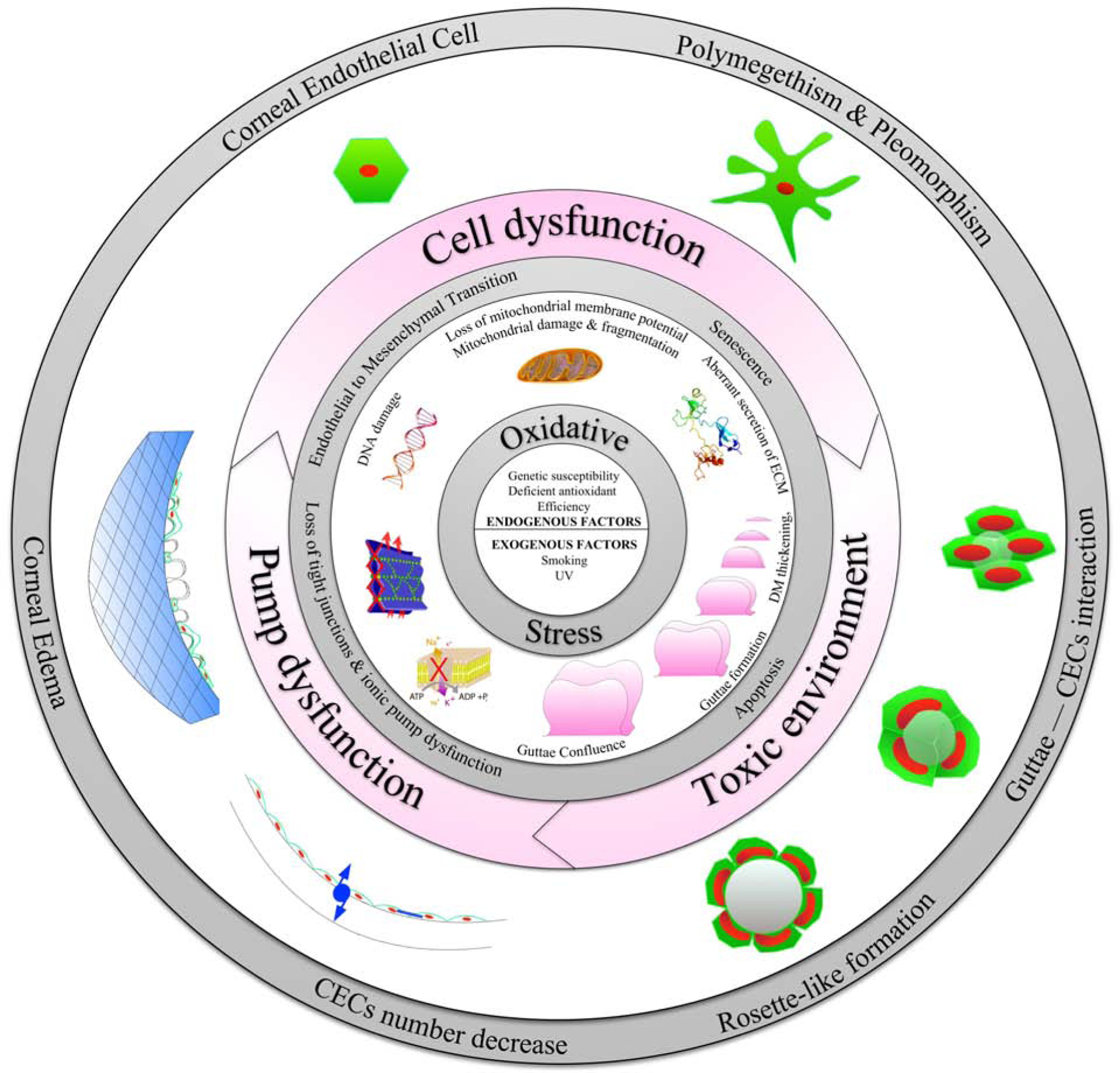
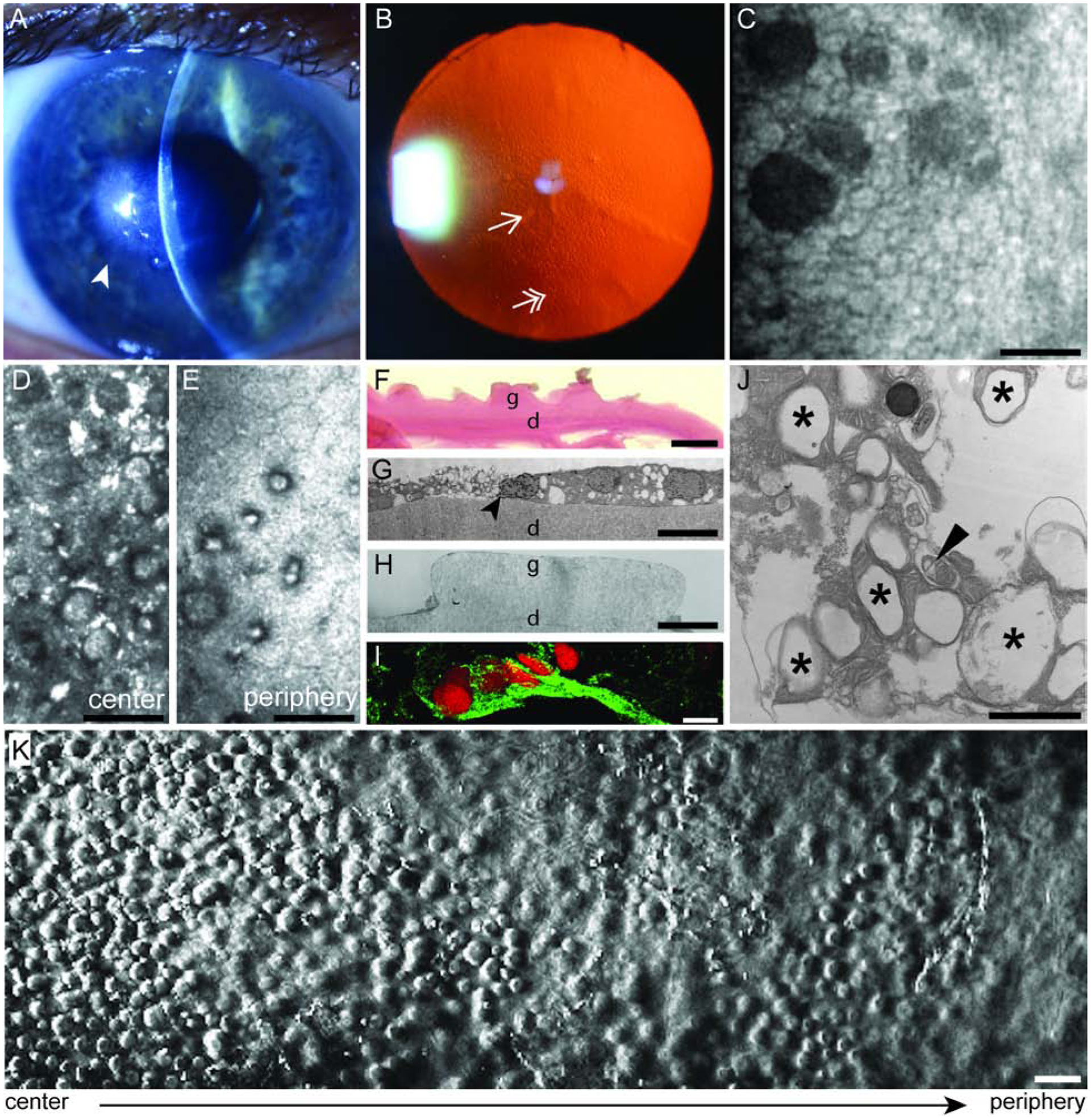
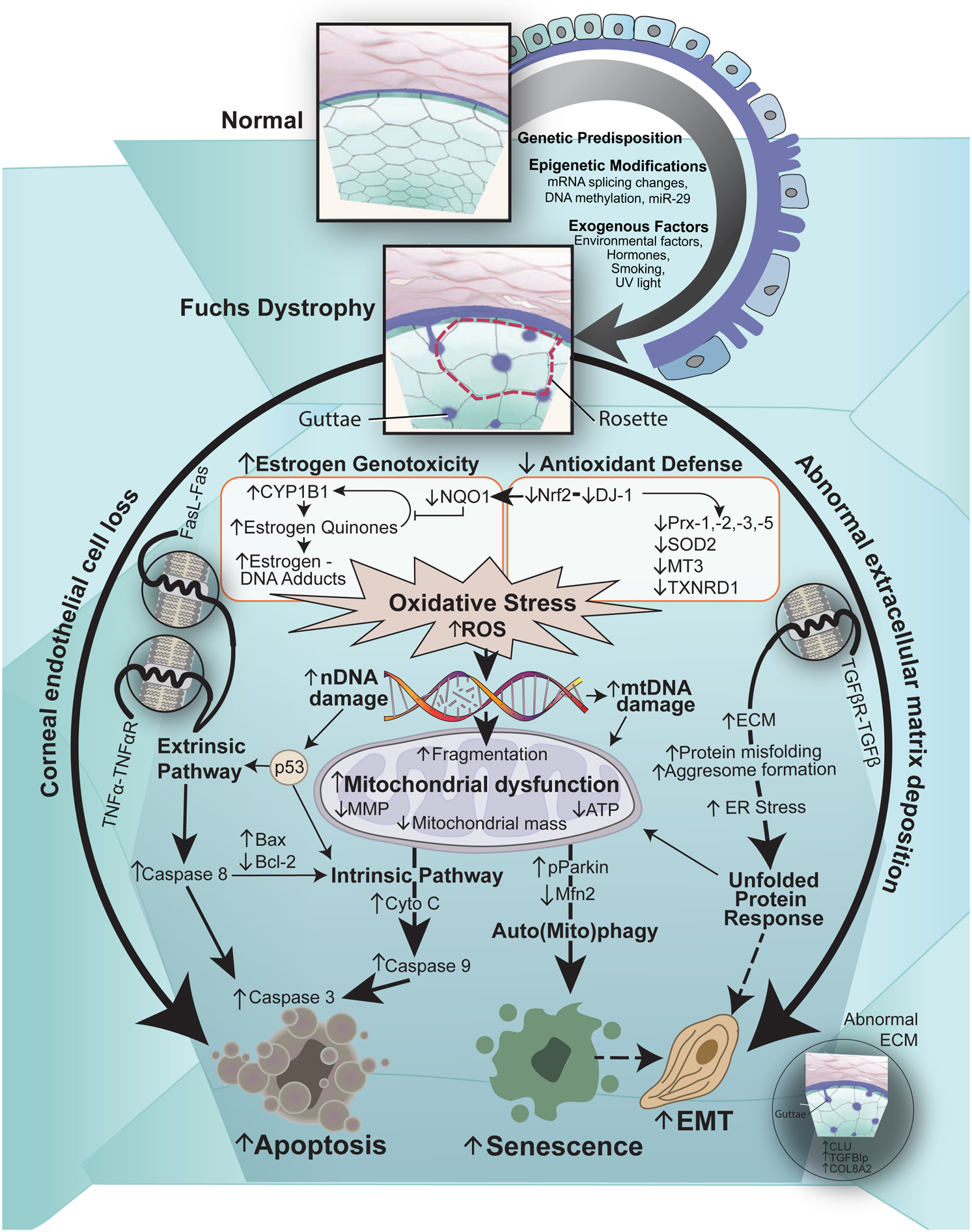
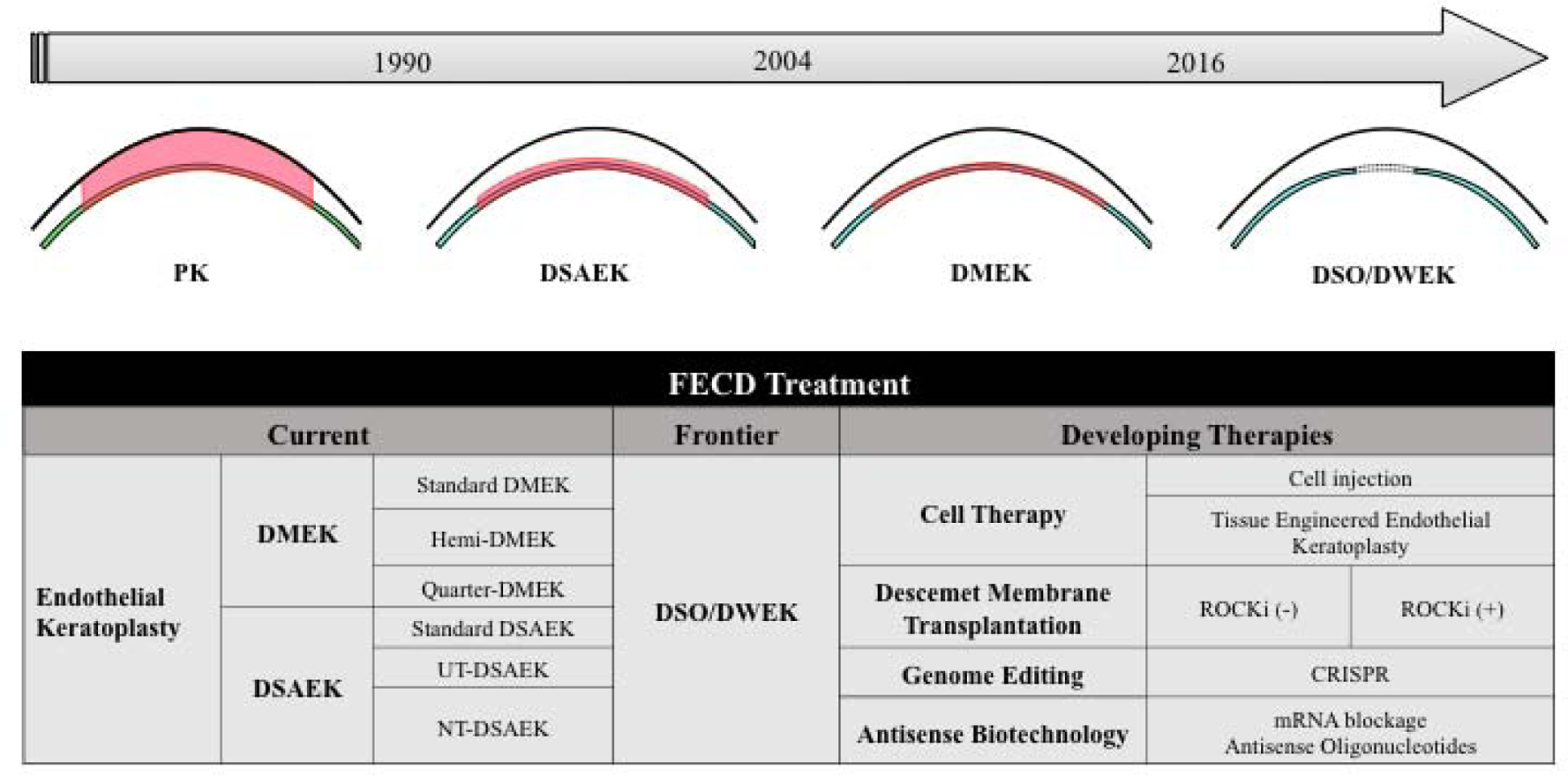
Fuchs endothelial corneal dystrophy (FECD) is the most common primary corneal endothelial dystrophy and the leading indication for corneal transplantation worldwide. FECD is characterized by the progressive decline of corneal endothelial cells (CECs) and the formation of extracellular matrix (ECM) excrescences in Descemet's membrane (DM), called guttae, that lead to corneal edema and loss of vision. FECD typically manifests in the fifth decades of life and has a greater incidence in women. FECD is a complex and heterogeneous genetic disease where interaction between genetic and environmental factors results in cellular apoptosis and aberrant ECM deposition. In this review, we will discuss a complex interplay of genetic, epigenetic, and exogenous factors in inciting oxidative stress, auto(mito)phagy, unfolded protein response, and mitochondrial dysfunction during CEC degeneration. Specifically, we explore the factors that influence cellular fate to undergo apoptosis, senescence, and endothelial-to-mesenchymal transition. These findings will highlight the importance of abnormal CEC-DM interactions in triggering the vicious cycle of FECD pathogenesis. We will also review clinical characteristics, diagnostic tools, and current medical and surgical management options for FECD patients. These new paradigms in FECD pathogenesis present an opportunity to develop novel therapeutics for the treatment of FECD.

Keywords: Apoptosis; Corneal endothelium; Fuchs endothelial corneal dystrophy; Guttae; Mitochondria; Oxidative stress.


Figures

Figure 1.
Figure 1.. The Vicious Cycle of Fuchs Endothelial Corneal Dystrophy (FECD).
Schematic diagram illustrating the complex interplay between genetic, epigenetic, and exogenous factors in inciting oxidative stress, endothelial to mesenchymal transition, senescence, mitochondrial dysfunction, and apoptosis in corneal endothelial cells (CECs) in FECD. Furthermore, guttae formation contributes to a toxic microenvironment resulting in abnormal CEC-Descemet’s membrane interactions triggering the vicious cycle of FECD pathogenesis.
Figure 2.
Figure 2.. Clinical Features of Fuchs Endothelial Corneal Dystrophy (FECD).
(A) Slit lamp photograph showing central cornea edema (arrowhead) in a patient with FECD. (B) Retroillumination photograph showing confluent central (single arrowhead) and non-confluent peripheral (double arrowhead) guttae. (C) Non-contact specular micrograph showing the corneal endothelium (white) and guttae (dark spaces) in a patient with FECD. Scale bar = 50 μm. (D) In vivo confocal microscopy showing the loss of corneal endothelial cells (CECs) and presence of guttae in the center and (E) periphery of a patient with advanced FECD. Scale bar = 50 μm. (F) Periodic Acid Schiff (PAS) staining of a cornea from a patient with FECD demonstrating excrescences called guttae (g) from Descemet’s membrane (d). Scale bar = 10 μm. (G) Transmission electron microscopy from a patient with FECD showing a CEC undergoing apoptosis (black arrowhead) on Descemet’s membrane (d) and (H) guttae (g). Scale bar = 10 μm. (I) Expression of endothelial mesenchymal marker in a FECD ex vivo specimen. Immunofluorescence distribution, observed by confocal microscopy, of Snail1 protein (green) in the cytoplasm of CECs on a FECD ex vivo specimen. Cells were stained with propidium iodide (red) to localize nuclei. Scale bar = 50 μm. (J) Transmission electron microscopy of FECD ex vivo specimen that demonstrate increased number of autophagic structures in the form of vacuoles (black stars) and autophagosome containing mitochondria (black arrowhead). Scale bar = 1 μm. (K) Phase contrast microscopy of an ex vivo corneal tissue specimen from a patient with FECD that underwent endothelial keratoplasty. A higher density of guttae is observed in the center of the cornea compared to the periphery. Scale bar = 50 μm.
Figure 3.
Figure 3.. The Pathogenesis of Fuchs Endothelial Corneal Dystrophy (FECD).
Schematic diagram illustrating our hypothesis on the vicious cycle of FECD pathogenesis. Cellular stress leading to corneal endothelial cell (CEC) loss and abnormal extracellular matrix (ECM) deposition, called guttae, and altered cellular-ECM interactions are the hallmarks of FECD. Genetic predisposition, epigenetic modifications and multiple exogenous factors leads to increased intracellular oxidative stress and reactive oxygen species (ROS) production, resulting in nuclear DNA (nDNA) and mitochondrial DNA (mDNA) damage, and mitochondrial dysfunction and alteration in the synthetic capacity of cells, which maintains the vicious cycle of propagating oxidant–antioxidant imbalance in the pathogenesis of FECD. Furthermore, CECs undergo apoptosis through both the extrinsic and intrinsic pathways, endothelial to mesenchymal transition (EMT) through the unfolded protein response and cellular senescence through auto(mito)phagy. Legend: mRNA=messenger RNA; miR-29= microRNA-29; UV=ultraviolet; TNFα= tumor necrosis factor alpha; TNFαR= tumor necrosis factor alpha receptor; FasL= Fas ligand; TGFβ= transforming growth factor beta; TGFβR= transforming growth factor beta receptor; Nrf= NF-E2 (nuclear factor erythroid 2) related factor; DJ-1= protein deglycase; NQO1= NAD(P)H: quinone oxidoreductase 1; Prx= peroxiredoxins; SOD2= superoxide dismutase 2; MT3= metallothionein-3; TXNRD1= thioredoxin reductase 1; MMP= ; ATP= ;ER= endoplasmic reticulum; pParkin= phosphoParkin; Mfn2= mitofusin 2; Cyto C= cytochrome C; Bax= Bcl-2-associated X protein; Bcl-2= B-cell lymphoma 2; CLU= clusterin; TGFBIp= transforming growth factor beta-induced protein.
Figure 4.
Figure 4.. Current, Frontier and Developing Therapies for Fuchs Endothelial Corneal Dystrophy (FECD).
Legend: PK= penetrating keratoplasty; DSAEK= Descemet stripping automated endothelial keratoplasty; DMEK= Descemet membrane endothelial keratoplasty; DWEK= Descemetorhexis without endothelial keratoplasty; DSO= Descemet stripping only; UT-DSAEK= ultrathin-DSAEK; NT-DSAEK= nanothin-DSAEK; ROCKi= Rho kinase inhibitor; TGFβ= transforming growth factor beta; EK= endothelial keratoplasty; DMT= Descemet membrane transfer; RNAi= RNA interference; miRNA= micro RNA; CRISPR= clustered regularly interspaced short palindromic repeats.

References

    1. Abrams GA, Schaus SS, Goodman SL, Nealey PF, Murphy CJ, 2000. Nanoscale topography of the corneal epithelial basement membrane and Descemet’s membrane of the human. Cornea 19, 57–64. - PubMed
    1. Adamis AP, Filatov V, Tripathi BJ, Tripathi RC, 1993. Fuchs’ endothelial dystrophy of the cornea. Surv Ophthalmol 38, 149–168. - PubMed
    1. Adamis AP, Molnar ML, Tripathi BJ, Emmerson MS, Stefansson K, Tripathi RC, 1985. Neuronal-specific enolase in human corneal endothelium and posterior keratocytes. Exp Eye Res 41, 665–668. - PubMed
    1. Afshari NA, Igo RP Jr., Morris NJ, Stambolian D, Sharma S, Pulagam VL, Dunn S, Stamler JF, Truitt BJ, Rimmler J, Kuot A, Croasdale CR, Qin X, Burdon KP, Riazuddin SA, Mills R, Klebe S, Minear MA, Zhao J, Balajonda E, Rosenwasser GO, Baratz KH, Mootha VV, Patel SV, Gregory SG, Bailey-Wilson JE, Price MO, Price FW Jr., Craig JE, Fingert JH, Gottsch JD, Aldave AJ, Klintworth GK, Lass JH, Li YJ, Iyengar SK, 2017. Genome-wide association study identifies three novel loci in Fuchs endothelial corneal dystrophy. Nat Commun 8, 14898. - PMC - PubMed
    1. Afshari NA, Li YJ, Pericak-Vance MA, Gregory S, Klintworth GK, 2009. Genome-wide linkage scan in fuchs endothelial corneal dystrophy. Investigative ophthalmology & visual science 50, 1093–1097. - PMC - PubMed

Publication types