. 2020 May 19;10(5):208.
doi: 10.3390/metabo10050208.

Tryptophan Metabolism, Inflammation, and Oxidative Stress in Patients with Neurovascular Disease

Affiliations

Tryptophan Metabolism, Inflammation, and Oxidative Stress in Patients with Neurovascular Disease

Martin Hajsl et al. Metabolites. .

Abstract

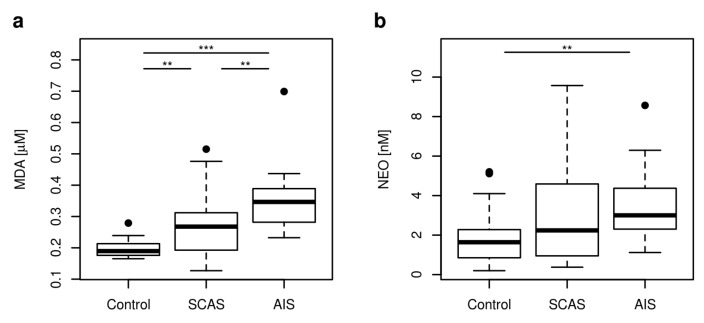
Atherosclerosis is a leading cause of major vascular events, myocardial infarction, and ischemic stroke. Tryptophan (TRP) catabolism was recognized as an important player in inflammation and immune response having together with oxidative stress (OS) significant effects on each phase of atherosclerosis. The aim of the study is to analyze the relationship of plasma levels of TRP metabolites, inflammation, and OS in patients with neurovascular diseases (acute ischemic stroke (AIS), significant carotid artery stenosis (SCAS)) and in healthy controls. Blood samples were collected from 43 patients (25 with SCAS, 18 with AIS) and from 25 healthy controls. The concentrations of twelve TRP metabolites, riboflavin, neopterin (NEO, marker of inflammation), and malondialdehyde (MDA, marker of OS) were measured by liquid chromatography-tandem mass spectrometry (LC-MS/MS). Concentrations of seven TRP metabolites (TRP, kynurenine (KYN), 3-hydroxykynurenine (3-HK), 3-hydroxyanthranilic acid (3-HAA), anthranilic acid (AA), melatonin (MEL), tryptamine (TA)), NEO, and MDA were significantly different in the studied groups. Significantly lower concentrations of TRP, KYN, 3-HAA, MEL, TA, and higher MDA concentrations were found in AIS compared to SCAS patients. MDA concentration was higher in both AIS and SCAS group (p < 0.001, p = 0.004, respectively) compared to controls, NEO concentration was enhanced (p < 0.003) in AIS. MDA did not directly correlate with TRP metabolites in the study groups, except for 1) a negative correlation with kynurenine acid and 2) the activity of kynurenine aminotransferase in AIS patients (r = -0.552, p = 0.018; r = -0.504, p = 0.033, respectively). In summary, TRP metabolism is clearly more deregulated in AIS compared to SCAS patients; the effect of TRP metabolites on OS should be further elucidated.

Keywords: acute ischemic stroke; atherosclerosis; carotid artery stenosis; inflammation; oxidative stress; tryptophan metabolism.


Conflict of interest statement

The authors declare no conflict of interest.

Figures

Figure A1
Figure A1
Correlation graphs of OS marker MDA with KA (a) and with 100 × [KA]/[KYN] (activity of KMO) (b) in plasma of AIS patients. Abbreviations: MDA: malondialdehyde; KA: kynurenic acid; KMO: kynurenine 3-monooxygenase.
Figure A2
Figure A2
Graphs of (a) 3-HK correlation with inflammatory marker NEO, (b) 3-HK correlation with inflammatory marker KTR, and (c) 3-HK correlation with QA in plasma of patients with SCAS (blue color) and AIS (red color). (d) Correlation of QA with KTR in plasma of patients with SCAS and AIS. Abbreviations: 3-HK: 3-hydroxykynurenine; QA: quinolinic acid; KTR: 1000 × [KYN]/[TRP].
Figure A3
Figure A3
Graph of (a) neurotoxin QA correlation with inflammatory marker NEO in plasma of SCAS patients (blue color) and (b) correlation of inflammatory markers KTR and NEO in plasma of SCAS patients and in plasma of all probands (black color). Abbreviations: QA: quinolinic acid; NEO: neopterin; KTR: 1000 × [KYN]/[TRP].
Figure 1
Figure 1
Simpified major metabolic pathways of tryptophan (TRP) in humans. Metabolites marked in bold were quantified by liquid chromatography–tandem mass spectrometry (LC-MS/MS) in the current study. The area enclosed by the dotted line indicates kynurenine pathway (KP).
Figure 2
Figure 2
Plasma concentrations of kynurenine pathway metabolites changed in neurovascular disease. The concentrations or ratios of metabolites in the plasma of the controls (control), patients with significant carotid artery stenosis (SCAS), or acute ischemic stroke (AIS) are represented as boxplots: (a) TRP: tryptophan; (b) KYN: kynurenine; (c) 3-HK: 3-hydroxykynurenine; (d) 3-HAA: 3-hydroxyanthranilic acid; (e) AA: anthranilic acid; (f) 3-HAA to AA ratio. Significance codes for post-hoc Dunn test: *** p < 0.001, ** p < 0.01, * p < 0.05. Vertical boxplot is constructed between first (Q1) and third (Q3) quartile, with horizontal median inside. The difference between Q3 and Q1 is called the interquartile range (IQR). Whiskers are drawn from Q1 and Q3 to minimal, respective maximal data point within range (Q1 – 1.5 IRQ) – (Q3 + 1.5 IRQ). Other data are outliers (black circles).
Figure 3
Figure 3
The boxplots of enzyme activities changed in neurovascular disease. (a) KTR: IDO activity defined as 1000 × [KYN]/[TRP]; (b) 100 × [3-HK]/[KYN]: KMO activity; (c) 100 × [AA]/[KYN]: KYNU A activity; (d) 100 × [3-HAA]/[3-HK]: KYNU B activity; (e) 100 × [QA]/[3-HAA]: 3-HAO activity; (f) 100 × [PA]/[3-HAA]: the composed 3-HAO and ACMSD activity. Significance codes for post-hoc Dunn test: *** p < 0.001, ** p < 0.01, * p < 0.05.
Figure 4
Figure 4
The boxplots of non KP metabolites concentrations changed in neurovascular disease. (a) MEL: melatonin; (b) TA: tryptamine. Significance codes for post-hoc Dunn test: *** p < 0.001, ** p < 0.01, * p < 0.05.
Figure 5
Figure 5
Plasma concentrations of MDA and NEO represented as boxplots. (a) MDA concentration was employed as a criterion of OS; (b) NEO concentration was employed as a marker of inflammation. Abbreviations: MDA: malondialdehyde; NEO: neopterin. Significance codes for post-hoc Dunn test: *** p < 0.001, ** p < 0.01.

References

    1. Wolf D., Ley K. Immunity and inflammation in atherosclerosis. Circ. Res. 2019;124:315–327. doi: 10.1161/CIRCRESAHA.118.313591. - DOI - PMC - PubMed
    1. Fatkhullina A.R., Peshkova I.O., Koltsova E.K. The Role of Cytokines in the Development of Atherosclerosis. Biochemistry. 2016;81:1358–1370. doi: 10.1134/S0006297916110134. - DOI - PMC - PubMed
    1. Forrester S.J., Kikuchi D.S., Hernandes M.S., Xu Q., Griendling K.K. Reactive Oxygen Species in Metabolic and Inflammatory Signaling. Circ. Res. 2018;122:877–902. doi: 10.1161/CIRCRESAHA.117.311401. - DOI - PMC - PubMed
    1. Miteva K., Madonna R., De Caterina R., Van Linthout S. Innate and adaptive immunity in atherosclerosis. Vascul. Pharmacol. 2018;107:67–77. doi: 10.1016/j.vph.2018.04.006. - DOI - PubMed
    1. Kattoor A.J., Pothineni N.V.K., Palagiri D., Mehta J.L. Oxidative Stress in Atherosclerosis. Curr. Atheroscler. Rep. 2017;19:42. doi: 10.1007/s11883-017-0678-6. - DOI - PubMed

LinkOut - more resources