. 2020 Jun;41(6):1001-1008.
doi: 10.3174/ajnr.A6547. Epub 2020 May 21.

Value of 3T Susceptibility-Weighted Imaging in the Diagnosis of Multiple Sclerosis

Affiliations

Value of 3T Susceptibility-Weighted Imaging in the Diagnosis of Multiple Sclerosis

M A Clarke et al. AJNR Am J Neuroradiol. 2020 Jun.

Abstract

Background and purpose: Previous studies have suggested that the central vein sign and iron rims are specific features of MS lesions. Using 3T SWI, we aimed to compare the frequency of lesions with central veins and iron rims in patients with clinically isolated syndrome and MS-mimicking disorders and test their diagnostic value in predicting conversion from clinically isolated syndrome to MS.

Materials and methods: For each patient, we calculated the number of brain lesions with central veins and iron rims. We then identified a simple rule involving an absolute number of lesions with central veins and iron rims to predict conversion from clinically isolated syndrome to MS. Additionally, we tested the diagnostic performance of central veins and iron rims when combined with evidence of dissemination in space.

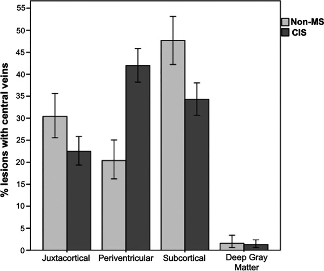
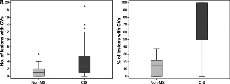
Results: We included 112 patients with clinically isolated syndrome and 35 patients with MS-mimicking conditions. At follow-up, 94 patients with clinically isolated syndrome developed MS according to the 2017 McDonald criteria. Patients with clinically isolated syndrome had a median of 2 central veins (range, 0-19), while the non-MS group had a median of 1 central vein (range, 0-6). Fifty-six percent of patients who developed MS had ≥1 iron rim, and none of the patients without MS had iron rims. The sensitivity and specificity of finding ≥3 central veins and/or ≥1 iron rim were 70% and 86%, respectively. In combination with evidence of dissemination in space, the 2 imaging markers had higher specificity than dissemination in space and positive findings of oligoclonal bands currently used to support the diagnosis of MS.

Conclusions: A single 3T SWI scan offers valuable diagnostic information, which has the potential to prevent MS misdiagnosis.


Figures

Fig 1.
Fig 1.
Patient flow diagram showing how patients were selected for the study and the reasons for exclusion. We excluded patients for the following reasons: 1) missing either their SWI or T2-FLAIR scan; 2) lesions <3 mm or infratentorial lesions that could not be identified on the SWI due to artifacts; 3) automated lesion masks not passing quality checks; and 4) diagnostic doubt by the end of the study follow-up. RRMS indicates relapsing-remitting multiple sclerosis.
Fig 2.
Fig 2.
Lesion appearance on axial T2-FLAIR (upper row) and the corresponding susceptibility-weighted (lower row) images in patients with Sjögren disease (A and C) and MS (B and D). The patient with Sjögren disease has no visible CVs or IRs on the SWI. The patient with MS has clearly visible IRs, which correspond to the lesion edges visible on the T2-FLAIR. CVs are also visible inside the lesions as hypointense dots or lines.
Fig 3.
Fig 3.
The location of lesions with central veins in the CIS and non-MS groups. Error bars represent standard error.
Fig 4.
Fig 4.
Summary of the incidence of lesions with CVs in the CIS and non-MS groups. A, The number of lesions with CVs (per patient) in the 2 groups. B, The percentage of lesions with CVs (per patient) in the 2 groups.
Fig 5.
Fig 5.
Summary of the incidence of lesions with iron rings in the CIS and non-MS groups. A, The number of lesions with iron rings (per patient) in the 2 groups. B, The location of lesions with iron rings in the CIS group.

References

    1. Thompson AJ, Banwell BL, Barkhof F, et al. . Diagnosis of multiple sclerosis: 2017 revisions of the McDonald criteria. Lancet Neurol 2018;17:162–73 10.1016/S1474-4422(17)30470-2 - DOI - PubMed
    1. Hyun JW, Kim W, Huh SY, et al. . Application of the 2017 McDonald diagnostic criteria for multiple sclerosis in Korean patients with clinically isolated syndrome. Mult Scler 2019;25:1488–95 10.1177/1352458518790702 - DOI - PubMed
    1. Gobbin F, Zanoni M, Marangi A, et al. . 2017 McDonald criteria for multiple sclerosis: earlier diagnosis with reduced specificity? Mult Scler Relat Disord 2019;29:23–25 10.1016/j.msard.2019.01.008 - DOI - PubMed
    1. Reichenbach JR, Venkatesan R, Schillinger DJ, et al. . Small vessels in the human brain: MR venography with deoxyhemoglobin as an intrinsic contrast agent. Radiology 1997;204:272–77 10.1148/radiology.204.1.9205259 - DOI - PubMed
    1. Haacke EM, Xu Y, Cheng YC, et al. . Susceptibility weighted imaging (SWI). Magn Reson Med 2004;52:612–18 10.1002/mrm.20198 - DOI - PubMed

Publication types