

Case Reports
doi: 10.3174/ajnr.A6595.
Epub 2020 May 21.
Hemorrhagic Posterior Reversible Encephalopathy Syndrome as a Manifestation of COVID-19 Infection
Affiliations
- PMID: 32439646
- PMCID: PMC7357664
- DOI: 10.3174/ajnr.A6595
Item in Clipboard
Case Reports
Hemorrhagic Posterior Reversible Encephalopathy Syndrome as a Manifestation of COVID-19 Infection
AJNR Am J Neuroradiol.
2020 Jul.
Abstract
We describe 2 hospitalized patients with confirmed coronavirus 19 (COVID-19) infection in whom brain imaging showed hemorrhagic posterior reversible encephalopathy syndrome, and we discuss the possible reasons for these findings and their relationship to the infection.
© 2020 by American Journal of Neuroradiology.
Figures
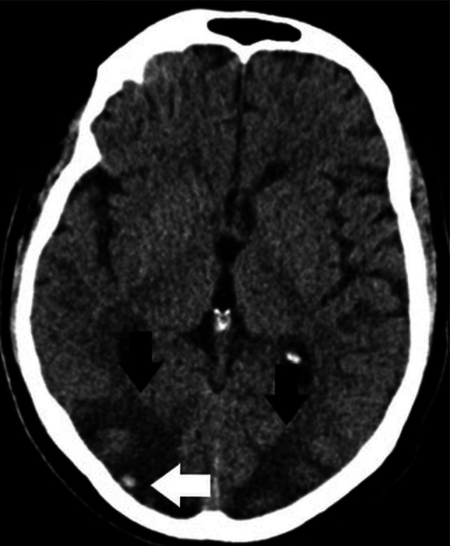
Axial noncontrast CT demonstrates edema in the posterior parieto-occipital regions (black arrows) with a superimposed small right-side hemorrhage (white arrow).
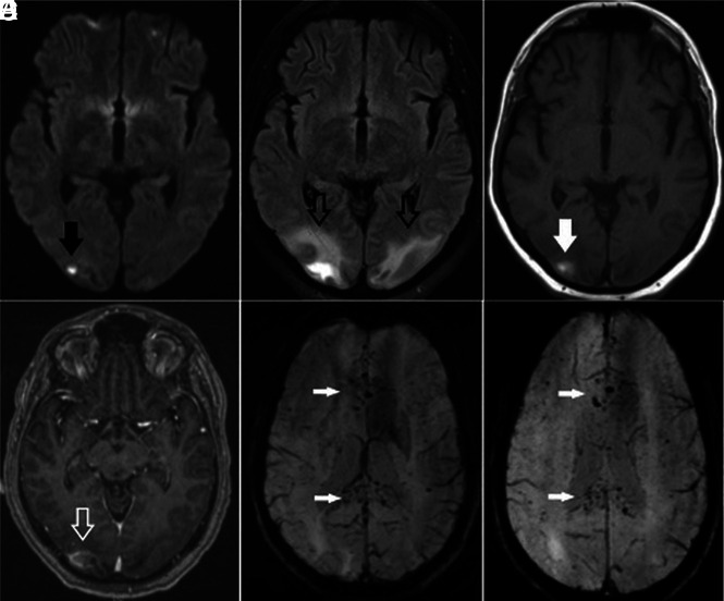
Brain axial DWI (A), FLAIR (B), precontrast T1-weighted (C), postcontrast T1-weighted (D), and susceptibility-weighted (E and F) images obtained 8 days after CT demonstrate a small infarct in the right occipital region (arrow, A), persistent edema in the posterior parieto-occipital regions (hollow black arrows, B), subacute blood products in the location of the infarction (solid white arrow, C), and some contrast enhancement (hollow white arrow, D). There are diffuse petechial hemorrhages on SWI throughout the corpus callosum (white arrows, E and F).
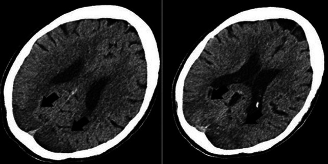
Axial noncontrast CT images demonstrate vasogenic/cytotoxic edema in the parieto-occipital regions suggestive of PRES (arrows).
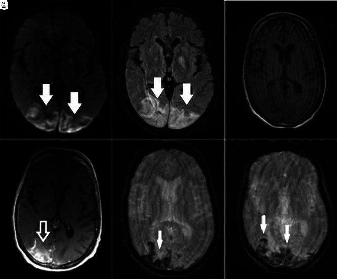
Brain axial DWI (A), FLAIR (B), precontrast T1-weighted (C), postcontrast T1-weighted (D) and SWI (E and F) obtained after CT demonstrate bilateral posterior infarctions (white arrows, A), edema in the posterior parieto-occipital regions (white arrows, B), and some contrast enhancement (hollow white arrow, D; compare with C). Selected SWI shows extensive blooming artifacts compatible with hemorrhages predominantly in the cortex (white arrows, E and F). Findings are more pronounced on the right side.
References
-
- Coronavirus disease (COVID-19) Pandemic. Geneva: World Health Organization. 2020. https://www.who.int/emergencies/diseases/novel-coronavirus-2019. Accessed April 15, 2020
-
- Jin Y, Cai L, Cheng Z, et al. ; for the Zhongnan Hospital of Wuhan University Novel Coronavirus Management and Research Team, Evidence-Based Medicine Chapter of China International Exchange and Promotive Association for Medical and Health Care (CPAM). A rapid advice guideline for the diagnosis and treatment of 2019 novel coronavirus (2019-nCoV) infected pneumonia (standard version). Mil Med Res 2020;7:4 10.1186/s40779-020-0233-6 - DOI - PMC - PubMed
Publication types
MeSH terms
LinkOut - more resources
Full Text Sources