. 2020 Jul 24;369(6502):433-436.
doi: 10.1126/science.abb5008. Epub 2020 May 21.

Structural basis for membrane insertion by the human ER membrane protein complex

Affiliations

Structural basis for membrane insertion by the human ER membrane protein complex

Tino Pleiner et al. Science. .

Abstract

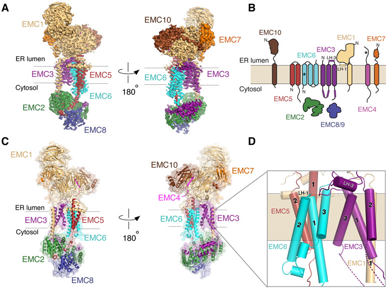
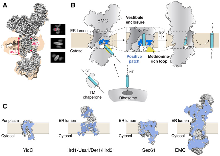
A defining step in the biogenesis of a membrane protein is the insertion of its hydrophobic transmembrane helices into the lipid bilayer. The nine-subunit endoplasmic reticulum (ER) membrane protein complex (EMC) is a conserved co- and posttranslational insertase at the ER. We determined the structure of the human EMC in a lipid nanodisc to an overall resolution of 3.4 angstroms by cryo-electron microscopy, permitting building of a nearly complete atomic model. We used structure-guided mutagenesis to demonstrate that substrate insertion requires a methionine-rich cytosolic loop and occurs via an enclosed hydrophilic vestibule within the membrane formed by the subunits EMC3 and EMC6. We propose that the EMC uses local membrane thinning and a positively charged patch to decrease the energetic barrier for insertion into the bilayer.


Figures

Figure 1.
Figure 1.. The structure of the human EMC.
(A) Two views of the sharpened ‘overall’ density map (fig. S3) from the perspective of the two intramembrane sides of the EMC colored by subunit. (B) Schematic representation of the topology of the nine EMC subunits as determined by the structure. EMC8 and 9 are functional paralogs, and their binding to EMC2 is mutually exclusive. For simplicity, we refer only to EMC8 throughout the text, though most observations will apply to both EMC8 and 9. Helices of EMC1 and 3 that are positioned in the lumenal plane of the membrane are labeled LH-1 and LH-3. Asterisks indicate newly determined topologies based on the structure and experimental data. Note, we cannot unambiguously define the topology of EMC4, but structural data is most consistent with it containing a single TM (fig. S7). (C) Atomic model of the EMC, in the same orientation as the density map in (A). (D) Close-up of the nine core TMs of the EMC and their subunit assignment.
Figure 2.
Figure 2.. Architecture of the cytosolic and lumenal regions of the EMC.
(A) View from the membrane of the cytosolic region of the EMC. (B) Close-up of the primary interfaces between the cytosolic subunits of the EMC indicated in (A). Dashed lines represent polar interactions, and asterisks indicate mutations that disrupt complex assembly (fig. S8). (C) 35S-methionine labeled wild type EMC2 or the indicated point mutants were translated in rabbit reticulocyte lysate (RRL) and tested for binding to FLAG-tagged EMC8, EMC3 or EMC5 by co-immunoprecipitation using anti-FLAG resin. (D) Side view of the EMC lumenal region. (E) Cartoon model of the globular N-termini of EMC1, 7 and 10. EMC1 and EMC4 together form one of the four-stranded blades of the bottom ß-propeller.
Figure 3.
Figure 3.. Substrate insertion by the EMC requires a positive patch in the bilayer and a flexible methionine-rich loop.
(A) Surface filling representation of the membrane-spanning region of the EMC colored with hydrophobic residues in grey and polar residues in blue. Displayed are the two sides of the complex: the ‘hydrophobic crevice’ (left) and the ‘hydrophilic vestibule’ (right) as in Fig. 1A and C. (B) Close-up view of the hydrophilic vestibule formed by EMC3 and 6, with polar residues shown in blue and displayed as sticks. Residues that were mutated in functional assays are highlighted with asterisks (fig. S13). (C) HEK293 cells were generated that stably expressed exogenous wild type or mutant EMC3, as well as the tail-anchored substrates RFP-squalene synthase (SQS; EMC-dependent) or RFP-VAMP2 (EMC-independent) (3). The relative RFP fluorescence, normalized to an internal expression control (GFP), is plotted as a histogram. (D) As in (C) but with the co-translational substrates Opioid Receptor Kappa 1 (OPRK1)-GFP (EMC-dependent) and TRAM2-GFP (EMC-independent). (E) As in (C), analysis of the role of positive charge in the hydrophilic vestibule.
Figure 4.
Figure 4.. Model for membrane protein insertion by the EMC.
(A) Unsharpened EM density maps are shown at low (tan) and high contour (grey) to highlight the thickness of the lipid nanodisc. Distances measured within the density are shown in red (fig. S14). Insets are representative 2D class averages that depict the local thinning of the lipid bilayer by the EMC. (B) Post- and co-translational EMC substrates are released from either a TM chaperone (e.g. calmodulin) or the ribosome, respectively. The flexible methionine-rich loop of EMC3 is positioned to capture substrates for insertion through the hydrophilic vestibule along the surface of EMC3 and 6. The EMC decreases the energetic barrier for insertion via local thinning of the membrane and a positively charged patch in the bilayer. The TMs of EMC4, 7, and 10 enclose the cytoplasmic vestibule and facilitate insertion. (C) Cut-away view of the space filling-models for the bacterial YidC (PDB 3WO6), the fungal Hrd1-Usa1/Der1/Hrd3 complex (6VJZ), mammalian Sec61 (3J7Q), and the human EMC. A hydrophilic conduit from the cytosol to the membrane is a general feature of evolutionarily diverse protein conducting channels.

Comment in

  • Membrane protein biogenesis by the EMC.
    Alvira S, Corey RA, Collinson I, Römisch K. Alvira S, et al. EMBO J. 2021 Jan 15;40(2):e107407. doi: 10.15252/embj.2020107407. Epub 2020 Dec 21. EMBO J. 2021. PMID: 33346928 Free PMC article.

References

    1. The UniProt Consortium, UniProt: the universal protein knowledgebase. Nucleic Acids Res 45, D158–D169 (2017). - PMC - PubMed
    1. Aviram N et al., The SND proteins constitute an alternative targeting route to the endoplasmic reticulum. Nature 540, 134–138 (2016). - PMC - PubMed
    1. Guna A, Volkmar N, Christianson JC, Hegde RS, The ER membrane protein complex is a transmembrane domain insertase. Science 359, 470–473 (2018). - PMC - PubMed
    1. Görlich D, Rapoport TA, Protein translocation into proteoliposomes reconstituted from purified components of the endoplasmic reticulum membrane. Cell 75, 615–630 (1993). - PubMed
    1. Mariappan M et al., The mechanism of membrane-associated steps in tail-anchored protein insertion. Nature 477, 61–66 (2011). - PMC - PubMed

Publication types

MeSH terms