. 2020 May 21;11(1):2551.
doi: 10.1038/s41467-020-16345-y.

FoxO1-Dio2 signaling axis governs cardiomyocyte thyroid hormone metabolism and hypertrophic growth

Affiliations

FoxO1-Dio2 signaling axis governs cardiomyocyte thyroid hormone metabolism and hypertrophic growth

Anwarul Ferdous et al. Nat Commun. .

Abstract

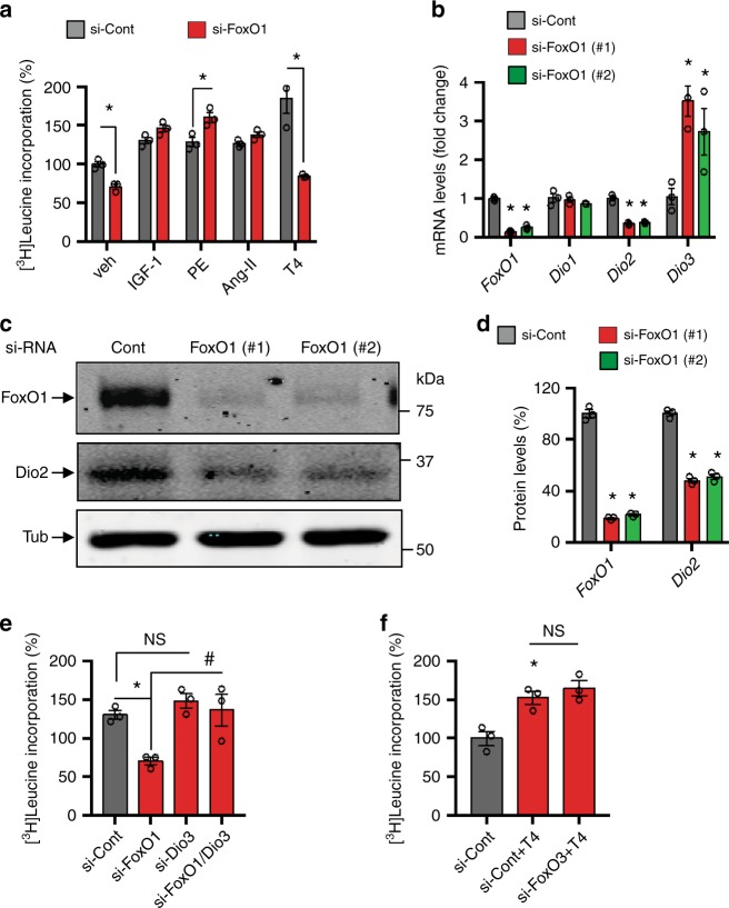
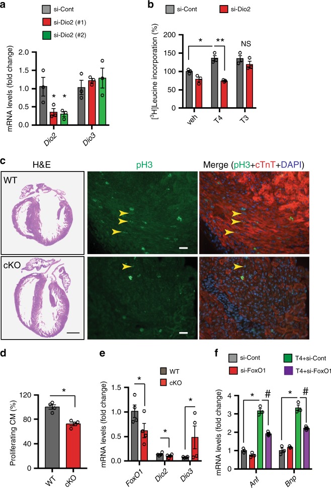
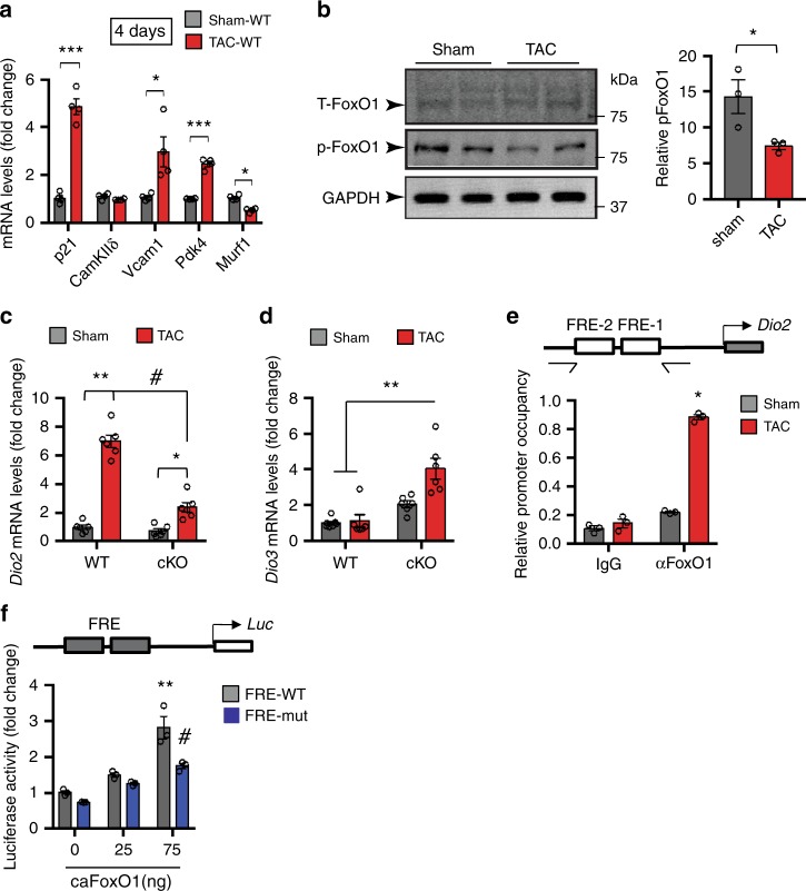
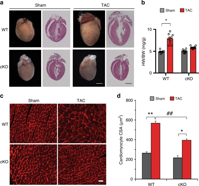
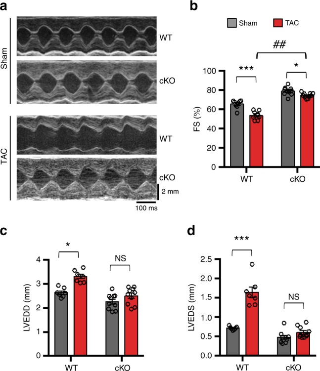
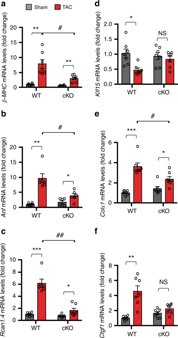
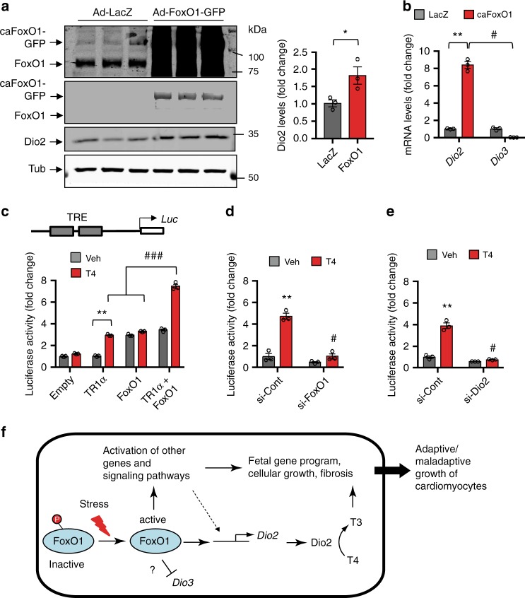
Forkhead box O (FoxO) proteins and thyroid hormone (TH) have well established roles in cardiovascular morphogenesis and remodeling. However, specific role(s) of individual FoxO family members in stress-induced growth and remodeling of cardiomyocytes remains unknown. Here, we report that FoxO1, but not FoxO3, activity is essential for reciprocal regulation of types II and III iodothyronine deiodinases (Dio2 and Dio3, respectively), key enzymes involved in intracellular TH metabolism. We further show that Dio2 is a direct transcriptional target of FoxO1, and the FoxO1-Dio2 axis governs TH-induced hypertrophic growth of neonatal cardiomyocytes in vitro and in vivo. Utilizing transverse aortic constriction as a model of hemodynamic stress in wild-type and cardiomyocyte-restricted FoxO1 knockout mice, we unveil an essential role for the FoxO1-Dio2 axis in afterload-induced pathological cardiac remodeling and activation of TRα1. These findings demonstrate a previously unrecognized FoxO1-Dio2 signaling axis in stress-induced cardiomyocyte growth and remodeling and intracellular TH homeostasis.


Conflict of interest statement

The authors declare no competing interests.

Figures

Fig. 1
Fig. 1. FoxO1 and Dio2/Dio3 transcriptional circuitry govern TH-induced NRVM growth in vitro.
a Selective knockdown of FoxO1 in NRVM specifically abrogated T4-induced hypertrophy but not the cellular growth response triggered by other stimuli. NRVM growth was evaluated by assessing radiolabeled leucine incorporation into protein following 24 h treatment, where NRVM growth in the control (Cont) siRNA- and vehicle (Veh)-treated cells was set to 100%. b Selective knockdown of FoxO1 in NRVM resulted in marked reduction of Dio2 mRNA levels and significantly induced Dio3 expression. c, d Immunoblotting (c) and quantitation (d) of FoxO1 and Dio2 levels in FoxO1-deficient NRVM. e T3-induced growth response of NRVM transfected with control, Dio3- and FoxO1-specific siRNAs alone or in combination. Note that the abrogation of T3-induced NRVM growth response in FoxO1-deficient cells was largely rescued in cells transfected with both Dio3- and FoxO1-specific siRNAs. f Selective knockdown of FoxO3 did not affect T4-induced growth response of NRVM. In all panels, data are depicted as mean ± SEM (n = 3 independent experiments). *p < 0.05 vs control; #p < 0.01 vs control, NS not statistically significant. Statistical analyses were conducted using a two-tailed, unpaired Student’s t-test.
Fig. 2
Fig. 2. FoxO1–Dio2 axis governs TH-induced hypertrophic growth of neonatal cardiomyocytes.
a qRT-PCR analyses of mRNA levels of the indicated genes in NRVM transfected with control or two sequence-independent Dio2-specific siRNAs. Note that efficient knockdown of Dio2 did not affect Dio3 expression (n = 3 independent experiments). b Selective knockdown of Dio2 in NRVM abrogated the T4-induced, but not T3-induced, cellular growth response (n = 3 independent experiments). c Histology (left) (bar = 2 mm) and co-immunostaining with anti-pH3 (green) (middle) and anti-cTnT (red) (right) antibodies of P3 FoxO1-WT (WT) and FoxO1-cKO (cKO) heart. Proliferating cardiomyocytes in LV free wall are shown (arrowhead), and DAPI was used for the nuclear staining (bar = 10 μm). d Quantitation of proliferating cardiomyocytes in WT and cKO hearts (n = 4), where total number of proliferating cells in WT hearts was considered to be 100%. Note the significant decrease in proliferating cells in cKO heart. e qRT-PCR analyses of ventricular mRNA levels of the indicated genes in P3 WT and cKO mice, and mRNA levels of ribosomal 18S was used as control (n = 4). f qRT-PCR analyses of mRNA levels of the indicated genes in control- and si-FoxO1-transfected NRVM with or without T4 treatment (n = 3 independent experiments). In all panels, data are depicted as mean ± SEM. *p < 0.05 vs control; **p < 0.01 vs control; #p < 0.001 vs control. Statistical analyses were conducted using a two-tailed, unpaired Student’s t-test.
Fig. 3
Fig. 3. Activation of FoxO1 in early post-TAC heart is required for reciprocal regulation of Dio2 and Dio3 genes expression.
a qRT-PCR analyses of mRNA levels of indicated genes in 4 day TAC- and sham-operated LVs (n = 4). b Immunoblotting (left) and quantitation (right) of total-FoxO1 (T-FoxO1) and phospho-FoxO1 (p-FoxO1) levels in 4 days TAC- and sham-operated LVs (n = 3). GAPDH was used as a loading control. Average of p-FoxO1/T-FoxO1 and p-FoxO1/GAPDH ratios were used to determine relative p-FoxO1 levels. c, d qRT-PCR analyses of mRNA levels of Dio2 (c) and Dio3 (d) in 4 days TAC- and sham-operated LVs of FoxO1-WT (WT, n = 12) and FoxO1-cKO (cKO, n = 12) mice. e Quantitative ChIP assays were conducted in 4 day sham- and TAC-operated ventricles of the WT mice (n = 3). Note the significant increase in FoxO1 occupancy at the Dio2 promoter in TAC hearts. Schematic of Dio2 illustrates that PCR amplification was performed using primers spanning the two FoxO-responsive elements (FREs) in the Dio2 promoter. f Schematic of Dio2 luciferase vector harboring two FREs. Note that co-transfection of a constitutively active FoxO1 (caFoxO1) elicited significant, concentration-dependent activation of reporter activity harboring WT, but not mutated, FREs. In all panels, data are depicted as mean ± SEM. *p < 0.05 vs sham/control; **p < 0.01 vs sham; ***p < 0.0001 vs sham, #p < 0.05 vs WT (f); #p < 0.001 vs control (c). Statistical analyses were conducted using a two-tailed, unpaired Student’s t-test.
Fig. 4
Fig. 4. FoxO1 is required for pathological cardiomyocyte growth.
a Male mice of respective genotypes, FoxO1-WT (WT) and FoxO1-cKO (cKO), were subjected to sham and TAC surgery. Whole mount (left) and histology (right) of representative 3-week post-sham and -TAC hearts are shown. Note that cardiomyocyte-specific silencing of FoxO1 (cKO) blunted TAC-induced hypertrophic growth of heart (bar = 2 mm). b Heart weight (HW)/body weight (BW) ratios indicate robust cardiac hypertrophy in WT (n = 16) mice 3-week post-TAC, which is attenuated in cKO mice (n = 18). *p < 0.001 vs sham. c, d Immunohistochemistry (c) and quantitation (d) of cross-sectional area (CSA) of cardiomyocytes of WT and cKO mice after 3-week sham and TAC surgery (n = 3). Data are depicted as mean ± SEM. *p < 0.05 vs sham; **p < 0.01 vs sham; ##p < 0.01 vs WT. Statistical analyses were conducted using a two-tailed, unpaired Student’s t-test.
Fig. 5
Fig. 5. FoxO1-cKO hearts manifest preserved contractile function in response to TAC.
a Representative M-mode tracings of 3-week sham- and TAC-operated hearts of FoxO1-WT (WT) and FoxO1-cKO (cKO) mice. b Percent of left ventricular fractional shortening (%FS) of WT (n = 16) and cKO (n = 20). c Left ventricular end-diastolic diameter (LVEDD, mm) of WT (n = 14) and cKO (n = 20), and d Left ventricular end-systolic diameter (LVESD, mm) of WT (n = 14) and cKO (n = 19) are shown. Note that cKO mice do not manifest contractile dysfunction when compared with WT littermates TAC heart. In all panels, data are depicted as mean ± SEM. *p < 0.01 vs sham; ***p < 0.001 vs sham; ##p < 0.0001 vs cKO; NS not statistically significant. Statistical analyses were conducted using a two-tailed, unpaired Student’s t-test.
Fig. 6
Fig. 6. Reactivation of fetal and fibrotic gene programs in response to TAC is blunted in cKO hearts.
a–f qRT-PCR analyses of mRNA levels of the indicated genes in 3-week TAC and sham LVs of FoxO1-WT [WT, n = 13 (a), 12 (b, c), and 14 (df)] and FoxO1-cKO [cKO, n = 13 (a), 12 (b, c), and 14 (df)] mice. mRNA levels of ribosomal 18S were used as control. In all panels, data are depicted as mean ± SEM. *p < 0.05 vs Sham; **p < 0.01 vs sham; ***p < 0.001 vs sham; #p < 0.05 vs WT, ##p < 0.001 vs WT; NS not statistically significant. Statistical analyses were conducted using a two-tailed, unpaired Student’s t-test.
Fig. 7
Fig. 7. FoxO1-dependent induction of Dio2 governs TH-responsive gene expression in hypertrophic cardiomyocytes.
a, b caFoxO1 overexpression in NRVM markedly induced Dio2 protein (a) and mRNA (b) levels but repressed Dio3 gene expression (b). c Schematic of thyroid hormone receptor (THR)-responsive reporter plasmid harboring two thyroid hormone-responsive elements (TREs). Note that co-expression of caFoxO1 and THRα1 (TRα1), but not either alone, in HEK293 cells resulted in robust and synergistic activation of reporter activity only in the presence of T4 (n = 3). d, e THR-responsive reporter activity in NRVM transfected with control or indicated gene-specific (FoxO1 and Dio2) siRNAs. Note that compared with control siRNA-treated cells, selective knockdown of FoxO1 (d) and Dio2 (e) significantly attenuated T4-dependent activation of luciferase activity. In all panels, data are depicted as mean ± SEM (n = 3 independent experiments). *p < 0.05 vs control; **p < 0.001 vs control; #p < 0.001 vs control; ###p < 0.0001 vs control. Statistical analyses were conducted using a two-tailed, unpaired Student’s t-test. f Working model for FoxO1’s role in adaptive and maladaptive hypertrophic growth of cardiomyocytes. In response to oxidative- and TAC-induced (Stress), activation of FoxO1 and FoxO1-dependent reciprocal regulation of Dio2 and Dio3 in cardiomyocytes is a key driver for subsequent activation of other transcriptional and signaling programs to activate fetal and fibrotic genes, which together ultimately lead to adaptive and maladaptive hypertrophic growth of neonatal and adult cardiomyocytes, respectively.

Similar articles

Cited by

References

    1. Benjamin EJ, et al. Heart disease and stroke statistics—2018 update: a report from the American Heart Association. Circulation. 2018;137:e67–e492. - PubMed
    1. Drazner MH. The progression of hypertensive heart disease. Circulation. 2011;123:327–334. - PubMed
    1. Burchfield JS, Xie M, Hill JA. Pathological ventricular remodeling: mechanisms: part 1 of 2. Circulation. 2013;128:388–400. - PMC - PubMed
    1. Xie M, Burchfield JS, Hill JA. Pathological ventricular remodeling: therapies: part 2 of 2. Circulation. 2013;128:1021–1030. - PMC - PubMed
    1. Hill JA. Braking bad hypertrophy. N. Engl. J. Med. 2015;372:2160–2162. - PubMed

Publication types

MeSH terms