Review
. 2020 May 5:12:67-73.
doi: 10.2147/OPTO.S183492. eCollection 2020.

A Review of Hypertensive Retinopathy and Chorioretinopathy

Affiliations
Review

A Review of Hypertensive Retinopathy and Chorioretinopathy

Mai Tsukikawa et al. Clin Optom (Auckl). .

Abstract

Hypertensive retinopathy and choroidopathy have important short- and long-term implications on patients' overall health and mortality. Eye care professionals should be familiar with the severity staging of these entities and be able to readily recognize and refer patients who are in need of systemic blood pressure control. This paper will review the diagnosis, staging, treatment, and long-term implications for vision and mortality of patients with hypertensive retinopathy and choroidopathy.

Keywords: hypertensive chorioretinopathy; hypertensive choroidopathy; hypertensive retinopathy.


Conflict of interest statement

The authors report no conflicts of interest in this work.

Figures

Figure 1
Figure 1
Grades of hypertensive retinopathy. (A) Mild hypertensive retinopathy (in an eye with an unrelated chorioretinal lesion) with arteriolar narrowing (white arrow), copper wiring (black star), and AV nicking (black arrow). (B) Moderate hypertensive retinopathy with features of mild hypertensive retinopathy as well as cotton wool spots (yellow arrow) and intraretinal hemorrhages (red arrow). (C) Severe hypertensive retinopathy with features of moderate hypertensive retinopathy and optic disc swelling (white star).
Figure 2
Figure 2
Optos wide-field color fundus photographs of the right eye (A) and left eye (B) of a patient with hypertensive chorioretinopathy. The photographs demonstrate copper wiring (black star), AV nicking (black arrow), intraretinal hemorrhages (red arrow), cotton wool spots (yellow arrow), Elschnig spots (white asterisk), and optic disc swelling (white star).
Figure 3
Figure 3
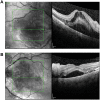
Optical coherence tomography of the macula of the right eye (A) and left eye (B) of a patient with hypertensive chorioretinopathy shows macular edema and serous retinal detachments. These serous retinal detachments improved after medical management of hypertension.
Figure 4
Figure 4
Fundus autofluorescence of the right eye (A) and left eye (B) of a patient with hypertensive chorioretinopathy. Magnification of the posterior pole reveals hypoautofluorescence of Elschnig spots, retinal hemorrhages and cotton wool spots.
Figure 5
Figure 5
Fluorescein angiography of the right eye (A) and left eye (B) of a patient with hypertensive chorioretinopathy shows patchy and delayed choroidal filling and areas of retinal capillary nonperfusion. There is also blockage from intraretinal hemorrhages and cotton wool spots, as well as optic disc leakage.

References

    1. The World Health Organization. A global brief on hypertension: silent killer, global public health crisis. Available from: http://apps.who.int/iris/bitstream/10665/79059/1/WHO_DCO_WHD_2013.2_eng.pdf. Accessed August 2019.
    1. Wong TY, Mitchell P. The eye in hypertension. Lancet. 2007;369:425–435. doi:10.1016/S0140-6736(07)60198-6 - DOI - PubMed
    1. Keith NM, Wagener HP, Barker NW. Some different types of essential hypertension: their course and prognosis. Am J Med Sci. 1939;197:332–343. doi:10.1097/00000441-193903000-00006 - DOI - PubMed
    1. Wong TY, Mitchell P. Hypertensive retinopathy. N Engl J Med. 2004;351:2310–2317. doi:10.1056/NEJMra032865 - DOI - PubMed
    1. Fraser-Bell S, Symes R, Vaze A. Hypertensive eye disease: a review. Clin Exp Ophthalmol. 2017;45(1):45–53. doi:10.1111/ceo.12905 - DOI - PubMed