

Two-dimensional Periapical, Panoramic Radiography Versus Three-dimensional Cone-beam Computed Tomography in the Detection of Periapical Lesion After Endodontic Treatment: A Systematic Review
- PMID: 32440383
- PMCID: PMC7237056
- DOI: 10.7759/cureus.7736
Two-dimensional Periapical, Panoramic Radiography Versus Three-dimensional Cone-beam Computed Tomography in the Detection of Periapical Lesion After Endodontic Treatment: A Systematic Review
Abstract
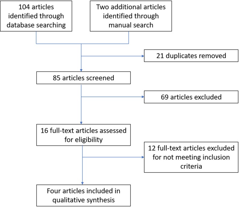
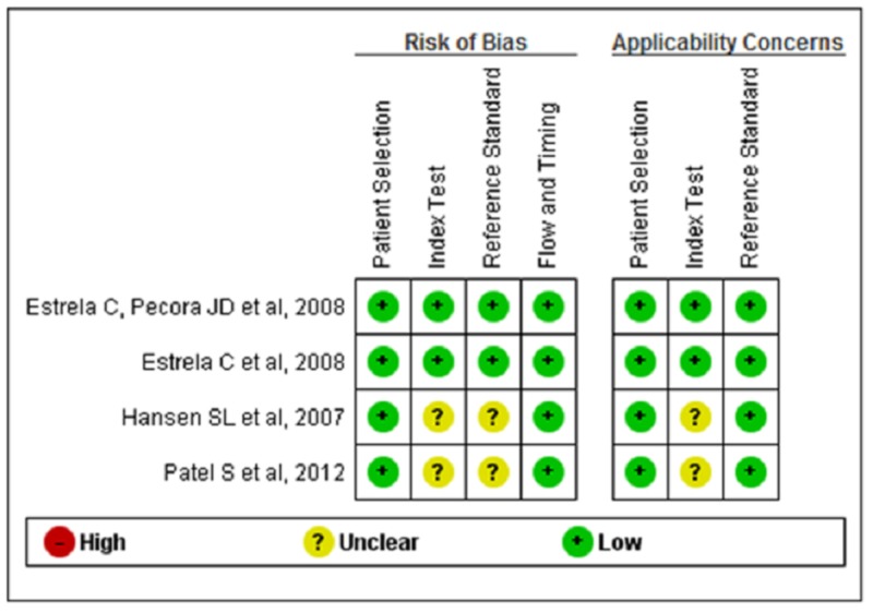
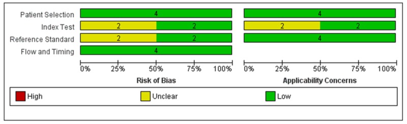
Radiographic imaging is a common resource for endodontic diagnosis, treatment, and prognosis. Two-dimensional (2D) periapical and digital panoramic radiographs often showed image distortion; this issue was resolved with the emergence of three-dimensional (3D) cone-beam computed tomography (CBCT). This review examines the accuracy of various radiographic techniques in the assessment of periapical lesion after endodontic treatment. Our goal was to determine whether a 2D radiograph (periapical and panoramic) is as accurate as a 3D radiograph (i.e., CBCT) in the assessment of periapical lesion after endodontic treatment. We searched the electronic databases Medline and Cochrane and trial registries for ongoing trials. We included both retrospective and prospective studies comparing the efficacy of periapical healing with various radiographic techniques after endodontic treatment. The outcome of interest was the percentage detection of periapical lesions and periapical healing assessment after endodontic treatment. All data were collected using a specially designed extraction form. We assessed the risk of bias in the studies using the Cochrane tool for diagnostic tests (QUADAS). We judged two studies to be at low risk and two to be at moderate risk of bias. Although there was a difference in the percentage detection of periapical healing efficacy by various radiographic techniques, all studies reported that CBCT had higher accuracy in the detection of periapical lesions compared to periapical and panoramic radiography. The next best choice is periapical radiographs, followed by panoramic radiographs as they provide better visualization and accuracy.
Keywords: cbct; endodontic treatment; panoramic radiograph; periapical; periapical lesion.
Copyright © 2020, Antony et al.
Conflict of interest statement
The authors have declared that no competing interests exist.
Figures
References
-
- The periapical index: a scoring system for radiographic assessment of apical periodontitis. Ørstavik D, Kerekes K, Eriksen HM. Endod Dent Traumatol. 1986;2:20–34. - PubMed
-
- Factors associated with endodontic treatment failures. Lin LM, Skribner JE, Gaengler P. J Endod. 1992;18:625–627. - PubMed
-
- Outcome of secondary root canal treatment: a systematic review of the literature. Ng YL, Mann V, Gulabivala K. Int Endod J. 2008;41:1026–1046. - PubMed
-
- Persistent periapical radiolucencies of root-filled human teeth, failed endodontic treatments, and periapical scars. Nair PNR, Sjögren U, Figdor D, Sundqvist G. Oral Surg Oral Med Oral Pathol Oral Radiol Endod. 1999;87:617–627. - PubMed
-
- Experimentally produced bone lesions. van der Stelt PF. Oral Surg Oral Med Oral Pathol. 1985;59:306–312. - PubMed
Publication types
LinkOut - more resources
Full Text Sources