. 2020 Mar 11;7(10):1903233.
doi: 10.1002/advs.201903233. eCollection 2020 May.

Small Protein Hidden in lncRNA LOC90024 Promotes "Cancerous" RNA Splicing and Tumorigenesis

Affiliations

Small Protein Hidden in lncRNA LOC90024 Promotes "Cancerous" RNA Splicing and Tumorigenesis

Nan Meng et al. Adv Sci (Weinh). .

Abstract

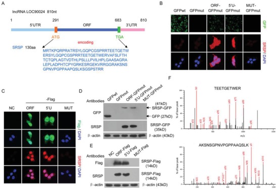
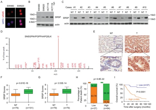
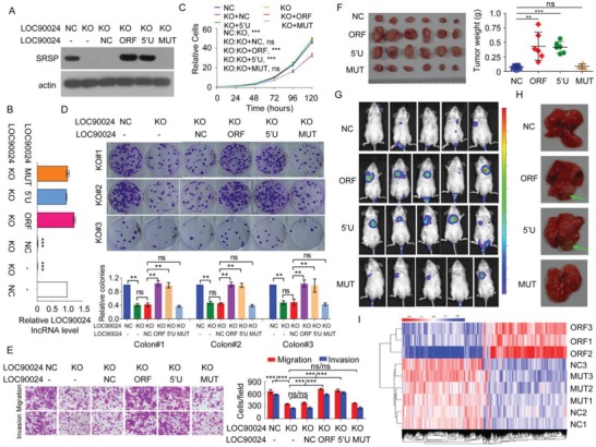
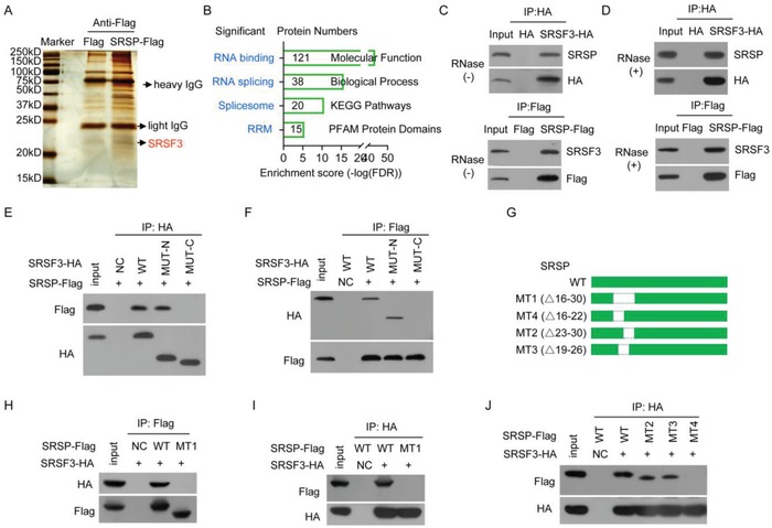
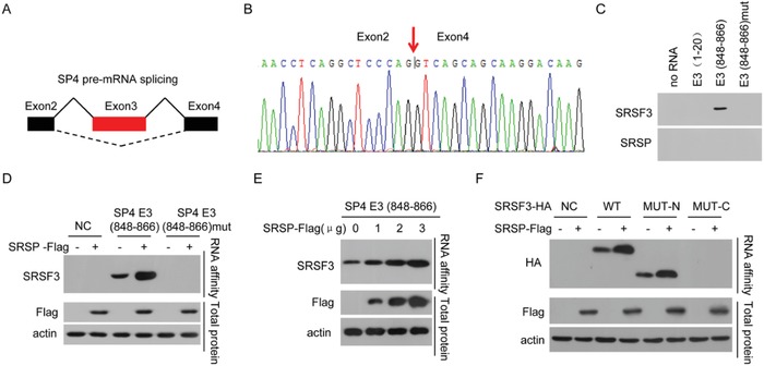
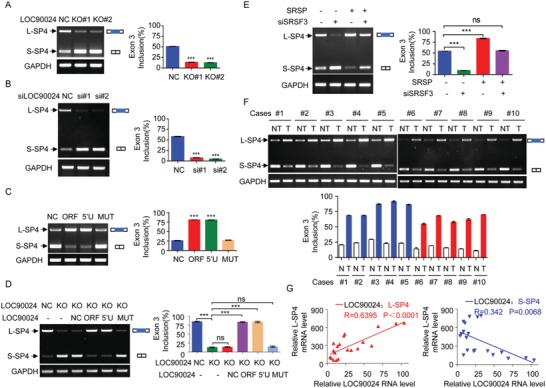
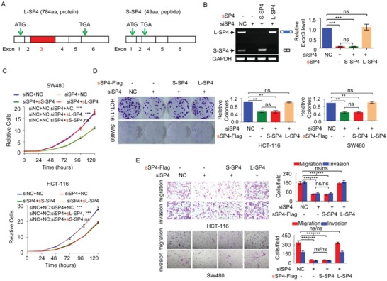
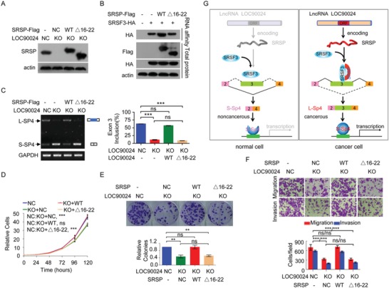
Conventional therapies for late-stage colorectal cancer (CRC) have limited effects because of chemoresistance, recurrence, and metastasis. The "hidden" proteins/peptides encoded by long noncoding RNAs (lncRNAs) may be a novel resource bank for therapeutic options for patients with cancer. Here, lncRNA LOC90024 is discovered to encode a small 130-amino acid protein that interacts with several splicing regulators, such as serine- and arginine-rich splicing factor 3 (SRSF3), to regulate mRNA splicing, and the protein thus is named "Splicing Regulatory Small Protein" (SRSP). SRSP, but not LOC90024 lncRNA itself, promotes CRC tumorigenesis and progression, while silencing of SRSP suppresses CRC tumorigenesis. Mechanistically, SRSP increases the binding of SRSF3 to exon 3 of transcription factor Sp4, resulting in the inclusion of Sp4 exon 3 to induce the formation of the "cancerous" long Sp4 isoform (L-Sp4 protein) and inhibit the formation of the "noncancerous" short Sp4 isoform (S-Sp4 peptide), which lacks the transactivation domain. The upregulated SRSP level is positively associated with malignant phenotypes and poor prognosis in patients with CRC. Collectively, the findings uncover that a lncRNA-encoded small protein SRSP induces "cancerous" Sp4 splicing variant formation and may be a potential prognostic biomarker and therapeutic target for patients with CRC.

Keywords: RNA splicing; colorectal cancer (CRC); lncRNA; prognostic biomarkers; small proteins; transcription factors.


Conflict of interest statement

The authors declare no conflict of interest.

Figures

Figure 1
Figure 1
LncRNA LOC90024 encodes a 130‐aa small protein, SRSP. A) 840 nt lncRNA LOC90024 contains a potential small protein‐encoding ORF, which may encode a 130‐aa small protein, SRSP. B,D) The LOC90024 ORF, 5′UTR‐ORF (5′U), or 5′UTR‐ORFmut (MUT, in which the start codon ATG of SRSP ORF was mutated to ATT) constructs fused with GFP, in which the start codon of GFP was mutated (GFPmut), were transfected into HeLa cells; B) GFP fluorescence and SRSP‐GFP immunostaining were detected using anti‐SRSP antibody and D) SRSP‐GFP fusion protein expression was detected by Western blotting with anti‐GFP and SRSP antibodies. C,E) The LOC90024 ORF, 5′U, or MUT constructs fused with a Flag tag were transfected into HeLa cells, and C) SRSP‐Flag immunostaining and E) expression were detected using anti‐Flag and ‐SRSP antibodies. F) Two unique peptides in the ectopically expressed SRSP‐GFP were identified using mass spectrometry.
Figure 2
Figure 2
The LOC90024‐encoded SRSP is endogenously, naturally expressed, and SRSP is upregulated in cancers and associated with the prognosis of patients with CRC. A) SRSP immunostaining between SW480 and SW620 cells was determined with an anti‐SRSP antibody. B) SRSP levels were increased in high‐metastatic CRC cells compared with their parental low‐metastatic CRC cells. C) SRSP levels were increased in tumor tissues (T) compared with their matched adjacent nontumoral tissues (NT). D) The unique peptide in the endogenous, natural SRSP in tumor tissues was identified using mass spectrometry. E) Representative IHC images of SRSP expression in CRC T and their matched NT. F) Differences in SRSP scores between T and NT are presented as a box plot. G) Differences in SRSP scores between T and their matched NT are presented as a box plot. H) CRC patients with high SRSP levels had a higher death rate than those with low SRSP levels. I) CRC patients with high SRSP levels had shorter survival times than those with low SRSP levels.
Figure 3
Figure 3
SRSP, but not LOC90024 lncRNA itself, stimulates CRC tumorigenesis and metastasis in vitro and in vivo. A–D) The indicated LOC90024 constructs were transfected into LOC90024 KO HeLa cells, A) SRSP expression, B) LOC90024 lncRNA level, C) cell growth, D) colony formation, E) migration and invasion were determined. F) The in vivo tumorigenesis of the indicated HCT‐116 cells stably expressing the indicated LOC90024 constructs was determined. The images and weights of the xenograft tumors are listed in the left and right panels, respectively (n = 6). G,H) The indicated Luc‐labeled HCT‐116 cells stably expressing the indicated LOC90024 constructs (2 × 10⁶ cells per mouse) were injected into NOD‐SCID mice via the tail vein; G) luciferase activities and H) metastases in mouse lung were visualized 50 d posttransplantation (n = 5). I) The cluster heatmap of gene expression in negative control (NC), LOC90024 ORF (ORF), and 5′UTR‐ORFmut (MUT) overexpression (n = 3). Data are represented as mean ± SD. **p < 0.01 or ***p < 0.001.
Figure 4
Figure 4
SRSP interacts with the splicing factor SRSF3. A) Proteins that interacted with SRSP were identified by interactomics analysis. B) The SRSP‐bound proteins were analyzed by GO. C,D) The SRSF3‐HA or SRSP‐Flag plasmids were transfected into HEK293T cells, cellular lysates were treated D) with or C) without RNase A; SRSF3‐HA or SRSP‐Flag complexes were co‐IPed with anti‐HA or Flag antibodies, and then the indicated proteins were detected. E,F) The wild type (WT), N‐terminal (MUT‐N), and C‐terminal (MUT‐C) mutants of SRSF3‐HA together with SRSP‐Flag vector were cotransfected into HEK293T cells, and E) SRSF3‐HA and F) SRSP‐Flag complexes were co‐IPed with anti‐HA and ‐Flag antibodies, respectively, to detect SRSP‐Flag and SRSF3‐HA. G) Diagram of wild‐type SRSP and its mutant constructs. H–J) The indicated SRSP‐Flag mutants together with the SRSF3‐HA vector were cotransfected into HEK293T cells; the interaction of the SRSP mutant with SRSF3 was determined.
Figure 5
Figure 5
SRSP promotes the binding of SRSF3 to exon 3 of Sp4. A,B) SRSP promotes the inclusion of Sp4 exon 3 (E3) using RNA‐seq analysis. C) The cellular nuclear extracts were affinity‐purified using the indicated biotin‐labeled RNAs, and SRSF3 and SRSP in purified complexes were detected. D) HCT‐116 cells were transfected with the SRSP‐Flag vector, RNA affinity purification was performed using the indicated biotin‐labeled RNAs, and SRSF3 was detected. E) SRSP‐Flag vector at the indicated doses was transfected into HCT‐116 cells, RNA affinity purification was performed using biotin‐labeled RNA E3 (848–866), and SRSF3 was detected. F) HCT‐116 cells were cotransfected with the indicated SRSF3‐HA mutants together with the SRSP‐Flag vector, RNA affinity purification was performed using biotin‐labeled RNA E3 (848–866), and SRSF3‐HA was detected.
Figure 6
Figure 6
SRSP promotes the inclusion of exon 3 of Sp4. A) L‐Sp4 and S‐Sp4 splicing variants were determined by RT‐PCR in LOC90024 KO HeLa cells. B) HCT‐116 cells were transfected with two anti‐LOC90024 siRNAs, and levels of L‐Sp4 and S‐Sp4 splicing variants were determined. C) The indicated LOC90024 constructs were transfected into HCT‐116 cells, and levels of L‐Sp4 and S‐Sp4 splicing variants were determined. D) The indicated LOC90024 constructs were transfected into LOC90024‐KO HeLa cells, and levels of L‐Sp4 and S‐Sp4 splicing variants were determined. E) HCT‐116 cells were transfected with SRSP vector together with anti‐SRSF3 siRNA, and levels of L‐Sp4 and S‐Sp4 splicing variants were determined. F) Levels of L‐Sp4 and S‐Sp4 splicing variants were determined between T and their matched NT. G) The relationships of LOC90024 lncRNA levels with L‐Sp4 (upper panel) and S‐Sp4 (low panel) mRNA levels were analyzed in clinical tissue samples. Data are represented as mean ± SD. ***p < 0.001.
Figure 7
Figure 7
The L‐Sp4 isoform exerts an oncogenic function, but the S‐Sp4 isoform does not. A) Schematic diagram of the L‐Sp4 and S‐Sp4 splicing variants, which encode a 784 aa protein and a 49 aa peptide without the transactivation domain of Sp4, respectively. B–E) CRC cells were cotransfected with anti‐Sp4 siRNA together with synonymously mutated S‐Sp4 or L‐Sp4 vector, which was resistant to anti‐Sp4 siRNA. B) The L‐Sp4 and S‐Sp4 splicing variant levels, C) cell growth, D) colony formation, and E) migration and invasion of these cells were determined. Data are represented as mean ± SD. **p < 0.01 or ***p < 0.001.
Figure 8
Figure 8
SRSP promotes the binding of SRSF3 to Sp4 exon 3, which facilitates exon 3 inclusion in Sp4 and tumorigenesis mainly by interacting with SRSF3. The wild‐type SRSP and its mutant Δ16–22 were re‐expressed in LOC90024 KO HeLa cells; A) SRSP protein level, B) the binding of SRSF3 to Sp4 exon 3, C) Sp4 splicing, D) cell growth, E) colony formation, and F) migration and invasion were analyzed. G) A regulatory model of LOC90024‐encoded small protein SRSP on tumorigenesis proposed in this study. Data are represented as mean ± SD. **p < 0.01 or ***p < 0.001.

References

    1. Wang F. W., Cao C. H., Han K., Zhao Y. X., Cai M. Y., Xiang Z. C., Zhang J. X., Chen J. W., Zhong L. P., Huang Y., Zhou S. F., Jin X. H., Guan X. Y., Xu R. H., Xie D., J. Clin. Invest. 2019, 129, 727. - PMC - PubMed
    1. Brody H., Nature 2015, 521, S1. - PubMed
    1. Bushweller J. H., Nat. Rev. Cancer 2019, 11, 019. - PMC - PubMed
    1. Lee T. I., Young R. A., Cell 2013, 152, 1237. - PMC - PubMed
    1. Papavassiliou K. A., Papavassiliou A. G., J. Cell. Biochem. 2016, 117, 2693. - PubMed