. 2020 May 15;6(20):eaba4311.
doi: 10.1126/sciadv.aba4311. eCollection 2020 May.

Dissolved oxygen from microalgae-gel patch promotes chronic wound healing in diabetes

Affiliations

Dissolved oxygen from microalgae-gel patch promotes chronic wound healing in diabetes

Huanhuan Chen et al. Sci Adv. .

Erratum in

Abstract

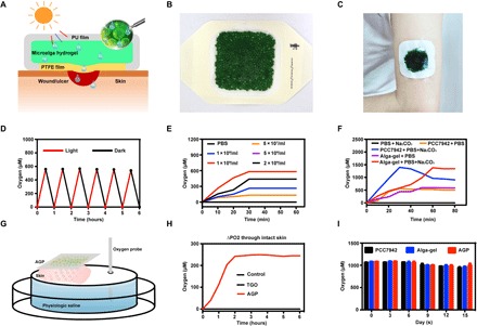
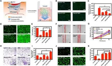
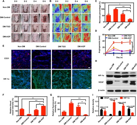
Chronic wounds in diabetes undergo a lifetime risk of developing into diabetic foot ulcers. Oxygen is crucial to wound healing by regulating cell proliferation, migration, and neovascularization. However, current oxygen therapies, including hyperbaric oxygen (HBO) and topical gaseous oxygen (TGO), mainly employ gaseous oxygen delivery, which is much less effective in penetrating the skin. Here, we introduce an oxygen-producing patch, made of living microalgae hydrogel, which can produce dissolved oxygen. The superior performance of the patch that results from its dissolved oxygen delivery is >100-fold much more efficient than TGO penetrating the skin. Further experiments indicate that the patch could promote cell proliferation, migration, and tube formation in vitro, and improve chronic wound healing and the survival of skin grafts in diabetic mice. We believe that the microalgae-gel patch can provide continuous dissolved oxygen to improve chronic wound healing.


Figures

Fig. 1
Fig. 1. AGP released dissolved oxygen through the intact skin.
(A) Schematic illustration of microalga-hydrogel patch (AGP) preparation through polyurethane film and polytetrafluoroethylene membrane to perform the light response dissolved oxygen release for chronic wound. (B) Images of the AGP. (C) Photograph of the AGP sticking on the arm. (D) Comparison of releasing dissolved oxygen of alga-gel under light or dark conditions. (E) Comparison of releasing dissolved oxygen of PCC7942 with different concentrations. (F) Comparison of releasing dissolved oxygen between the PCC7942 solution and alga-gel (1 × 10⁹ cells/ml) supplement with or without 500 μM Na2CO3. (G) Diagram of apparatus for measuring delivery of dissolved oxygen. (H) Transfer of dissolved oxygen through mice intact skin into saline at 37°C. (I) Comparison of releasing dissolved oxygen during the storage of PCC7942 solution, alga-gel beads, and AGP (1 × 10⁹ cells/ml) at days 0, 5, 10, and 15. Photo credit (B and C): Huanhuan Chen (Medical School and School of Life Sciences, Nanjing University).
Fig. 2
Fig. 2. Alga-gel activates cells against hypoxia and high glucose in vitro.
(A) Illustration of the wound-healing process and design scheme with alga-gel. (B and C) Alga-gel reduced HIF-1α on high glucose–induced HSF (n = 3). Scale bars, 200 μm. (D and E) HSF cell proliferation with 33 mM glucose and 6 hours of hypoxia in different groups (n = 3). Scale bars, 200 μm. (F and G) Representative images and quantification of HaCaT cell migration (n = 3). Scale bars, 100 μm. (H and I) Representative images and quantitative analysis of transwell migration assay in HUVECs (n = 3). Scale bars, 200 μm. (J and K) Representative images and quantification of HUVECs’ tube formation (n = 3). Scale bars, 200 μm. Significantly different (one-way ANOVA): *P < 0.05, **P < 0.01, and ***P < 0.001.
Fig. 3
Fig. 3. AGP accelerated diabetic wounds by turning chronic into acute.
(A) Representative images of the wound area by different treatments on days 0, 3, 6, and 12 after operation (n = 6). (B) Fractions of wounds healed by different treatments at days 3, 6, and 12 (n = 6). (C) Summary of the complete wound-closure times (n = 6). (D) H&E and Masson staining of the wound area reflected the regenerated skin in different groups at day 12 (n = 3). Scale bars, 100 μm. (E) Immunohistochemical images of the blood vessel CD31-positive endothelial cells (n = 3). Scale bar, 50 μm. Quantification of the epithelial gap (F), granulation tissue (G), and collagen deposition (H) in different groups (n = 3). (I) The average microvessel densities in different groups (n = 3). (J) Gene expression levels of quantitative PCR show that AGP accelerated diabetic wounds by turning chronic into acute (n = 3). Significantly different (one-way ANOVA): *P < 0.05, **P < 0.01, and ***P < 0.001. Photo credit (A): Huanhuan Chen (Medical School and School of Life Sciences, Nanjing University).
Fig. 4
Fig. 4. AGP promotes skin flap regeneration and angiogenesis.
(A and B) Photographic images of the skin flaps and the laser speckle contrast imaging captured real-time blood flow images from the Non-DM, DM-Control, DM-TGO, and DM-AGP groups 6 days after operation (n = 7). (C) Flap necrosis area percentages of different groups (n = 6). (D) Flap angiogenesis length of different groups (n = 6). (E) Immunofluorescence images of flaps highlighting blood vessel CD31-positive endothelial cells (red) and HIF-1α (green) (n = 3). Scale bars, 50 μm. (F and G) The average microvessel and HIF-1α densities in different groups (n = 3). (H and I) Western blotting data showing the levels of angiogenesis-related growth factors and hypoxia (n = 3). Significantly different (one-way ANOVA): *P < 0.05, **P < 0.01, and ***P < 0.001. Photo credit (A): Huanhuan Chen (Medical School and School of Life Sciences, Nanjing University).

References

    1. International Diabetes Federation, IDF Diabetes Atlas (IDF, ed. 8, 2017).
    1. Hart T., Milner R., Cifu A., Management of a diabetic foot. JAMA 318, 1387–1388 (2017). - PubMed
    1. Botusan I. R., Sunkari V. G., Savu O., Catrina A. I., Grünler J., Lindberg S., Pereira T., Ylä-Herttuala S., Poellinger L., Brismar K., Catrina S. B., Stabilization of HIF-1α is critical to improve wound healing in diabetic mice. Proc. Natl. Acad. Sci. U.S.A. 105, 19426–19431 (2008). - PMC - PubMed
    1. Thangarajah H., Vial I. N., Grogan R. H., Yao D., Shi Y., Januszyk M., Galiano R. D., Chang E. I., Galvez M. G., Glotzbach J. P., Wong V. W., Brownlee M., Gurtner G. C., HIF-1α dysfunction in diabetes. Cell Cycle 9, 75–79 (2010). - PubMed
    1. Thangarajah Y. D., Yao D., Chang E. I., Shi Y., Jazayeri L., Vial I. N., Galiano R. D., Du X. L., Grogan R., Galvez M. G., Januszyk M., Brownlee M., Gurtner G. C., The molecular basis for impaired hypoxia-induced VEGF expression in diabetic tissues. Proc. Natl. Acad. Sci. U.S.A. 106, 13505–13510 (2009). - PMC - PubMed

Publication types

LinkOut - more resources