Review
. 2020 Jun;78(2):165-180.
doi: 10.1007/s12013-020-00913-6. Epub 2020 May 22.

Photoprotective Properties of Vitamin D and Lumisterol Hydroxyderivatives

Affiliations
Review

Photoprotective Properties of Vitamin D and Lumisterol Hydroxyderivatives

Andrzej T Slominski et al. Cell Biochem Biophys. 2020 Jun.

Abstract

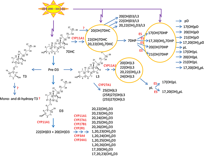
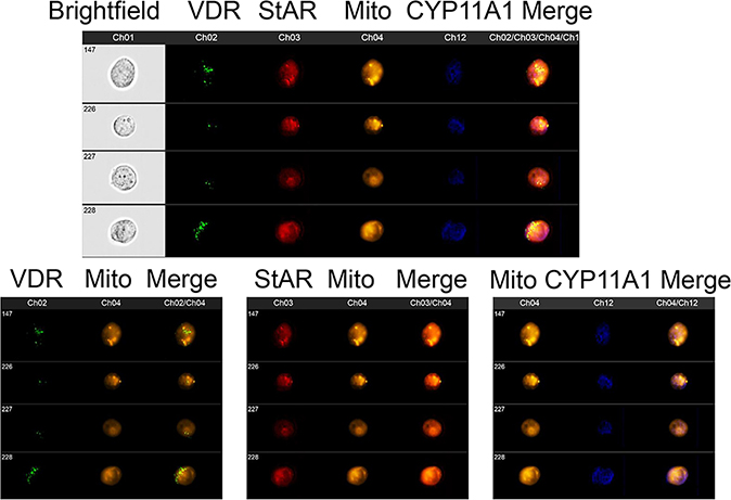
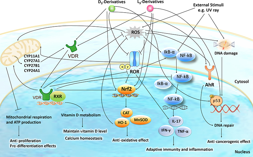
We have previously described new pathways of vitamin D3 activation by CYP11A1 to produce a variety of metabolites including 20(OH)D3 and 20,23(OH)2D3. These can be further hydroxylated by CYP27B1 to produce their C1α-hydroxyderivatives. CYP11A1 similarly initiates the metabolism of lumisterol (L3) through sequential hydroxylation of the side chain to produce 20(OH)L3, 22(OH)L3, 20,22(OH)2L3 and 24(OH)L3. CYP11A1 also acts on 7-dehydrocholesterol (7DHC) producing 22(OH)7DHC, 20,22(OH)27DHC and 7-dehydropregnenolone (7DHP) which can be converted to the D3 and L3 configurations following exposure to UVB. These CYP11A1-derived compounds are produced in vivo and are biologically active displaying anti-proliferative, anti-inflammatory, anti-cancer and pro-differentiation properties. Since the protective role of the classical form of vitamin D3 (1,25(OH)2D3) against UVB-induced damage is recognized, we recently tested whether novel CYP11A1-derived D3- and L3-hydroxyderivatives protect against UVB-induced damage in epidermal human keratinocytes and melanocytes. We found that along with 1,25(OH)2D3, CYP11A1-derived D3-hydroxyderivatives and L3 and its hydroxyderivatives exert photoprotective effects. These included induction of intracellular free radical scavenging and attenuation and repair of DNA damage. The protection of human keratinocytes against DNA damage included the activation of the NRF2-regulated antioxidant response, p53-phosphorylation and its translocation to the nucleus, and DNA repair induction. These data indicate that novel derivatives of vitamin D3 and lumisterol are promising photoprotective agents. However, detailed mechanisms of action, and the involvement of specific nuclear receptors, other vitamin D binding proteins or mitochondria, remain to be established.

Keywords: DNA damage; Lumisterol; Oxidative stress; Skin; Ultraviolet B; Vitamin D.


Conflict of interest statement

Conflict of Interest:

The authors declare that they have no conflict of interest.

Figures

Figure 1.
Figure 1.
Noncanonical pathways of vitamin D3 and lumisterol (L3) activation. D3, L3 and 7DHC are substrates for CYP11A1 activity that by itself or in cooperation with other CYPs enzymes produces the corresponding hydroxyderivatives. In the case of L3 and 7HDC, the side chain can be cleaved by CYP11A1 to produce 7DHP or pL that can be further metabolized by steroidogenic enzymes (ES). In the skin UVB acting on 5,7-dienes can lead to production of D3, L3 and T3 derivatives with a full-length side chain, and pD, pL and pT derivatives with a shortened side chain. While the cut-off for UVC/UVB is 280 nm, we show the range 290–315 nm because wavelengths below 290 nm are filtered by the ozone layer and no additional pre-D3 is produced above 315 nm .
Figure 2.
Figure 2.
Vitamin D3 and L3 target receptors and mitochondria for regulation of the cell phenotype and homeostatic activities. D3 and L3 precursors are hydroxylated by either microsomal or mitochondrial CYPs to generate (OH)nD3, (OH)nL3 and classical 1,25(OH)2D3, which can bind to the A or G site of VDR, or to RORs, AhR or 1,25D3 MARRS to activate genomic or non-genomic signal transduction pathways. The mitochondrion is also a target for these hydroxyderivatives.
Figure 3.
Figure 3.
Co-localization of VDR, StAR and CYP11A1 with mitochondria in human keratinocytes. Fixed and permeabilized HaCaT cells (human keratinocytes) were stained for expression of VDR (Ch2:green), StAR (Ch3:red), mitochondria (Ch4:Orange) and CYP11A1 (Ch12:blue) and analyzed using an ImageStream II (Amms, Seattle, WA, USA) cytometer as described previously. The composite image of four different cells (upper panel) shows that all of the StAR colocalizes with mitochondria. VDR and CYP11A1 have different subcellular distributions but both of them are partially found colocalized with mitochondria. This is observed with greater clarity when the same four cells are analyzed for VDR and mitochondria (lower left panel), StAR and mitochondria (lower middle panel) and CYP11A1 and mitochondria (lower right panel). HaCaT cells were detached and processed as previously described. The cells were fixed, permeabilized and stained with antibodies to VDR (Santa Cruz; Dallas, TX, USA), CYP11A1 (Cell signaling technology; Danvers, MA, USA), StAR (Santa Cruz; Dallas, TX, USA), and Mitotracker Red (10 nM - CMX Ros Invitrogen; Carlsbad, CA, USA) as described previously. Data were analyzed using IDEAS software (Amms, Seattle, WA, USA).
Figure 4.
Figure 4.
The effects of vitamin D3 derivatives on mitochondrial function. A, Representative traces of mitochondrial oxygen consumption rates (OCR) and, extracellular acidification rates (ECAR) in control (vehicle), 100n M 20(OH)D3 or 1,25(OH)2D3-treated HaCaT cells. B, Indices of mitochondrial function include basal, ATP-linked, maximal, reserve capacity, proton leak, and nonmitochondrial oxygen consumption rates. Data are presented as mean ± SD, n = 2. *P < 0.05 (Student’s t-test). A Seahorse XFe24 Analyzer (Agilent Technologies, Inc., Santa Clara, CA) was used to determine ATP production rates, extracellular acidification rates (ECAR) and oxygen consumption rates (OCR). An XF Real-Time ATP Rate Assay Kit and an XF Cell Mito Stress Test Kits were used according to the manufacture’s protocol. HaCaT cells were cultured on a XF Cell Culture Microplate in DMEM media containing 5% charcoal-stripped FBS for 24 h followed by treatment with vitamin D3 derivatives, as indicated, for 24 h. Next, cells were washed with assay media and incubated for 60 min prior to OCR and ECAR assays.
Figure 5.
Figure 5.
The effects of vitamin D3 derivatives on OXPHOS and glycolytic flux. A, Energetic map indicates that there is a shift from mitochondrial to glycolytic metabolism after treatment HaCaT cells with 100 nM 20(OH)D3 or 1,25(OH)2D3 for 24 h. The effects of vitamin D3 derivatives on ATP production by mitochondria (B), glycolysis (C), and total amounts (cytoplasm) (D) are shown. Data are means ± SD, n = 2). *P < 0.05 (Student’s t-test).
Figure 6.
Figure 6.
The intracellular action of vitamin D3 (D3)- and lumisterol (L3)-hydroxyderivatives in photoprotection against UVR. Signal transduction includes activation of nuclear receptors including the VDR, RORα/γ, and AhR and the action of D3- and L3-hydroxyderivatives on mitochondrial processes. The nuclear receptors activities are linked with the transcriptional master regulators Nrf2, p53 and NFκB to coordinate anti-oxidative, DNA repair, anti-inflammatory, and anti-proliferative as well as anti-carcinogenesis mechanisms.
Figure 7.
Figure 7.
Binding modes for 1α, 20S(OH)2D3(red), 1α,25(OH)2D3 (yellow), 20S(OH)D3 (green) and 20S,23R(OH)2D3 (cyan), indirubin (native ligand, blue) and indole acetic acid (native ligand, magenta) to the ligand binding domain (LBD) of AhR (in white, Homology model from previous study). The light blue meshing area shown in the figure is the hydrophobic binding pocket in AhR. Vitamin D3 derivatives share the same ligand binding pocket with the corresponding native ligand in the LBDs for AhR.

Similar articles

Cited by

References

    1. Holick MF & Clark MB The photobiogenesis and metabolism of vitamin D. Fed Proceed 37, 2567–2574 (1978). - PubMed
    1. Holick MF Vitamin D: A millenium perspective. J Cell Biochem 88, 296–307, doi:10.1002/jcb.10338 (2003). - DOI - PubMed
    1. Bikle DD Vitamin D: an ancient hormone. Exp Dermatol 20, 7–13, doi:10.1111/j.1600-0625.2010.01202.x (2011). - DOI - PubMed
    1. Jenkinson C The vitamin D metabolome: An update on analysis and function. Cell Biochem Funct, doi:10.1002/cbf.3421 (2019). - DOI - PubMed
    1. Tuckey RC, Cheng CYS & Slominski AT The serum vitamin D metabolome: What we know and what is still to discover. J Steroid Biochem Mol Biol 186, 4–21, doi:10.1016/j.jsbmb.2018.09.003 (2019). - DOI - PMC - PubMed

MeSH terms