Review
. 2020 Nov;11(6):e1598.
doi: 10.1002/wrna.1598. Epub 2020 May 22.

Unraveling the structure and biological functions of RNA triple helices

Affiliations
Review

Unraveling the structure and biological functions of RNA triple helices

Jessica A Brown. Wiley Interdiscip Rev RNA. 2020 Nov.

Abstract

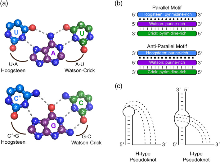
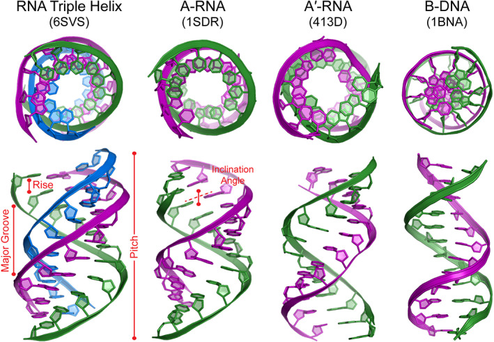
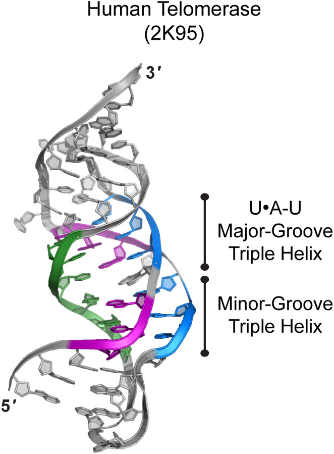
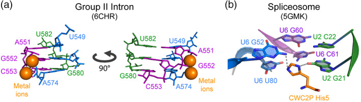
It has been nearly 63 years since the first characterization of an RNA triple helix in vitro by Gary Felsenfeld, David Davies, and Alexander Rich. An RNA triple helix consists of three strands: A Watson-Crick RNA double helix whose major-groove establishes hydrogen bonds with the so-called "third strand". In the past 15 years, it has been recognized that these major-groove RNA triple helices, like single-stranded and double-stranded RNA, also mediate prominent biological roles inside cells. Thus far, these triple helices are known to mediate catalysis during telomere synthesis and RNA splicing, bind to ligands and ions so that metabolite-sensing riboswitches can regulate gene expression, and provide a clever strategy to protect the 3' end of RNA from degradation. Because RNA triple helices play important roles in biology, there is a renewed interest in better understanding the fundamental properties of RNA triple helices and developing methods for their high-throughput discovery. This review provides an overview of the fundamental biochemical and structural properties of major-groove RNA triple helices, summarizes the structure and function of naturally occurring RNA triple helices, and describes prospective strategies to isolate RNA triple helices as a means to establish the "triplexome". This article is categorized under: RNA Structure and Dynamics > RNA Structure and Dynamics RNA Structure and Dynamics > RNA Structure, Dynamics and Chemistry RNA Structure and Dynamics > Influence of RNA Structure in Biological Systems.

Keywords: RNA stability element; RNA triple helix; base triples; catalytic triplex; riboswitch; telomerase; triplexome.


Conflict of interest statement

The author has declared no conflicts of interest for this article.

Figures

FIGURE 1
FIGURE 1
Composition and structural arrangement of major‐groove RNA triple helices. Hydrogen‐bonding interactions (gray dashed lines) are shown for the two canonical major‐groove base triples: (a) U•A‐U and C+•G‐C (ball‐and‐stick representation of base triples from PDB 6SVS; Ruszkowska, Ruszkowski, Hulewicz, Dauter, & Brown, 2020). Interactions are shown along with the Hoogsteen and Watson–Crick faces. (b) Schematics are shown for strand polarity and nucleotide composition in parallel and anti‐parallel motifs. Interbase interactions are represented as a circle (•) for Hoogsteen base pair, a single dash (−) for Watson–Crick base pair, and a square (■) for reverse Hoogsteen base pair. (c) Major‐groove RNA triple helices are commonly found in H‐type or I‐type pseudoknot structures. The three strands of triple helix are identified by the following tricolor scheme: Hoogsteen strand is blue, Watson strand is purple, and Crick strand is green
FIGURE 2
FIGURE 2
Structural views of triple and double helices. The top and side views are shown for X‐ray crystal structures (cartoon representation) solved for an RNA triple helix, A‐RNA double helix, A′‐RNA double helix, and B‐DNA double helix. Hoogsteen strand is blue, Watson (or purine‐rich) strand is purple, and Crick (or pyrimidine‐rich) strand is green. Select structural parameters are defined by red text and pictures. The PDB IDs for displayed structures are listed below each name
FIGURE 3
FIGURE 3
Triple helix in telomerase RNA. Telomerase RNA forms a nearly continuous triple helix (tricolor cartoon representation) in which a U•A‐U major‐groove triple helix is adjacent to a minor‐groove triple helix
FIGURE 4
FIGURE 4
Catalytic triplexes involved in splicing. (a) The catalytic triplex of both the group II self‐splicing intron (tricolor stick representation) and the spliceosome (not shown) coordinate two catalytic metal ions (orange spheres) via electrostatic interactions with phosphate backbone. Two views are shown. (b) His5 of CWC2P (orange stick representation) may form hydrogen bonds (gray dashed lines) with two of the three base triples (tricolor sticks) in the catalytic triplex observed for the S. cerevisiae spliceosome
FIGURE 5
FIGURE 5
Structural basis of ligand recognition by an RNA triple helix. The 3D structures are shown for six ligand‐bound riboswitches: (a) SAM‐II, SAM‐V, (b) PreQ1‐II, PreQ1‐III, (c) c‐di‐GMP‐II, and (d) guanidine‐III. For each riboswitch, there is a panel that displays the entire experimentally determined structure with PDB in parenthesis (left panel, cartoon representation), the triple helix interacting with ligand (middle panel, cartoon representation), and noncovalent interactions of ligand and nucleotides (right panel, ball‐and‐stick representation). The ligand is shown in orange and triple helix components are shown in tricolor scheme
FIGURE 6
FIGURE 6
Structures of triple helices that function as RNA stability elements. (a) A generalized schematic diagram is shown for a single‐domain stability element alongside the X‐ray crystal structure of the KSHV PAN RNA triple helix (cartoon representation). Important structural regions are labeled. (b) A generalized schematic diagram is shown for the predicted structure of a double‐domain stability element: TWIFB1_Osa. Gray circles (•) and dashed lines (−) indicate putative nucleotide interactions. (c) A generalized schematic diagram and X‐ray crystal structure is shown for a blunt‐ended RNA triple helix: Human MALAT1 (cartoon representation). The tricolor scheme of strands and symbols for their interactions are described in Figure 1. The PDB IDs are listed below each RNA label
FIGURE 7
FIGURE 7
Chemical structures of triplex‐binding small molecules. Chemical structures are shown for small molecules that (a) broadly recognize RNA triple helices and (b) bind to the MALAT1 RNA triple helix

References

    1. Abu Almakarem, A. S. , Petrov, A. I. , Stombaugh, J. , Zirbel, C. L. , & Leontis, N. B. (2012). Comprehensive survey and geometric classification of base triples in RNA structures. Nucleic Acids Research, 40(4), 1407–1423. 10.1093/nar/gkr810 - DOI - PMC - PubMed
    1. Abulwerdi, F. A. , Xu, W. , Ageeli, A. A. , Yonkunas, M. J. , Arun, G. , Nam, H. , … Le Grice, S. F. J. (2019). Selective small‐molecule targeting of a triple helix encoded by the long noncoding RNA, MALAT1. ACS Chemical Biology, 14(2), 223–235. 10.1021/acschembio.8b00807 - DOI - PMC - PubMed
    1. Adams, P. L. , Stahley, M. R. , Kosek, A. B. , Wang, J. , & Strobel, S. A. (2004). Crystal structure of a self‐splicing group I intron with both exons. Nature, 430(6995), 45–50. 10.1038/nature02642 - DOI - PubMed
    1. Ageeli, A. A. , McGovern‐Gooch, K. R. , Kaminska, M. M. , & Baird, N. J. (2019). Finely tuned conformational dynamics regulate the protective function of the lncRNA MALAT1 triple helix. Nucleic Acids Research, 47(3), 1468–1481. 10.1093/nar/gky1171 - DOI - PMC - PubMed
    1. Arnott, S. , & Bond, P. J. (1973). Structures for poly(U)•poly(A)‐poly(U) triple‐stranded polynucleotides. Nature: New Biology, 244(134), 99–101. 10.1038/newbio244099a0 - DOI - PubMed

Publication types

LinkOut - more resources