Endemic Chromoblastomycosis Caused Predominantly by Fonsecaea nubica, Madagascar1

Tahinamandranto Rasamoelina et al. Emerg Infect Dis. 2020 Jun.

Abstract

Chromoblastomycosis is an implantation fungal infection. Twenty years ago, Madagascar was recognized as the leading focus of this disease. We recruited patients in Madagascar who had chronic subcutaneous lesions suggestive of dermatomycosis during March 2013-June 2017. Chromoblastomycosis was diagnosed in 50 (33.8%) of 148 patients. The highest prevalence was in northeastern (1.47 cases/100,000 persons) and southern (0.8 cases/100,000 persons) Madagascar. Patients with chromoblastomycosis were older (47.9 years) than those without (37.5 years) (p = 0.0005). Chromoblastomycosis was 3 times more likely to consist of leg lesions (p = 0.003). Molecular analysis identified Fonsecaea nubica in 23 cases and Cladophialophora carrionii in 7 cases. Of 27 patients who underwent follow-up testing, none were completely cured. We highlight the persistence of a high level of chromoblastomycosis endemicity, which was even greater at some locations than 20 years ago. We used molecular tools to identify the Fonsecaea sp. strains isolated from patients as F. nubica.

Keywords: Fonseca pedrosoi; Fonsecaea nubica; Madagascar; chromoblastomycosis; clinical outcome; clinical presentation; dermatomycosis; epidemiology; fungal infections; fungi; molecular diagnosis; prevalence.


Figures

Figure 1
Figure 1
Recruitment of patients for study of chromoblastomycosis and prevalence by region, Madagascar, March 2013–June 2017. A) Recruitment sites (green triangles). Region of Sava: 1) Centre Hospitalier de Référence Régionale, Sambava District; 2) Centre Hospitalier de District and Hôpital Adventiste, Andapa District, Analamanga Region; 3) Centre de Santé de Base Alakamisy-Anjozorobe, Anjozorobe District; 4) Centre Hospitalier Universitaire Joseph Ravoahangy Befelatanana, Antananarivo District; 5) Centre de Santé de Base, Andramasina District, Vatovavy Fitovinany Region; 6) Fondation Médicale Ampasimanjeva, Manakara District; Anosy Region; 7) Centre Médical Tolagnaro, Centre Hospitalier de Référence Régionale Tolagnaro and Hôpital Luthérien Manambaro, Tolagnaro District. B) Geographic origin of patients recruited. Regions from north to south: D, Diana; S, Sava; I, Itasy; A, Analamanga; V, Vakinankaratra; B, Bongolava; So, Sofia; Bo, Boeny; Be, Betsiboka; Me, Melaky; Al, Alaotra-Mangoro; At, Atsinanana, An, Analanjirofo; Am, Amoron’I Mania; H, Haute Matsiatra; Va, Vatovavy-Fitovinany; Ato, Atsimo-Atsinanana; Ih, Ihorombe; Mb, Menabe; Ats, Atsimo Andrefana; And, Androy; Ano, Anôsy. No. patients recruited; dark purple, >6; medium purple, 3–5; light purple, <3; white, missing (none). C) Geographic distribution of chromoblastomycosis cases and causative fungal agents. Prevalence is no. cases/100,000 persons: dark purple, >0.5; medium purple, 0.1–0.5; light purple, <0.1; white, missing (none). Causative agent distribution: yellow dots, Fonsecaea nubica; black dots, Cladophialophora carrionii; green dots, Fonsecaea sp.
Figure 2
Figure 2
Clinical forms of chromoblastomycosis caused by Fonsecaea sp., Madagascar. A) Plaque; B) mixed: tumorous and cicatricial; C) nodular; D) raised plaque; E) plaque; F) cicatricial; G) tumorous caused by Cladophialophora carrionii; H) mixed: cicatricial and modified by previous therapy.
Figure 3
Figure 3
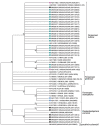
Phylogenetic tree of internal transcribed spacers sequences of fungal isolates from patients with chromoblastomycosis, Madagascar. Tree was constructed by using MEGA7.0 software (https://www.megasoftware.net) and applying the maximum-likelihood method based on the Kimura 2-parameter model (100 bootstrap replicates). Numbers along branches are bootstrap values. GenBank accession numbers are provided. Detailed information for strains is available (Appendix Table). Sporpthrix schenckii was used as the outgroup. Dark blue squares, Fonsecaea nubica sequences isolated in this study; black squares, Cladophialophora carrionii isolated in this study; pink squares, F. nubica previously identified as F. pedrosoi; light blue squares, F. monophora previously identified as F. pedrosi.

References

    1. Queiroz-Telles F, de Hoog S, Santos DW, Salgado CG, Vicente VA, Bonifaz A, et al. Chromoblastomycosis. Clin Microbiol Rev. 2017;30:233–76. 10.1128/CMR.00032-16 - DOI - PMC - PubMed
    1. Rasamoelina T, Raharolahy O, Rakotozandrindrainy N, Ranaivo I, Andrianarison M, Rakotonirina B, et al. Chromoblastomycosis and sporotrichosis, two endemic but neglected fungal infections in Madagascar. J Mycol Med. 2017;27:312–24. 10.1016/j.mycmed.2017.08.003 - DOI - PubMed
    1. Badali H, Bonifaz A, Barrón-Tapia T, Vázquez-González D, Estrada-Aguilar L, Oliveira NM, et al. Rhinocladiella aquaspersa, proven agent of verrucous skin infection and a novel type of chromoblastomycosis. Med Mycol. 2010;48:696–703. 10.3109/13693780903471073 - DOI - PubMed
    1. Bonifaz A, Carrasco-Gerard E, Saúl A. Chromoblastomycosis: clinical and mycologic experience of 51 cases. Mycoses. 2001;44:1–7. 10.1046/j.1439-0507.2001.00613.x - DOI - PubMed
    1. Queiroz-Telles F, Esterre P, Perez-Blanco M, Vitale RG, Salgado CG, Bonifaz A. Chromoblastomycosis: an overview of clinical manifestations, diagnosis and treatment. Med Mycol. 2009;47:3–15. 10.1080/13693780802538001 - DOI - PubMed

Publication types

Substances

Supplementary concepts

LinkOut - more resources