Case Reports
. 2020 Jul;34(4):1587-1593.
doi: 10.1111/jvim.15780. Epub 2020 May 22.

Antiviral treatment using the adenosine nucleoside analogue GS-441524 in cats with clinically diagnosed neurological feline infectious peritonitis

Affiliations
Case Reports

Antiviral treatment using the adenosine nucleoside analogue GS-441524 in cats with clinically diagnosed neurological feline infectious peritonitis

Peter J Dickinson et al. J Vet Intern Med. 2020 Jul.

Abstract

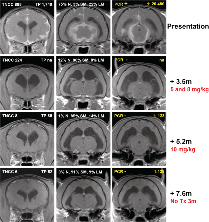
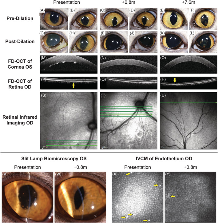
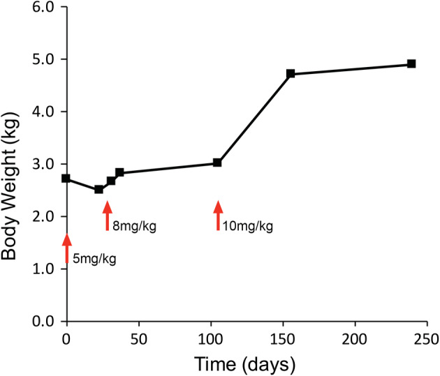
Feline infectious peritonitis (FIP) is caused by a mutant biotype of the feline enteric coronavirus. The resulting FIP virus (FIPV) commonly causes central nervous system (CNS) and ocular pathology in cases of noneffusive disease. Over 95% of cats with FIP will succumb to disease in days to months after diagnosis despite a variety of historically used treatments. Recently developed antiviral drugs have shown promise in treatment of nonneurological FIP, but data from neurological FIP cases are limited. Four cases of naturally occurring FIP with CNS involvement were treated with the antiviral nucleoside analogue GS-441524 (5-10 mg/kg) for at least 12 weeks. Cats were monitored serially with physical, neurologic, and ophthalmic examinations. One cat had serial magnetic resonance imaging (MRI), cerebrospinal fluid (CSF) analysis (including feline coronavirus [FCoV]) titers and FCoV reverse transcriptase [RT]-PCR) and serial ocular imaging using Fourier-domain optical coherence tomography (FD-OCT) and in vivo confocal microscopy (IVCM). All cats had a positive response to treatment. Three cats are alive off treatment (528, 516, and 354 days after treatment initiation) with normal physical and neurologic examinations. One cat was euthanized 216 days after treatment initiation following relapses after primary and secondary treatment. In 1 case, resolution of disease was defined based on normalization of MRI and CSF findings and resolution of cranial and caudal segment disease with ocular imaging. Treatment with GS-441524 shows clinical efficacy and may result in clearance and long-term resolution of neurological FIP. Dosages required for CNS disease may be higher than those used for nonneurological FIP.

Keywords: antiviral; cat; corona virus; ophthalmology.


Conflict of interest statement

Authors declare no conflict of interest.

Figures

FIGURE 1
FIGURE 1
Sequential magnetic resonance imaging from Case 4. Rows represent selected postcontrast (gadolinium) T1‐weighted transverse images of the brain acquired in a single imaging sequence. Routine analysis from cerebrospinal fluid analysis at the time of imaging is presented in white for each imaging time point: TNCC = CSF total nucleated cell count (cells/μL); TP = CSF total protein (mg/dL); N = neutrophils, SM = small mononuclear, LM = large mononuclear. Characteristic neutrophilic pleocytosis resolved over the course of the treatment. Additional CSF analyses relating to FCoV detection are presented in yellow for each time point: PCR = FCoV RT‐PCR result [positive (+) or negative (−)]; Dilution ratio = cerebrospinal fluid FCoV antibody titer. Time points and doses of GS‐1441524 delivered before imaging time points are described for each imaging sequence. Initial pronounced meningeal contrast enhancement resolves after GS‐144524 treatment and does not recur after cessation of treatment. Ventriculomegaly that is present after initial response to treatment, resolved slowly on subsequent imaging. Decreasing abnormalities in CSF analysis findings paralleled decreased abnormalities on MR imaging
FIGURE 2
FIGURE 2
Sequential multimodal imaging of the cranial and caudal segments from Case 4. At presentation (A, B) predilatation and (G, H) postdilatation cranial segment photographs showing mild diffuse corneal edema, pigmented keratic precipitates, rubeosis iridis, obscured detail of the iris because of aqueous flare, and incomplete dilatation OU; dyscoria with incomplete pupillary dilatation because of caudal synechia OS (H) was also observed. Keratic precipitates were also visualized OS with slit lamp biomicroscopy (V), corneal FD‐OCT (M), and IVCM of the endothelium (X, arrows); increased corneal thickness was also observed with FD‐OCT (X). Imaging of the retina and choroid with FD‐OCT revealed cellular infiltrate in the choroid (P, arrow) that was visible as a hyporeflective lesion with infrared photography (S). At 0.8 months after initiation of GS‐441524 treatment, pre‐ (C,D) and postdilatation (I,J) cranial segment photographs demonstrated clear corneas and cranial chambers OU, isocoria, decreased rubeosis iridis, and complete pupillary dilatation OS. A marked decrease in pigmented keratic precipitates was noted with slit lamp biomicroscopy (W), corneal FD‐OCT (N), and IVCM of the endothelium (Y, arrow). Normal retinal and choroidal morphology is observed with FD‐OCT (Q) although the hyporeflective lesions remain with infrared imaging (T). At 7.6 months, pre‐ (E,F) and postdilatation (K, L) cranial segment photographs demonstrated clear corneas and cranial chambers OU, isocoria, normal iris morphology, and postinflammatory pigment on the cranial lens capsule OS. Keratic precipitates are absent with corneal FD‐OCT (O). With FD‐OCT and infrared imaging, thinning of the dorsal peripheral retina was present (R, arrow) with loss of the normal layering but no cellular infiltrate or retinal separation (U)
FIGURE 3
FIGURE 3
Case 4 body weight plotted with respect to time postinitiation of GS‐1441524 treatment. Changes in drug dose are marked with red arrows. Increased body weight was seen after increases in drug dose beyond initial 5 mg/kg dose, and was accompanied by resolution of clinical signs

References

    1. Murphy BG, Perron M, Murakami E, et al. The nucleoside analog GS‐441524 strongly inhibits feline infectious peritonitis (FIP) virus in tissue culture and experimental cat infection studies. Vet Microbiol. 2018;219:226‐233. - PMC - PubMed
    1. Pedersen NC, Perron M, Bannasch M, et al. Efficacy and safety of the nucleoside analog GS‐441524 for treatment of cats with naturally occurring feline infectious peritonitis. J Feline Med Surg. 2019;21:271‐281. - PMC - PubMed
    1. Pedersen NC. An update on feline infectious peritonitis: diagnostics and therapeutics. Vet J. 2014;201:133‐141. - PMC - PubMed
    1. Pedersen NC. A review of feline infectious peritonitis virus infection: 1963‐2008. J Feline Med Surg. 2009;11:225‐258. - PMC - PubMed
    1. Pedersen NC, Kim Y, Liu H, et al. Efficacy of a 3C‐like protease inhibitor in treating various forms of acquired feline infectious peritonitis. J Feline Med Surg. 2018;20:378‐392. - PMC - PubMed

Publication types

MeSH terms