. 2020 Jul 1;319(1):H109-H122.
doi: 10.1152/ajpheart.00114.2020. Epub 2020 May 22.

Cardiac mesenchymal cells from failing and nonfailing hearts limit ventricular dilation when administered late after infarction

Affiliations

Cardiac mesenchymal cells from failing and nonfailing hearts limit ventricular dilation when administered late after infarction

Timothy N Audam et al. Am J Physiol Heart Circ Physiol. .

Abstract

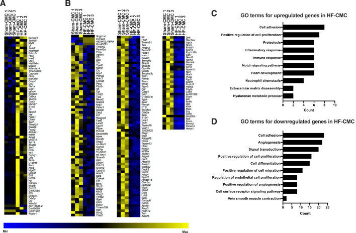
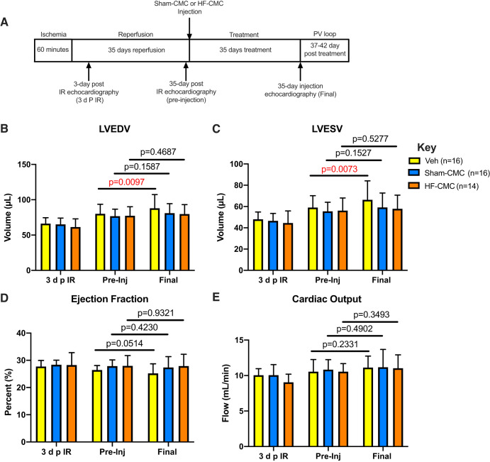
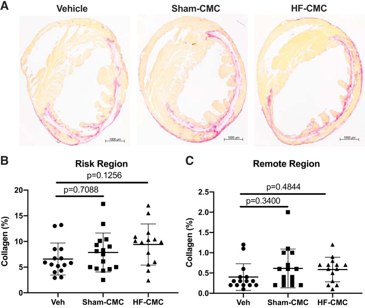
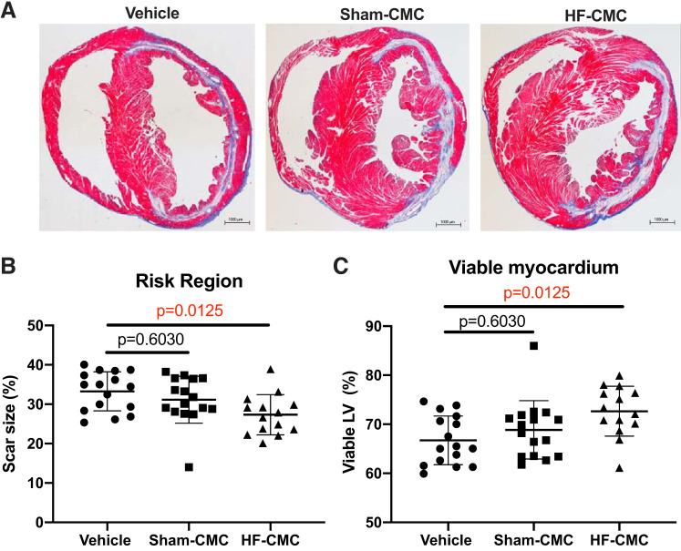
Although cell therapy-mediated cardiac repair offers promise for treatment/management of heart failure, lack of fundamental understanding of how cell therapy works limits its translational potential. In particular, whether reparative cells from failing hearts differ from cells derived from nonfailing hearts remains unexplored. Here, we assessed differences between cardiac mesenchymal cells (CMC) derived from failing (HF) versus nonfailing (Sham) hearts and whether the source of donor cells (i.e., from HF vs. Sham) limits reparative capacity, particularly when administered late after infarction. To determine the impact of the donor source of CMCs, we characterized the transcriptional profile of CMCs isolated from sham (Sham-CMC) and failing (HF-CMC) hearts. RNA-seq analysis revealed unique transcriptional signatures in Sham-CMC and HF-CMC, suggesting that the donor source impacts CMC. To determine whether the donor source affects reparative potential, C57BL6/J female mice were subjected to 60 min of regional myocardial ischemia and then reperfused for 35 days. In a randomized, controlled, and blinded fashion, vehicle, HF-CMC, or Sham-CMC were injected into the lumen of the left ventricle at 35 days post-MI. An additional 5 weeks later, cardiac function was assessed by echocardiography, which indicated that delayed administration of Sham-CMC and HF-CMC attenuated ventricular dilation. We also determined whether Sham-CMC and HF-CMC treatments affected ventricular histopathology. Our data indicate that the donor source (nonfailing vs. failing hearts) affects certain aspects of CMC, and these insights may have implications for future studies. Our data indicate that delayed administration of CMC limits ventricular dilation and that the source of CMC may influence their reparative actions.NEW & NOTEWORTHY Most preclinical studies have used only cells from healthy, nonfailing hearts. Whether donor condition (i.e., heart failure) impacts cells used for cell therapy is not known. We directly tested whether donor condition impacted the reparative effects of cardiac mesenchymal cells in a chronic model of myocardial infarction. Although cells from failing hearts differed in multiple aspects, they retained the potential to limit ventricular remodeling.

Keywords: cardiac repair; cell therapy; fibrosis; ventricular remodeling.


Conflict of interest statement

M.W. has pending intellectual property related to cell therapy. None of the other authors have any relevant conflicts to declare.

Figures

Fig. 1.
Fig. 1.
Donor condition impacts transcriptional signature of cardiac mesenchymal cells (CMCs). Total RNA was isolated from CMCs derived from sham (Sham-CMC; n = 3) and heart failure mice (HF-CMC; n = 3) and was subjected to unbiased RNA-seq analysis to characterize global transcriptional changes. A: heat map analysis showing upregulated genes. B: heat map analysis showing downregulated genes. C: Gene Ontology (GO) enrichment analysis showing top upregulated genes. D: GO enrichment analysis showing top downregulated genes. A test statistic was used to determine significance of differentially expressed genes in Sham-CMC and HF-CMC groups. A Fisher’s exact test was used to determine significantly enriched GO terms.
Fig. 2.
Fig. 2.
Delayed administration of cardiac mesenchymal cells limits ventricular dilation. Female C57BL6/J mice subjected to 60 min of regional myocardial ischemia and allowed to reperfuse for 35 days were treated with either saline (vehicle; n = 16) or cardiac mesenchymal cells (CMCs) isolated from sham (Sham-CMC; n = 16) and heart failure (HF-CMC; n = 14) mice. On day 35, treated mice were subjected to final echocardiography. A: schematic illustrating timeline for ischemia-reperfusion (I/R) procedure, CMC injection, and echocardiography time points [at 3 days post-I/R (3 d p I/R), 35 days post-I/R (preinjection), and 35 days postinjection (final)]. B: left ventricular end diastolic volume (LVEDV) from preinjection to final for hearts treated with vehicle, Sham-CMC, and HF-CMC. C: left ventricular end-systolic volume (LVESV) from preinjection (pre-Inj) to final for hearts treated with vehicle, Sham-CMC, and HF-CMC. D: ejection fraction (EF) from pre-Inj to final for hearts treated with vehicle, Sham-CMC, and HF-CMC. E: cardiac output (CO) from pre-Inj to final for hearts treated with vehicle, Sham-CMC, and HF-CMC. Data are represented as means ± SD. Differences in the outcomes by treatment groups and pre-Inj vs. postinjection were tested using repeated-measures ANOVA. Interaction terms between groups and pre-/postinjection were included in models to determine whether pre-/postdifferences were modified by group type.
Fig. 3.
Fig. 3.
Collagen content is not changed in cardiac mesenchymal cell (CMC)-treated hearts. Sections from saline [vehicle (Veh); n = 15]-, sham (Sham-CMC; n = 16)-, and heart failure (HF-CMC; n = 14)-treated hearts were stained with picrosirius red to determine total collagen content. A: representative images of Veh-, Sham-CMC-, and HF-CMC-treated hearts. B: quantification of total collagen content in the risk region of Veh, Sham-CMC, and HF-CMC treated hearts. C: quantification of total collagen content in the remote region of Veh-, Sham-CMC-, and HF-CMC-treated hearts. Data are represented as means ± SD. One-way ANOVA followed by Sidak’s multiple-comparison test was used to determine significant differences between Sham-CMC- or HF-CMC-treated hearts compared with vehicle.
Fig. 4.
Fig. 4.
Heart failure-derived cardiac mesenchymal cells (HF-CMC) significantly reduced scar size and limited loss of viable myocardium. Sections from saline (vehicle; n = 16)-, sham (Sham-CMC; n = 16)-, and heart failure (HF-CMC; n = 14)-treated hearts were stained with Masson’s trichrome to determine scar size. A: representative images of vehicle, Sham-CMC, and HF-CMC treated hearts. B: quantification of scar size in vehicle-, Sham-CMC-, and HF-CMC-treated hearts. C: quantification of viable myocardium in vehicle, Sham-CMC, and HF-CMC treated hearts. Data are represented as means ± SD. One-way ANOVA followed by Sidak’s multiple-comparison test was used to determine significant differences between Sham-CMC- or HF-CMC-treated hearts compared with vehicle.
Fig. 5.
Fig. 5.
Capillary density is not changed in sham heart-derived cardiac mesenchymal cell (Sham-CMC)- or heart failure-derived cardiac mesenchymal cell (HF-CMC)-treated hearts. Sections from saline (vehicle; n = 9)-, sham (Sham-CMC; n = 9)-, and heart failure (HF-CMC; n = 9)-treated hearts were stained with isolectin B4 to assess capillary density. A: representative images of isolectin B4-stained vehicle-, Sham-CMC-, and HF-CMC-treated hearts. B: quantification of capillary density (green) in the border zone (BZ), infarct zone (IZ), and remote zone (RZ) of treated hearts. Data are represented as means ± SD. Two-way ANOVA followed by Dunnett’s multiple-comparison test was used to determine significant differences between Sham-CMC- or HF-CMC-treated hearts compared with vehicle in the BZ, IZ, and RZ.
Fig. 6.
Fig. 6.
Cardiomyocyte size is not changed in sham heart-derived cardiac mesenchymal cell (Sham-CMC)- or heart failure-derived cardiac mesenchymal cell (HF-CMC)-treated hearts. Sections from saline (vehicle; n = 9)-, sham (Sham-CMC; n = 9)-, and heart failure (HF-CMC; n = 9)-treated hearts were stained with wheat germ agglutinin (WGA) to assess cardiomyocyte size. A: representative images of WGA-stained vehicle, Sham-CMC, and HF-CMC treated hearts. B: quantification of cardiomyocyte size in the border zone (BZ), infarct zone (IZ), and remote zone (RZ) of treated hearts. Data are represented as means ± SD. Two-way ANOVA followed by Dunnett’s multiple-comparison test was used to determine significant differences between Sham-CMC or HF-CMC treated hearts compared with vehicle in the BZ, IZ, and RZ.
Fig. 7.
Fig. 7.
Administration of heart-derived cardiac mesenchymal cells (Sham-CMCs) reduced immune cell abundance in the infarct zone of the heart. Sections from saline (vehicle; n = 9)-, sham (Sham-CMC; n = 9)-, and heart failure (HF-CMC; n = 9)-treated hearts were stained with CD45 antigen to assess presence of leukocytes. A: representative images of vehicle-, Sham-CMC-, and HF-CMC-treated hearts. B: quantification of CD45 (red) cells in the border zone (BZ), infarct zone (IZ), and remote zone (RZ) of treated hearts. Data are represented as means ± SD. Two-way ANOVA followed by Tukey’s multiple-comparison test was used to determine significant differences between Sham-CMC- or HF-CMC-treated hearts compared with vehicle in the BZ, IZ, and RZ.
Fig. 8.
Fig. 8.
Administration of heart-derived cardiac mesenchymal cells (Sham-CMCs) reduced hyaluronan (HA) levels in treated hearts. HA metabolism was characterized in CMCs derived from sham (Sham-CMC) and heart failure (HF-CMC) mice by quantitative PCR, agarose gel electrophoresis, and fluorescent microscopy. A: expression of Has2 mRNA in Sham-CMC (n = 3) and HF-CMC (n = 4). B: HA sizing by agarose gel from conditioned media of Sham-CMC (n = 3) and HF-CMC (n = 3). C: quantification of HA sizing by agarose gel from conditioned media of Sham-CMC (n = 3) and HF-CMC (n = 3). D: hyaluronan levels according to hyaluronan-binding protein (HABP; red) staining in vehicle (n = 15)-, Sham-CMC (n = 15)-, and HF-CMC (n = 13)-treated hearts. E: quantification of hyaluronan levels according to HABP (red) staining in vehicle (n = 15)-, Sham-CMC (n = 15)-, and HF-CMC (n = 13)-treated hearts. Data are represented as means ± SD. For 2 groups, an unpaired 2-tailed Student’s t test was used to determine significance between Sham-CMC and HF-CMC. For 3 groups, a 1-way ANOVA followed by Sidak’s multiple-comparison test was used to determine significance between Sham-CMC- or HF-CMC-treated hearts compared with vehicle.

References

    1. Bolger AM, Lohse M, Usadel B. Trimmomatic: a flexible trimmer for Illumina sequence data. Bioinformatics 30: 2114–2120, 2014. doi:10.1093/bioinformatics/btu170. - DOI - PMC - PubMed
    1. Brainard RE, Watson LJ, Demartino AM, Brittian KR, Readnower RD, Boakye AA, Zhang D, Hoetker JD, Bhatnagar A, Baba SP, Jones SP. High fat feeding in mice is insufficient to induce cardiac dysfunction and does not exacerbate heart failure. PLoS One 8: e83174, 2013. [Erratum in: PLoS One 9: e113944, 2014.] 10.1371/journal.pone.0083174. - DOI - PMC - PubMed
    1. Cheng K, Malliaras K, Smith RR, Shen D, Sun B, Blusztajn A, Xie Y, Ibrahim A, Aminzadeh MA, Liu W, Li TS, De Robertis MA, Marbán L, Czer LS, Trento A, Marbán E. Human cardiosphere-derived cells from advanced heart failure patients exhibit augmented functional potency in myocardial repair. JACC Heart Fail 2: 49–61, 2014. doi:10.1016/j.jchf.2013.08.008. - DOI - PMC - PubMed
    1. Condorelli G, Roncarati R, Ross J Jr, Pisani A, Stassi G, Todaro M, Trocha S, Drusco A, Gu Y, Russo MA, Frati G, Jones SP, Lefer DJ, Napoli C, Croce CM. Heart-targeted overexpression of caspase3 in mice increases infarct size and depresses cardiac function. Proc Natl Acad Sci USA 98: 9977–9982, 2001. doi:10.1073/pnas.161120198. - DOI - PMC - PubMed
    1. Dassanayaka S, Brainard RE, Watson LJ, Long BW, Brittian KR, DeMartino AM, Aird AL, Gumpert AM, Audam TN, Kilfoil PJ, Muthusamy S, Hamid T, Prabhu SD, Jones SP. Cardiomyocyte Ogt limits ventricular dysfunction in mice following pressure overload without affecting hypertrophy. Basic Res Cardiol 112: 23, 2017. doi:10.1007/s00395-017-0612-7. - DOI - PMC - PubMed

Publication types