. 2020 Jul 2;48(W1):W509-W514.
doi: 10.1093/nar/gkaa407.

TIMER2.0 for analysis of tumor-infiltrating immune cells

Affiliations

TIMER2.0 for analysis of tumor-infiltrating immune cells

Taiwen Li et al. Nucleic Acids Res. .

Abstract

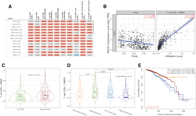
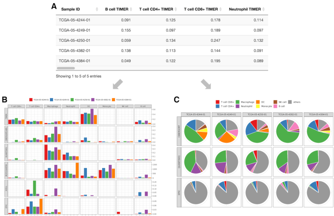
Tumor progression and the efficacy of immunotherapy are strongly influenced by the composition and abundance of immune cells in the tumor microenvironment. Due to the limitations of direct measurement methods, computational algorithms are often used to infer immune cell composition from bulk tumor transcriptome profiles. These estimated tumor immune infiltrate populations have been associated with genomic and transcriptomic changes in the tumors, providing insight into tumor-immune interactions. However, such investigations on large-scale public data remain challenging. To lower the barriers for the analysis of complex tumor-immune interactions, we significantly improved our previous web platform TIMER. Instead of just using one algorithm, TIMER2.0 (http://timer.cistrome.org/) provides more robust estimation of immune infiltration levels for The Cancer Genome Atlas (TCGA) or user-provided tumor profiles using six state-of-the-art algorithms. TIMER2.0 provides four modules for investigating the associations between immune infiltrates and genetic or clinical features, and four modules for exploring cancer-related associations in the TCGA cohorts. Each module can generate a functional heatmap table, enabling the user to easily identify significant associations in multiple cancer types simultaneously. Overall, the TIMER2.0 web server provides comprehensive analysis and visualization functions of tumor infiltrating immune cells.


Figures

Figure 1.
Figure 1.
An illustration of immune component outputs. Upon a query to the immune component, TIMER2.0 evaluates associations between immune infiltrates and genetic or clinical features and displays the results as a functional heatmap (A). Upon a user clicking on the heatmap, each module subsequently generates figures (B–E) showing the detailed information about the corresponding relationship. (A) An example of functional heatmap table generated by the ‘Gene Module’ shows the association between PDCD1 expression and immune infiltration level of multiple CD8+ T cell types estimated by all six algorithms across TCGA cancer types. The red indicates a statistically significant positive association, and the blue indicates a statistically significant negative association. Gray denotes a non-significant result. (B) An example of scatter plots from the ‘Gene Module’. Correlation of PDCD1 expression with tumor purity (left) and with the infiltration level of CD8 T cell estimated by TIMER (right) in lung squamous carcinoma. (C) An example of the violin plot from the ‘Mutation Module’ displays the difference in TIMER-estimated CD8 T cell infiltration levels between tumors with mutant or wild-type TP53 in bladder cancer. (D) An example of the violin plot from the ‘sCNA Module’ visualizes the difference of CD8 T cell infiltration level estimate among tumors with different sCNA status of PIK3CA gene in head neck cancer. (E) An example of the Kaplan–Meier plot from the ‘Outcome Module’ shows the difference of overall survival among patients stratified by both the estimated infiltration level of CD8+ T cell and PDCD1 expression level in breast cancer.
Figure 2.
Figure 2.
An illustration of outputs for the estimation component. TIMER2.0 provides estimations of immune infiltration levels for user-provided tumor profiles using six state-of-the-art algorithms. An illustration of expression profiles (TPM-normalized) of five randomly selected TCGA LUAD samples into the estimation component. (A) The data table presents the different immune cell type infiltration estimated by multiple algorithms. (B) For the eight immune cell types for which all six algorithms can estimate the abundance, TIMER2.0 draws a multi-panel bar plot showing differences of their infiltration level estimated by different algorithms among samples. Of the six algorithms, three algorithms (CIBERSORT in original mode, quanTIseq and EPIC) generate values comparable within the same sample. Based on estimations from these algorithms, TIMER2.0 also presents (C) a multi-panel pie plot showing the proportion of immune cell types in each sample. The eight previously described immune cell types are highlighted in different colors.

References

    1. Angelova M., Charoentong P., Hackl H., Fischer M.L., Snajder R., Krogsdam A.M., Waldner M.J., Bindea G., Mlecnik B., Galon J.. Characterization of the immunophenotypes and antigenomes of colorectal cancers reveals distinct tumor escape mechanisms and novel targets for immunotherapy. Genome Biol. 2015; 16:64. - PMC - PubMed
    1. Lee N., Zakka L.R., Mihm M.C. Jr, Schatton T.. Tumour-infiltrating lymphocytes in melanoma prognosis and cancer immunotherapy. Pathology. 2016; 48:177–187. - PubMed
    1. Denkert C., von Minckwitz G., Darb-Esfahani S., Lederer B., Heppner B.I., Weber K.E., Budczies J., Huober J., Klauschen F., Furlanetto J.. Tumour-infiltrating lymphocytes and prognosis in different subtypes of breast cancer: a pooled analysis of 3771 patients treated with neoadjuvant therapy. Lancet Oncol. 2018; 19:40–50. - PubMed
    1. Galon J., Bruni D.. Approaches to treat immune hot, altered and cold tumours with combination immunotherapies. Nat. Rev. Drug Discov. 2019; 18:197–218. - PubMed
    1. Zhao Y., Schaafsma E., Gorlov I.P., Hernando E., Thomas N.E., Shen R., Turk M.J., Berwick M., Amos C.I., Cheng C.. A leukocyte infiltration score defined by a gene signature predicts melanoma patient prognosis. Mol. Cancer Res. 2019; 17:109–119. - PMC - PubMed

Publication types