. 2020 Jun 4;106(6):830-845.
doi: 10.1016/j.ajhg.2020.04.015. Epub 2020 May 21.

De Novo SOX6 Variants Cause a Neurodevelopmental Syndrome Associated with ADHD, Craniosynostosis, and Osteochondromas

Collaborators, Affiliations

De Novo SOX6 Variants Cause a Neurodevelopmental Syndrome Associated with ADHD, Craniosynostosis, and Osteochondromas

Dara Tolchin et al. Am J Hum Genet. .

Abstract

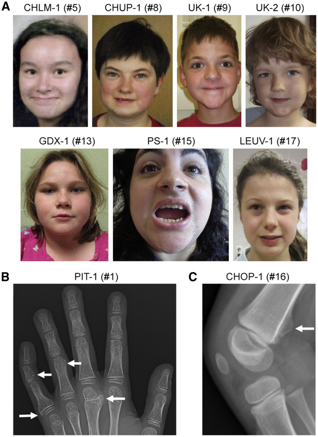
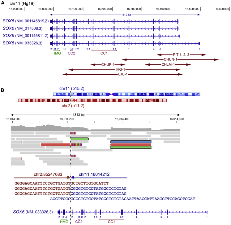
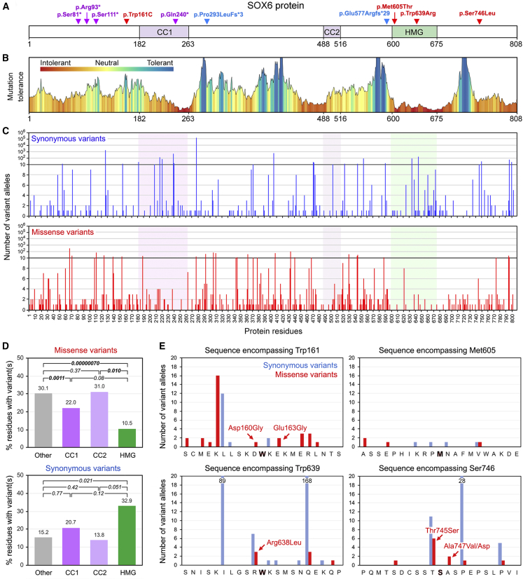
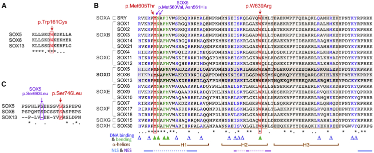
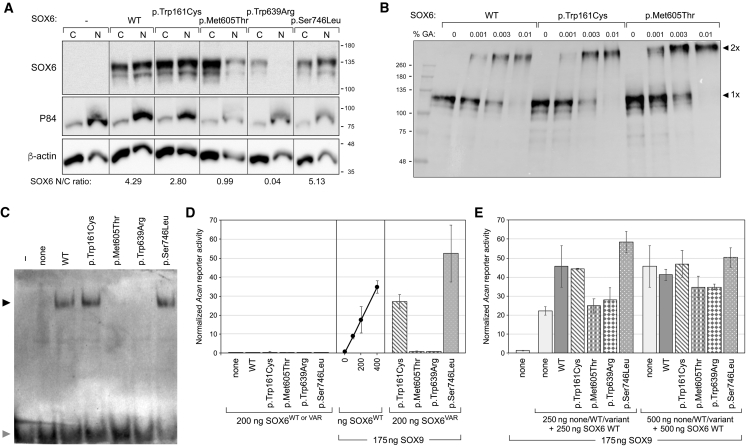
SOX6 belongs to a family of 20 SRY-related HMG-box-containing (SOX) genes that encode transcription factors controlling cell fate and differentiation in many developmental and adult processes. For SOX6, these processes include, but are not limited to, neurogenesis and skeletogenesis. Variants in half of the SOX genes have been shown to cause severe developmental and adult syndromes, referred to as SOXopathies. We here provide evidence that SOX6 variants also cause a SOXopathy. Using clinical and genetic data, we identify 19 individuals harboring various types of SOX6 alterations and exhibiting developmental delay and/or intellectual disability; the individuals are from 17 unrelated families. Additional, inconstant features include attention-deficit/hyperactivity disorder (ADHD), autism, mild facial dysmorphism, craniosynostosis, and multiple osteochondromas. All variants are heterozygous. Fourteen are de novo, one is inherited from a mosaic father, and four offspring from two families have a paternally inherited variant. Intragenic microdeletions, balanced structural rearrangements, frameshifts, and nonsense variants are predicted to inactivate the SOX6 variant allele. Four missense variants occur in residues and protein regions highly conserved evolutionarily. These variants are not detected in the gnomAD control cohort, and the amino acid substitutions are predicted to be damaging. Two of these variants are located in the HMG domain and abolish SOX6 transcriptional activity in vitro. No clear genotype-phenotype correlations are found. Taken together, these findings concur that SOX6 haploinsufficiency leads to a neurodevelopmental SOXopathy that often includes ADHD and abnormal skeletal and other features.

Keywords: ADHD; SOX6; SOXopathy; craniosynostosis; developmental delay; dysmorphism; genetic variant; human disease; intellectual disability; osteochondroma.


Conflict of interest statement

A.B., C.F., L.B.H., and P.R. are employees of GeneDx. All other authors declare no competing interests.

Figures

Figure 1
Figure 1
Clinical Findings in Subjects with SOX6 Variants (A) Photos of seven subjects showing mild, nonspecific facial dysmorphism. (B) X-ray showing multiple osteochondromas (marked by arrows) in the right hand of affected individual PIT-1. (C) X-ray showing an osteochondroma (marked by arrow) at the right distal femur of affected individual CHOP-1.
Figure 2
Figure 2
Structural Variants Detected in Affected Individuals 1–8 and 19 (A) Location of CNV variants. The main SOX6 transcript isoforms are schematized; taller vertical lines correspond to coding sequences, smaller vertical lines correspond to 5′ and 3′ untranslated sequences, and arrowheads in introns point to the transcriptional direction. NCBI accession numbers are indicated. The GenBank: NM_033326.3 coding exons are labeled 1–16. Coordinates of chromosomal region 11p15 are shown above the schematics. Double-arrowed lines depict the microdeletions identified in subjects 1–8. CC1, primary coiled-coil domain; CC2, secondary coiled-coil domain; HMG, HMG domain. (B) Location of the breakpoint of the 46,XY,t(2;11)(p11.2;p15.2)-balanced reciprocal translocation identified in affected individual 19. From top to bottom are schematics of chromosomes 2 and 11, in which a vertical dotted line indicates the breakpoint involving 11p15.2 and 2p11.2; reads (BAM file) aligned by the Integrative Genomics Viewer (IGV); the sequence of the breakpoint, in which red and blue lines show the normal sequences of chromosomes 2 and 11, respectively, and red sequences followed by blue sequences show the sequence junction; and the same representation of SOX6 as in (A).
Figure 3
Figure 3
Analysis of SOX6 SNVs in Affected Individuals 9–18 and in gnomAD Individuals (A) Location of study subjects’ SNVs in the SOX6 isoform encoded by the GenBank: NM_033326.3 transcript. The protein and domain residue boundaries are indicated underneath the schematic. CC1, primary coiled-coil domain; CC2, secondary coiled-coil domain; HMG, HMG domain. Red represents missense variants, purple represents nonsense variants, and blue represents frameshift variants. (B) Plot of the mutation tolerance of SOX6 residues downloaded from MetaDome. (C) Counts and distribution of SOX6 synonymous and missense variants found in gnomAD individuals. (D) Percentages of residues carrying at least one missense or synonymous variant in the functional and other domains of SOX6 in gnomAD individuals. We performed paired t tests to calculate the statistical significance of differences between domains. p values are indicated. (E) Numbers of synonymous and missense variants detected in gnomAD individuals in the sequences encompassing the missense variants identified in four study subjects. The nature of the missense variants closest to the residues altered in the four affected individuals is indicated.
Figure 4
Figure 4
Evolutionary Conservation of SOX6 Residues Altered in Affected Individuals (A) Alignment of human SOXD sequences encompassing Trp161. Asterisks represent fully conserved residues, and dots represent semi-conserved residues. (B) Alignment of the HMG domain sequences of all human SOX proteins: residues altered in SOX6 in affected individuals in our study are shown in red, and residues altered in SOX5 in affected individuals with LAMSHF syndrome are shown in purple. Triangles represent residues mediating DNA binding (blue) and bending (green). Brackets represent H1, H2, and H3 α helices. Lines linked with dots represent key amino acids in nuclear localization signal sequences (NLSs) and nuclear export signal sequences (NESs). (C) Alignment of human SOXD sequences encompassing Ser746 with indication of the position of SOX5 and SOX6 variants identified in affected individuals in our study.
Figure 5
Figure 5
Functional Tests of SOX6 Missense Variants (A) Stability and intracellular distribution of SOX6 variants. COS-1 cells were transfected with plasmids encoding 3FLAG-tagged SOX6 WT and variant proteins, as indicated. Cytoplasmic (C) and nuclear (N) extracts were tested via immunoblotting with a FLAG antibody. As expected, the P84 protein was enriched in nuclear extracts, and β-actin was enriched in cytoplasmic extracts. The distribution of SOX6 relative to β-actin in the nuclear versus cytoplasmic compartment is indicated underneath the blots. The migration of protein markers (Mr in k values) is indicated. (B) Ability of SOX6 variants to homodimerize. Lysates of COS-1 cells, transfected as described in (A), were incubated without (0) or with 0.001%, 0.03%, or 0.01% glutaraldehyde. SOX6 was visualized via immunoblotting with a FLAG antibody. The migration of protein standards (Mr in k values) and SOX6 monomers (1×) and homodimers (2×) is indicated. (C) Ability of SOX6 variants to bind DNA. An Acan enhancer probe was used in EMSA with no protein extract (−) or with extracts from COS-1 cells transfected with expression plasmids for no protein (none), SOX6 WT, or variant proteins, as indicated. SOX6-probe complexes (indicated with a black arrowhead) and free probes (indicated with a gray arrowhead) were resolved by electrophoresis. (D) Ability of SOX6 variants to synergize with SOX9 in transactivation. HEK293 cells were transfected with an Acan reporter, a control reporter, and plasmids encoding no SOX (none), SOX9, SOX6 WT, or SOX6 variant proteins, as indicated. The amounts of SOX plasmids are indicated too. Acan reporter activities were normalized for transfection efficiency. They are presented as the mean ± standard deviation obtained for triplicates in an experiment representative of five independent ones. The arrowhead in the middle panel points to the amount of WT SOX6 plasmid (200 ng) that was also used for variant plasmids in the right panel. (E) Interference of SOX6 variant proteins with WT SOX6 in transactivation. HEK293 cells were transfected as described in (D) with the indicated types and amounts of plasmids encoding no protein (none) or SOX proteins. Acan reporter activities were normalized for transfection efficiency. They are presented as the mean ± standard deviation obtained for triplicates in an experiment representative of four independent ones.
Figure 6
Figure 6
Analysis of SOX6 Expression in the Human Brain (A) SOX6 RNA expression measured by RNA-seq in various regions of the developing brain of a representative human fetus 9 weeks after conception. (B) SOX6 RNA expression measured by RNA-seq in the amygdaloid complex, hippocampus, striatum, and cerebellar cortex of multiple individuals whose ages are within the range spanning the three trimesters of gestation in utero and the first four decades of life. Data are presented as averages with standard deviation for each age category (n = 1–10 per group). The four brain structures were selected because their SOX6 RNA expression is higher than that of other structures.

References

    1. Kamachi Y., Kondoh H. Sox proteins: regulators of cell fate specification and differentiation. Development. 2013;140:4129–4144. - PubMed
    1. Angelozzi M., Lefebvre V. SOXopathies: growing family of developmental disorders due to SOX mutations. Trends Genet. 2019;35:658–671. - PMC - PubMed
    1. Gubbay J., Collignon J., Koopman P., Capel B., Economou A., Münsterberg A., Vivian N., Goodfellow P., Lovell-Badge R. A gene mapping to the sex-determining region of the mouse Y chromosome is a member of a novel family of embryonically expressed genes. Nature. 1990;346:245–250. - PubMed
    1. Sinclair A.H., Berta P., Palmer M.S., Hawkins J.R., Griffiths B.L., Smith M.J., Foster J.W., Frischauf A.M., Lovell-Badge R., Goodfellow P.N. A gene from the human sex-determining region encodes a protein with homology to a conserved DNA-binding motif. Nature. 1990;346:240–244. - PubMed
    1. Kashimada K., Koopman P. Sry: the master switch in mammalian sex determination. Development. 2010;137:3921–3930. - PubMed

Publication types

MeSH terms

Substances