. 2020 Aug;77(2):186-197.
doi: 10.1111/his.14160. Epub 2020 Jul 24.

Pulmonary and systemic involvement in COVID-19 patients assessed with ultrasound-guided minimally invasive autopsy

Affiliations

Pulmonary and systemic involvement in COVID-19 patients assessed with ultrasound-guided minimally invasive autopsy

Amaro N Duarte-Neto et al. Histopathology. 2020 Aug.

Abstract

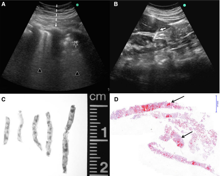
Aims: Brazil ranks high in the number of coronavirus disease 19 (COVID-19) cases and the COVID-19 mortality rate. In this context, autopsies are important to confirm the disease, determine associated conditions, and study the pathophysiology of this novel disease. The aim of this study was to assess the systemic involvement of COVID-19. In order to follow biosafety recommendations, we used ultrasound-guided minimally invasive autopsy (MIA-US), and we present the results of 10 initial autopsies.

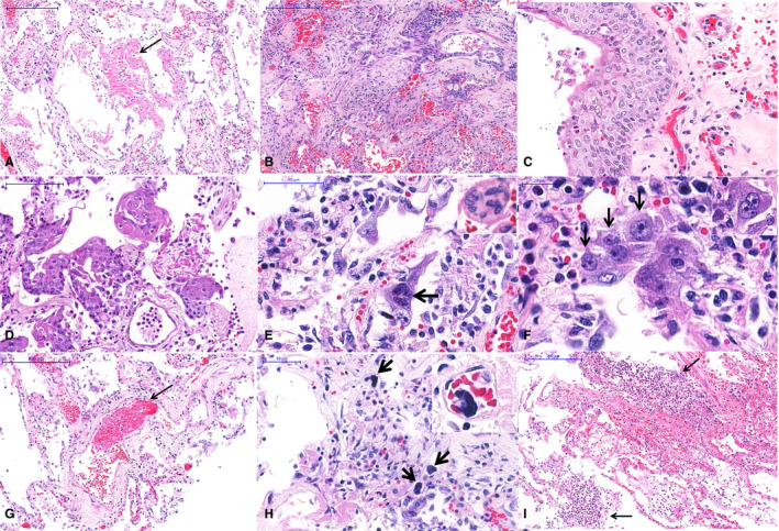
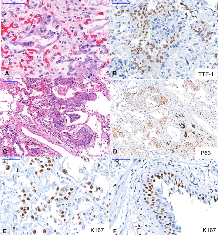
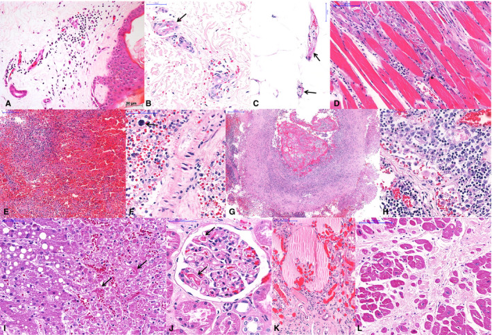
Methods and results: We used MIA-US for tissue sampling of the lungs, liver, heart, kidneys, spleen, brain, skin, skeletal muscle and testis for histology, and reverse transcription polymerase chain reaction to detect severe acute respiratory syndrome coronavirus 2 RNA. All patients showed exudative/proliferative diffuse alveolar damage. There were intense pleomorphic cytopathic effects on the respiratory epithelium, including airway and alveolar cells. Fibrinous thrombi in alveolar arterioles were present in eight patients, and all patients showed a high density of alveolar megakaryocytes. Small thrombi were less frequently observed in the glomeruli, spleen, heart, dermis, testis, and liver sinusoids. The main systemic findings were associated with comorbidities, age, and sepsis, in addition to possible tissue damage due to the viral infection, such as myositis, dermatitis, myocarditis, and orchitis.

Conclusions: MIA-US is safe and effective for the study of severe COVID-19. Our findings show that COVID-19 is a systemic disease causing major events in the lungs and with involvement of various organs and tissues. Pulmonary changes result from severe epithelial injury and microthrombotic vascular phenomena. These findings indicate that both epithelial and vascular injury should be addressed in therapeutic approaches.

Keywords: COVID-19; SARS-CoV-2; autopsy; diffuse alveolar damage; lung pathology; minimally invasive autopsy.


Conflict of interest statement

The authors have no conflicts of interest.

Figures

Figure 1
Figure 1
Ultrasound‐guided minimally invasive autopsy (MIA‐US) in fatal cases of coronavirus disease 19 (COVID‐19). A, Lung ultrasonographic image from a COVID‐19 case. The dark lines represent the interference of ribs with the ultrasound waves (dashed line). The pulmonary parenchyma shows different degrees of consolidation, in this case ranging from severe (triangles) up to intense consolidated areas (star), characterised by an uneven echogenic pattern. In this situation, it is possible to orient the sampling to areas showing distinct degrees of pulmonary involvement. B, Ultrasonographic aspect of the liver and kidney and the point of entrance of the Tru‐cut needle (white arrow). This image shows that even a retroperitoneal organ can be assessed via the anterior abdominal wall. This increases the safety of MIA‐US, as it avoids the dislocation of the body to a lateral position, reducing the contact with a potentially infected body surface. C, Macroscopy of lung biopsy samples. D, Pulmonary tissue samples obtained by MIA‐US, showing consolidation and few haemorrhagic areas (arrows, haematoxylin and eosin, low magnification).
Figure 2
Figure 2
Pulmonary histological features of 10 fatal cases of coronavirus disease 19 (COVID‐19), autopsied by the use of ultrasound‐guided minimally invasive autopsy. A, Exudative diffuse alveolar damage with hyaline membranes in the alveolar space (arrow). B, Proliferative diffuse alveolar damage. C, Squamous metaplasia of the respiratory epithelium. D, Alveolar epithelium with squamous metaplasia. E, Alveolar cells with cytopathic changes, including enlarged cells and multinucleation (arrow and inset). F, Morphological changes in alveolar cells in COVID‐19 pneumonia with large single (black arrows) or multinucleated (inset) cells with eosinophilic central nucleoli, resembling cytomegalovirus cytopathic effects. G, A fibrinous thrombus within a septal arteriole (arrow). H, Numerous megakaryocytes within septal vessels are common in COVID‐19 pneumonia (arrows and inset). I, Suppurative bronchopneumonia associated with COVID‐19 viral pneumonia.
Figure 3
Figure 3
Epithelial markers in pulmonary tissue. AB, Intense cytopathic effects in thyroid transcription factor‐1‐positive (B) alveolar cells. CD, Alveolar squamous metaplasia. The positive p63 staining (D) indicates that the metaplasia is probably the result of bronchiolar basal cell proliferation in response to epithelial insult. EF, Ki67 expression in alveolar (E) and bronchiolar (F) cells indicates a high index of epithelial cell proliferation.
Figure 4
Figure 4
Extrapulmonary histological features of 10 fatal cases of coronavirus disease 19 (COVID‐19), autopsied by the use of ultrasound‐guided minimally invasive autopsy. AC, Skin collected with a punch needle, showing a perivascular mononuclear infiltrate at the superficial dermis (A), purpura (B), and fibrinoid alteration in small vessels of the dermis (B) and hypodermis (C). D, Thoracic skeletal muscle with myositis and myolisis. EG, Spleen showing red pulp haemorrhage and lymphoid hypoplasia (E), splenitis and extramedullary haematopoiesis (F, arrow), and thrombosis and vasculitis in a large artery (G). H, A thoracic lymph node with hyperplasia of sinusoidal histocytes, haemophagocytosis, and activated lymphocytes. I, Liver with macrovesicular steatosis, coagulative necrosis in the central area, and sinusoidal congestion with fibrin thrombi (arrows). JK, Kidney with fibrin thrombi in the capillary tuft (J, arrows), and a collapsed tuft and interstitial fibrosis (K). L, Heart with hypertrophy of cardiomyocytes and extensive myocardial fibrosis (previous infarction).

References

    1. World Health Organization . Coronavirus disease 2019 (COVID‐19) situation Report—101. Available at: https://www.who.int/docs/default‐source/coronaviruse/situation‐reports/2... (accessed 1 May 2020).
    1. Zhou F, Yu T, Du R et al. Clinical course and risk factors for mortality of adult inpatients with COVID‐19 in Wuhan, China: a retrospective cohort study. Lancet 2020; 395; 1054–1062. - PMC - PubMed
    1. Ministério da Saúde‐Secretaria de Vigilância em Saúde . Boletim Epidemiológico Especial—14|SE 18—26 de abril de 2020. Available at: https://portalarquivos.saude.gov.br/images/pdf/2020/April/27/2020‐04‐27‐... (accessed 29 April 2020).
    1. Lighter J, Phillips M, Hochman S et al. Obesity in patients younger than 60 years is a risk factor for Covid‐19 hospital admission. Clin. Infect. Dis. 2020; ciaa415. 10.1093/cid/ciaa415 - DOI - PMC - PubMed
    1. Wang W, Xu Y, Gao R et al. Detection of SARS‐CoV‐2 in different types of clinical specimens. JAMA 2020; 323; 1843–1844. - PMC - PubMed