. 2020 May 18;13(5):99.
doi: 10.3390/ph13050099.

Upregulated and Hyperactivated Thalamic Connexin 43 Plays Important Roles in Pathomechanisms of Cognitive Impairment and Seizure of Autosomal Dominant Sleep-Related Hypermotor Epilepsy with S284L-Mutant α4 Subunit of Nicotinic ACh Receptor

Affiliations

Upregulated and Hyperactivated Thalamic Connexin 43 Plays Important Roles in Pathomechanisms of Cognitive Impairment and Seizure of Autosomal Dominant Sleep-Related Hypermotor Epilepsy with S284L-Mutant α4 Subunit of Nicotinic ACh Receptor

Kouji Fukuyama et al. Pharmaceuticals (Basel). .

Abstract

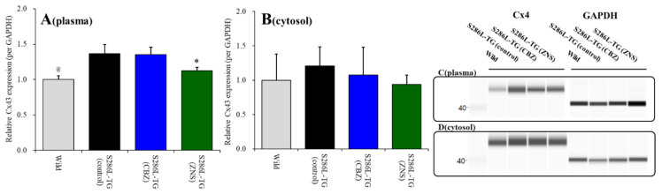
To understand the pathomechanism and pathophysiology of autosomal dominant sleep-related hypermotor epilepsy (ADSHE), we studied functional abnormalities of glutamatergic transmission in thalamocortical pathway from reticular thalamic nucleus (RTN), mediodorsal thalamic nucleus (MDTN) to orbitofrontal cortex (OFC) associated with S286L-mutant α4β2-nicotinic acetylcholine receptor (nAChR), and connexin43 (Cx43) hemichannel of transgenic rats bearing rat S286L-mutant Chrna4 gene (S286L-TG), corresponding to the human S284L-mutant CHRNA4 gene using simple Western analysis and multiprobe microdialysis. Cx43 expression in the thalamic plasma membrane fraction of S286L-TG was upregulated compared with that of wild-type. Subchronic administrations of therapeutic-relevant doses of zonisamide (ZNS) and carbamazepine (CBZ) decreased and did not affect Cx43 expression of S286L-TG, respectively. Upregulated Cx43 enhanced glutamatergic transmission during both resting and hyperexcitable stages in S286L-TG. Furthermore, activation of GABAergic transmission RTN-MDTN pathway conversely enhanced, but not inhibited, l-glutamate release in the MDTN via upregulated/activated Cx43. Local administration of therapeutic-relevant concentration of ZNS and CBZ acutely supressed and did not affect glutamatergic transmission in the thalamocortical pathway, respectively. These results suggest that pathomechanisms of ADSHE seizure and its cognitive deficit comorbidity, as well as pathophysiology of CBZ-resistant/ZNS-sensitive ADSHE seizures of patients with S284L-mutation.

Keywords: carbamazepine; cognition; connexin; hemichannel; idiopathic epilepsy; zonisamide.


Conflict of interest statement

The authors state no conflict of interest.

Figures

Figure 1
Figure 1
Effects of subcutaneously subchronic administration of therapeutic-relevant doses of carbamazepine (CBZ) and zonisamide (ZNS) (25 mg/kg/day) for seven days on connexin 43 (Cx43) expression in the thalamic plasma membrane (panel A) and cytosol (panel B) fractions. The pseudo-gel images using simple Western results using anti-glyceraldehyde-3-phosphate dehydrogenase (GAPDH) and anti-Cx43 antibody for blotting of plasma membrane (panel C) and cytosol (panel D) fractions. In panels 2A and 2B, ordinate: mean ± SD (n = 6) of relative protein level of Cx43. @ p < 0.05 relative to control by student’s t-test, and * p < 0.05 relative to control by one-way analysis of variance (ANOVA) with Tukey’s post hoc test.
Figure 2
Figure 2
Effects of local administration of 100 μM carbenoxolone (CBX: non-selective hemichannel inhibitor: red circles) and therapeutic-relevant concentration of 100 μM carbamazepine (CBZ: blue circles, estimated concentration in brain tissue is 22 μM) and 500 μM zonisamide (ZNS: green circles, estimated concentration brain tissue is 98 μM) into the mediodorsal thalamic nucleus (MDTN) on pre amino-3-(3-hydroxy-5-methyl-isoxazol-4-yl)propanoic acid (AMPA)-evoked (panel A: pre) and post-AMPA-evoked (panel B: post) l-glutamate release (before and after hemichannel activation induced by perfusion with Ca2+-free with 100 mM K+ containing modified Ringer’s solution (FCHK-MRS) into the MDTN for 20 min) in the orbitofrontal cortex (OFC) of wild-type. Ordinates indicate mean extracellular l-glutamate level (μM) (n = 6), and abscissas indicate time after AMPA-evoked stimulations (min). Blue bars indicate the perfusion with CBX, CBZ, or ZNS into the MDTN. Gray bars indicate the perfusion with 100 μM AMPA into the MDTN (AMPA-evoked stimulation). (Panel C) indicates the area under curve (AUC) value of extracellular l-glutamate level (nmol) before (basal extracellular l-glutamate level) and after AMPA-evoked stimulation (from 20 to 180 min) of panels A and B. Opened columns indicate the AUC values of basal extracellular levels of l-glutamate in panel A and B. * p < 0.05; relative to control by multivariate ANOVA (MANOVA) with Tukey’s post hoc test.
Figure 3
Figure 3
Effects of local administration of 100 μM CBX (red circles) and therapeutic-relevant concentration of CBZ (100 μM: blue circles) and ZNS (500 μM: green circles) into the MDTN on pre-AMPA-evoked (panel A) and post-AMPA-evoked (panel B) l-glutamate release (before and after hemichannel activation induced by perfusion with FCHK-MRS into the MDTN for 20 min) in the OFC of S286L-TG. Ordinates indicate mean extracellular l-glutamate level (μM) (n = 6), and abscissas indicate time after AMPA-evoked stimulations (min). Blue bars indicate the perfusion with CBX, CBZ, or ZNS into the MDTN. Gray bars indicate the perfusion with 100 μM AMPA into the MDTN. (Panel C) indicates AUC value of extracellular l-glutamate level (nmol) before (basal extracellular l-glutamate level) and after AMPA-evoked stimulation (from 20 to 180 min) of panels A and B. Opened columns indicate the AUC values of basal extracellular levels of l-glutamate in panel A and B. * p < 0.05; relative to control by MANOVA with Tukey’s post hoc test.
Figure 4
Figure 4
Effects of local administration of 100 μM (E)-N-Methyl-4-(3-pyridinyl)-3-buten-1-amine oxalate (RJR2403) (RJR: selective α4β2-nAChR agonist: closed circles) into the RTN on pre-AMPA-evoked (panel A) and post-AMPA-evoked (panel B) l-glutamate release (before and after hemichannel activation induced by perfusion with FCHK-MRS into the MDTN for 20 min) in the OFC of wild-type. Ordinates indicate mean extracellular l-glutamate level (μM) (n = 6), and abscissas indicate time after AMPA-evoked stimulations (min). Closed bars indicate the perfusion with RJR2403 into the RTN. Gray bars indicate the perfusion with 100 μM AMPA into the MDTN. (Panel C) indicates the AUC value of the extracellular l-glutamate level (nmol) before (basal extracellular l-glutamate level) and after AMPA-evoked stimulation (from 20 to 180 min) of panels (A,B). Opened columns indicate the AUC values of basal extracellular levels of l-glutamate in panel (A,B). * p < 0.05; relative to control by MANOVA with Tukey’s post hoc test. The control data were the same data in study 1 (Figure 2).
Figure 5
Figure 5
Effects of local administration of 100 μM RJR2403 (RJR: selective α4β2-nAChR agonist: closed circles) into the RTN on pre-AMPA-evoked (panel A) and post-AMPA-evoked (panel B) l-glutamate release (before and after hemichannel activation induced by perfusion with FCHK-MRS into the MDTN for 20 min) in the OFC of S286L-TG. Ordinates indicate mean extracellular l-glutamate level (μM) (N = 6), and abscissas indicate time after AMPA-evoked stimulations (min). Closed bars indicate the perfusion with RJR2403 into the RTN. Gray bars indicate the perfusion with 100 μM AMPA into the MDTN. (Panel C) indicates the AUC value of the extracellular l-glutamate level (nmol) before (basal extracellular l-glutamate level) and after AMPA-evoked stimulation (from 20 to 180 min) of panels (A,B). Opened columns indicate the AUC values of basal extracellular levels of l-glutamate in panel (A,B). * p < 0.05; relative to control by MANOVA with Tukey’s post hoc test. The control data were the same data in Figure 3.
Figure 6
Figure 6
Effects of local administration of 100 μM CBX (non-selective hemichannel inhibitor) and 1 μM muscimol (MUS: GABAA receptor agonist) into the MDTN on pre-RJR- (panel A) and post-RJR-evoked (panel B) l-glutamate release (before and after hemichannel activation induced by perfusion with FCHK-MRS into the MDTN for 20 min) in the MDTN of wild-type. Ordinates indicate mean extracellular l-glutamate level (μM) (N = 6), and abscissas indicate time after RJR-evoked stimulations (min). Gray bars indicate the perfusion with 100 μM RJR2403 into the RTN. Red bars indicate the perfusion with 100 μM CBX or 1 μM MUS into the MDTN. (Panel C) indicates the AUC value of the extracellular l-glutamate level (nmol) before (basal extracellular l-glutamate level) and after RJR-evoked stimulation (from 20 to 180 min) of panels (A,B). Opened columns indicate the AUC values of basal extracellular levels of l-glutamate in panel (A,B). * p < 0.05 relative to control, @ p < 0.05 relative to before hemichannel activation (pre) by MANOVA with Tukey’s post hoc test.
Figure 7
Figure 7
Effects of local administration of 100 μM CBX (non-selective hemichannel inhibitor) and 1 μM MUS (GABAA receptor agonist) into the MDTN on pre-RJR- (panel A) and post-RJR-evoked (panel B) l-glutamate release (before and after hemichannel activation induced by perfusion with FCHK-MRS into the MDTN for 20 min) in the MDTN of S286L-TG. Ordinates indicate mean the extracellular l-glutamate level (μM) (N = 6), and abscissas indicate time after RJR-evoked stimulations (min). Gray bars indicate the perfusion with 100 μM RJR2403 into the RTN. Red bars indicate the perfusion with 100 μM CBX or 1 μM MUS into the MDTN. (Panel C) indicates the AUC value of the extracellular l-glutamate level (nmol) before (basal extracellular l-glutamate level) and after RJR-evoked stimulation (from 20 to 180 min) of panels (A,B). Opened columns indicate the AUC values of basal extracellular levels of l-glutamate in panel A and B. * p < 0.05 relative to control, @ p < 0.05 relative to before hemichannel activation (pre) by MANOVA with Tukey’s post hoc test.
Figure 8
Figure 8
Effects of local administration of 100 μM CBX (non-selective hemichannel inhibitor) into the MDTN on pre-RJR- (panels A,C) and post-RJR-evoked (panels B,D) GABA release (before and after hemichannel activation induced by perfusion with FCHK-MRS into the MDTN for 20 min) in the MDTN of wild-type (panels A,B) and S286L-TG (panels C,D). Ordinates indicate mean extracellular GABA level (μM) (N = 6), and abscissas indicate time after RJR-evoked stimulations (min). Gray bars indicate the perfusion with 100 μM RJR2403 into the RTN. Red bars indicate the perfusion with 100 μM CBX into the MDTN.
Figure 9
Figure 9
Proposed hypothesis of pathomechanisms and pathophysiology of S286L-TG. Proposed hypothesis for functional abnormalities of glutamatergic transmission in the thalamocortical pathways in wild-type (panel A) and S286L-TG (panel B) during the resting stage, as well as in wild-type (panel C) and S286L-TG (panel D) during the hemichannel activation stage. RTN mainly projects GABAergic terminals to MDTN. Activation of α4β2-nAChR in the RTN enhances GABAergic transmission in the RTN-MDTN pathways of wild-type (panel A). MDTN project glutamatergic terminals to the OFC. In the MDTN, both α4β2-nAChR and AMPA/glutamate receptor activate glutamatergic transmission to the OFC (panel A). Wild-type α4β2-nAChR suppresses Cx43 expression in astroglial plasma membrane in the thalamus (panel A). Contrary to wild-type, in S286L-TG, loss-of-function S286L-mutant α4β2-nAChR attenuates the stimulatory and inhibitory effects on respective GABAergic transmission (RTN-MDTN) and Cx43 expression, resulting in upregulated Cx43 in the thalamus (panel B). After the activation of hemichannel in the MDTN induced by calcium-free with high potassium stimulation, α4β2-nAChR induced GABA release (RTN-MDTN) is not affected in both wild-type and S286L-TG (panels C,D). Enhanced astroglial glutamatergic transmission associated with activated hemichannel in the MDTN leads to apparent impairment of GABAergic inhibition in wild-type, similar to S286L-TG during the resting stage (panel C). In S286L-TG, propagation of neuronal excitabilities from the RTN to MDTN enhances thalamocortical glutamatergic transmission GABAA receptor independently via increased extracellular potassium and reduced extracellular calcium levels as hemichannel activation signalling (panel D). A therapeutic-relevant concentration of ZNS chronically supresses Cx43 expression in the plasma membrane and acutely inhibits hemichannel activity (panel D).
Figure 10
Figure 10
Schematic experimental designs of microdialysis. MRS: modified Ringer’s solution (gray columns), FCHK-MRS: Ca2+-free with 100 mM K+ containing MRS (closed columns), RJR2403 (100 μM: selective α4β2-nAChR agonist): (E)-N-Methyl-4-(3-pyridinyl)-3-buten-1-amine oxalate, AMPA (100 μM: AMPA/glutamate receptor agonist): amino-3-(3-hydroxy-5-methyl-isoxazol-4-yl) propanoic acid, CBX (100 μM: non-selective hemichannel inhibitor): carbenoxolone, CBZ (100 μM): carbamazepine, MDTN: mediodorsal thalamic nuclei, RTN: reticular thalamic nucleus, OFC: orbitofrontal cortex, and ZNS (500 μM): zonisamide.

References

    1. Steinlein O.K. Animal models for autosomal dominant frontal lobe epilepsy: On the origin of seizures. Expert Rev. Neurother. 2010;10:1859–1867. doi: 10.1586/ern.10.130. - DOI - PubMed
    1. Scheffer I.E., Bhatia K.P., Lopes-Cendes I., Fish D.R., Marsden C.D., Andermann F., Andermann E., Desbiens R., Cendes F., Manson J.I., et al. Autosomal dominant frontal epilepsy misdiagnosed as sleep disorder. Lancet. 1994;343:515–517. doi: 10.1016/S0140-6736(94)91463-X. - DOI - PubMed
    1. Tinuper P., Bisulli F., Cross J.H., Hesdorffer D., Kahane P., Nobili L., Provini F., Scheffer I.E., Tassi L., Vignatelli L., et al. Definition and diagnostic criteria of sleep-related hypermotor epilepsy. Neurology. 2016;86:1834–1842. doi: 10.1212/WNL.0000000000002666. - DOI - PMC - PubMed
    1. Okada M., Zhu G., Yoshida S., Kaneko S. Validation criteria for genetic animal models of epilepsy. Epilepsy Seizure. 2010;3:109–120. doi: 10.3805/eands.3.109. - DOI
    1. Nobili L., Proserpio P., Combi R., Provini F., Plazzi G., Bisulli F., Tassi L., Tinuper P. Nocturnal frontal lobe epilepsy. Curr. Neurol. Neurosci. Rep. 2014;14:424. doi: 10.1007/s11910-013-0424-6. - DOI - PubMed

LinkOut - more resources