

Nanoparticulate Gels for Cutaneous Administration of Caffeic Acid
- PMID: 32443503
- PMCID: PMC7279527
- DOI: 10.3390/nano10050961
Nanoparticulate Gels for Cutaneous Administration of Caffeic Acid
Abstract
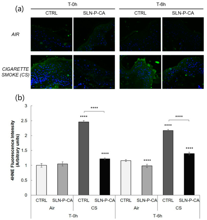
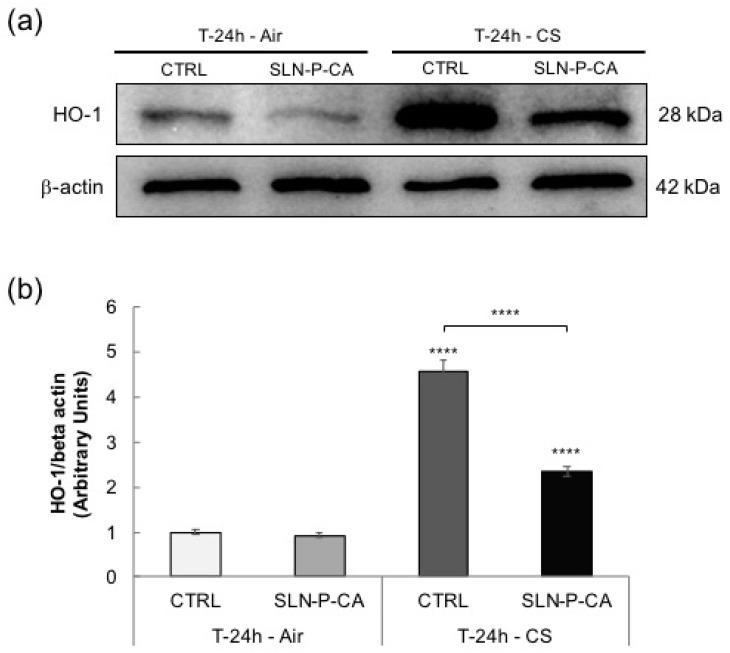
Caffeic acid is a natural antioxidant, largely distributed in plant tissues and food sources, possessing anti-inflammatory, antimicrobial, and anticarcinogenic properties. The object of this investigation was the development of a formulation for caffeic acid cutaneous administration. To this aim, caffeic acid has been loaded in solid lipid nanoparticles by hot homogenization and ultrasonication, obtaining aqueous dispersions with high drug encapsulation efficiency and 200 nm mean dimension, as assessed by photon correlation spectroscopy. With the aim to improve the consistence of the aqueous nanodispersions, different types of polymers have been considered. Particularly, poloxamer 407 and hyaluronic acid gels containing caffeic acid have been produced and characterized by X-ray and rheological analyses. A Franz cell study enabled to select poloxamer 407, being able to better control caffeic acid diffusion. Thus, a nanoparticulate gel has been produced by addition of poloxamer 407 to nanoparticle dispersions. Notably, caffeic acid diffusion from nanoparticulate gel was eight-fold slower with respect to the aqueous solution. In addition, the spreadability of nanoparticulate gel was suitable for cutaneous administration. Finally, the antioxidant effect of caffeic acid loaded in nanoparticulate gel has been demonstrated by ex-vivo evaluation on human skin explants exposed to cigarette smoke, suggesting a protective role exerted by the nanoparticles.
Keywords: caffeic acid; cigarette smoke; poloxamer; small angle X-ray scattering; solid lipid nanoparticles.
Conflict of interest statement
The authors declare no conflict of interest. The funders had no role in the design of the study; in the collection, analyses, or interpretation of data; in the writing of the manuscript, or in the decision to publish the results.
Figures
Similar articles
-
The Potential of Caffeic Acid Lipid Nanoparticulate Systems for Skin Application: In Vitro Assays to Assess Delivery and Antioxidant Effect.Nanomaterials (Basel). 2021 Jan 12;11(1):171. doi: 10.3390/nano11010171. Nanomaterials (Basel). 2021. PMID: 33445433 Free PMC article.
-
Production and Characterization of Nanoparticle Based Hyaluronate Gel Containing Retinyl Palmitate for Wound Healing.Curr Drug Deliv. 2018;15(8):1172-1182. doi: 10.2174/1567201815666180518123926. Curr Drug Deliv. 2018. PMID: 29779480
-
Design and Characterization of Ethosomes for Transdermal Delivery of Caffeic Acid.Pharmaceutics. 2020 Aug 6;12(8):740. doi: 10.3390/pharmaceutics12080740. Pharmaceutics. 2020. PMID: 32781717 Free PMC article.
-
Gallic acid loaded poloxamer gel as new adjuvant strategy for melanoma: A preliminary study.Colloids Surf B Biointerfaces. 2020 Jan 1;185:110613. doi: 10.1016/j.colsurfb.2019.110613. Epub 2019 Oct 31. Colloids Surf B Biointerfaces. 2020. PMID: 31715454
-
The clinical efficacy of cosmeceutical application of liquid crystalline nanostructured dispersions of alpha lipoic acid as anti-wrinkle.Eur J Pharm Biopharm. 2014 Feb;86(2):251-9. doi: 10.1016/j.ejpb.2013.09.008. Epub 2013 Sep 18. Eur J Pharm Biopharm. 2014. PMID: 24056055
Cited by
-
Niosomes for Topical Application of Antioxidant Molecules: Design and In Vitro Behavior.Gels. 2023 Jan 26;9(2):107. doi: 10.3390/gels9020107. Gels. 2023. PMID: 36826277 Free PMC article.
-
Potential Unlocking of Biological Activity of Caffeic Acid by Incorporation into Hydrophilic Gels.Gels. 2024 Dec 4;10(12):794. doi: 10.3390/gels10120794. Gels. 2024. PMID: 39727552 Free PMC article.
-
Recent Advances of Hyaluronan for Skin Delivery: From Structure to Fabrication Strategies and Applications.Polymers (Basel). 2022 Nov 10;14(22):4833. doi: 10.3390/polym14224833. Polymers (Basel). 2022. PMID: 36432961 Free PMC article. Review.
-
Lipid-Based Nano-Sized Cargos as a Promising Strategy in Bone Complications: A Review.Nanomaterials (Basel). 2022 Mar 30;12(7):1146. doi: 10.3390/nano12071146. Nanomaterials (Basel). 2022. PMID: 35407263 Free PMC article. Review.
-
Ultraviolet Light Protection: Is It Really Enough?Antioxidants (Basel). 2022 Jul 29;11(8):1484. doi: 10.3390/antiox11081484. Antioxidants (Basel). 2022. PMID: 36009203 Free PMC article. Review.
References
LinkOut - more resources
Full Text Sources
