. 2020 May 18;21(10):3556.
doi: 10.3390/ijms21103556.

The Zinc-Finger Domain Containing Protein ZC4H2 Interacts with TRPV4, Enhancing Channel Activity and Turnover at the Plasma Membrane

Affiliations

The Zinc-Finger Domain Containing Protein ZC4H2 Interacts with TRPV4, Enhancing Channel Activity and Turnover at the Plasma Membrane

Laura Vangeel et al. Int J Mol Sci. .

Abstract

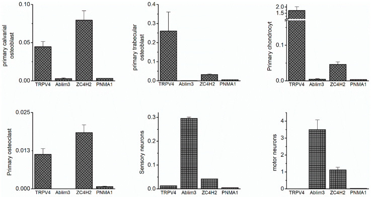
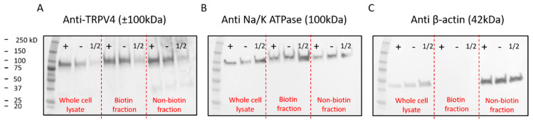
The Ca2+-permeable Transient Receptor Potential channel vanilloid subfamily member 4 (TRPV4) is involved in a broad range of physiological processes, including the regulation of systemic osmotic pressure, bone resorption, vascular tone, and bladder function. Mutations in the TRPV4 gene are the cause of a spectrum of inherited diseases (or TRPV4-pathies), which include skeletal dysplasias, arthropathies, and neuropathies. There is little understanding of the pathophysiological mechanisms underlying these variable disease phenotypes, but it has been hypothesized that disease-causing mutations affect interaction with regulatory proteins. Here, we performed a mammalian protein-protein interaction trap (MAPPIT) screen to identify proteins that interact with the cytosolic N terminus of human TRPV4, a region containing the majority of disease-causing mutations. We discovered the zinc-finger domain-containing protein ZC4H2 as a TRPV4-interacting protein. In heterologous expression experiments, we found that ZC4H2 increases both the basal activity of human TRPV4 as well as Ca2+ responses evoked by ligands or hypotonic cell swelling. Using total internal reflection fluorescence (TIRF) microscopy, we further showed that ZC4H2 accelerates TRPV4 turnover at the plasma membrane. Overall, these data demonstrate that ZC4H2 is a positive modulator of TRPV4, and suggest a link between TRPV4 and ZC4H2-associated rare disorders, which have several neuromuscular symptoms in common with TRPV4-pathies.

Keywords: Ca2+ signaling; TRPV4; ZC4H2; channel turnover; protein–protein interaction.


Conflict of interest statement

The authors declare no conflict of interest. The funders had no role in the design of the study; in the collection, analyses, or interpretation of data; in the writing of the manuscript, or in the decision to publish the results.

Figures

Figure A1
Figure A1
Initial evaluation of other MAPPIT hits. mRNA expression of TRPV4 and the indicated MAPPIT hits in the indicated mouse tissues. Values were normalized to the internal control HPRT. PNMA1 expression was low in all samples, while abLIM3 was primarily found in tissues with low TRPV4 expression.
Figure A2
Figure A2
Expression levels of TRPV4 and ZC4H2 in HEK-293T cells. mRNA expression of TRPV4 and ZC4H2 in non-transfected (NT) cells and cells co-transfected with TRPV4-GFP and ZC4H2-mCherry.
Figure A3
Figure A3
Effect of ZC4H2 on TRPV4 function is not stimulus specific. Representative time course traces of the intracellular Ca2+ concentration ([Ca2+]i) (mean ± SEM) in non-transfected (NT) HEK-293T cells (n = 30), and cells co-transfected with TRPV4 and mCherry or TRPV4 and ZC4H2-mCherry, upon stimulation with (A) HTS and (B) 4αPDD (10µM).
Figure A4
Figure A4
Biotinylation experiments did not reveal differences in TRPV4 membrane expression. Example blot probed for (A) TRPV4, (B) the membrane marker Na/K ATPase, and (C) β-actin. The protein ladder (left) is followed by three lanes of whole-cell lysis samples, middle three lanes demonstrate the biotinylated membrane fraction, while the last three lanes consist of the non-biotinylated fraction. Samples are, from left to right; (‘+’) HEK-293T cells co-transfected with TRPV4 and ZC4H2, (‘−’) HEK-293T cells co-transfected with TRPV4 and control, (‘1/2’) HEK-293T cells transfected with a half concentration of TRPV4 (0.5 µM).
Figure 1
Figure 1
MAPPIT and co-IP show an interaction of ZC4H2 and TRPV4. (A) Volcano plot of the whole protein library tested in an initial MAPPIT screen, with the p-value in the function of the MAPPIT signal. Interesting ‘hits’ are located at the upper right corner. (B) Luciferase read-out of the top 17 hits identified in the MAPPIT screening. Baits cloned in the pSEL and pCLG vector background were tested in parallel and contained either the human N-terminus TRPV4 or an irrelevant protein (E. coli dihydrofolate reductase (eDHFR) or myelin and lymphocyte protein (MAL)). Additionally, an empty prey plasmid was used as a negative control. Positive assay controls are RRAD and GEM Like GTPase 2 (REM2) and EF-hand domain family member A1 (EFHA1), two proteins known to bind the cytokine receptor complex bait independently. (C) Human embryonic kidney (HEK)-293T cells were co-transfected with TRPV4-GFP and ZC4H2-mCherry (+) or TRPV4-GFP and mCherry (−). SDS-PAGE was performed for three conditions: the whole cell lysate, the fraction bound to GFP-Trap® beads, and the unbound (wash) fraction. Staining was done using specific antibodies for human TRPV4 (98 kDa), ZC4H2 (26 kDa), and β-actin (42 kDa). Note that in these experiments, TRPV4 and ZC4H2 are coupled to green fluorescent protein (GFP) and mCherry, respectively, which increases the molecular weights of the detected proteins by 27 kDa. In two independent experiments, we confirmed that there was negligible binding of mCherry and ZCH2-mCherry to the GFP-Trap® beads.
Figure 2
Figure 2
Effect of ZC4H2 on TRPV4 channel activity. (A) Time course of the intracellular Ca2+ concentration ([Ca2+]i) (mean ± SEM) in non-transfected (NT) HEK-293T cells (n = 30), and cells co-transfected with TRPV4 and mCherry (control, n = 79) or ZC4H2-mCherry (n = 80), upon stimulation with arachidonic acid (AA; 10 µM). (B) Mean baseline [Ca2+]i in experiments as in (A). (C) Normalized [Ca2+]i amplitudes in response to the TRPV4-activating stimuli AA (10 µM), 4α-PDD (10mM), or hypotonic solution. Values are normalized to the response of cells expressing TRPV4 and mCherry. Representative traces are shown in Figure A3. (D) Co-expression of ZC4H2 is without effect on baseline [Ca2+]i in cells expressing TRPV3 (n = 144 for ZC4H2 co-transfected, 183 for control, n = 26 for non-transfected). (E) Normalized [Ca2+]i amplitudes in TRPV3-expressing cells in response to the agonist 2-APB (25 µM). Values are normalized to the response of cells expressing TRPV3 and mCherry. p-values < 0.05 were considered significant (*).
Figure 3
Figure 3
ZC4H2 does not affect the expression or subcellular localization of TRPV4. (A) Representative TIRF images showing the localization of heterologously expressed TRPV4-GFP and ZC4H2-mCherry. (B) Relative mRNA expression of TRPV4 in cells co-transfected with ZC4H2 or mCherry control. Data are normalized to the housekeeping gene Glyceraldehyde-3-Phosphate Dehydrogenase (GAPDH). (n = 11) (C) Mean TRPV4-GFP fluorescence (EPImode). (n = 245 for ZC4H2, 220 for control) (D) Mean TRPV4 expression in whole cell lysate after SDS-PAGE. (n = 14) (E) Mean TRPV4 expression in the biotinylated fraction, normalized to the NaK-ATPase (plasma membrane marker). (n = 8) (F) Representative images showing TRPV4 GFP fluorescence measured in epifluorescence (EPI) mode and TIRF mode in cells co-transfected with ZC4H2 or mCherry control. (G) Ratio of TRPV4-GFP fluorescence in TIRF mode versus EPI mode, as an estimate of the distribution of TRPV4-GFP between the bulk cell and the perimembrane area in close vicinity of the coverslip. (n = 24 for ZC4H2, n = 21 control). Values are mean ± SEM.
Figure 4
Figure 4
Effect of ZC4H2 on TRPV4 turnover at the plasma membrane assayed using TIR-FRAP. (A) TIRF images of TRPV4-GFP in HEK-293T cells co-expressing mCherry (control) or ZC4H2-mCherry. Images were taken before bleaching, and at 0 and 800s after bleaching. Scale bar = 10 µm. (B,C) Time course of the decay of TRPV4-GFP fluorescence during the bleaching process (B) and the recovery of the fluorescence following bleaching (C) in representative cells expressing ZC4H2 or the control. Dotted and solid lines in C represent mono- and bi-exponential fits, respectively. (D) Mean basal TRPV4-GFP fluorescence before bleaching, in control and ZC4H2-expressing cells. (E) Mean TRPV4-GFP fluorescence at 0 and 800s after bleaching. (F,G) Time constants and corresponding relative amplitudes obtained from exponential fits to recovery time courses as in (C). In control cells, a mono-exponential fit was generally sufficient to describe the recovery process. In ZC4H2-expressing cells, adequate fitting required a second faster kinetic component. Number of cells in (DG): control: n = 16; ZC4H2: n = 14. p-values < 0.05 were considered significant (*).

References

    1. Nilius B., Owsianik G., Voets T., Peters J.A. Transient receptor potential cation channels in disease. Physiol. Rev. 2007;87:165–217. doi: 10.1152/physrev.00021.2006. - DOI - PubMed
    1. White J.P., Cibelli M., Urban L., Nilius B., McGeown J.G., Nagy I. TRPV4: Molecular Conductor of a Diverse Orchestra. Physiol. Rev. 2016;96:911–973. doi: 10.1152/physrev.00016.2015. - DOI - PubMed
    1. Auer-Grumbach M., Olschewski A., Papic L., Kremer H., McEntagart M.E., Uhrig S., Fischer C., Frohlich E., Balint Z., Tang B., et al. Alterations in the ankyrin domain of TRPV4 cause congenital distal SMA, scapuloperoneal SMA and HMSN2C. Nat. Genet. 2010;42:160–164. doi: 10.1038/ng.508. - DOI - PMC - PubMed
    1. Deng H.X., Klein C.J., Yan J., Shi Y., Wu Y., Fecto F., Yau H.J., Yang Y., Zhai H., Siddique N., et al. Scapuloperoneal spinal muscular atrophy and CMT2C are allelic disorders caused by alterations in TRPV4. Nat. Genet. 2010;42:165–169. doi: 10.1038/ng.509. - DOI - PMC - PubMed
    1. Landoure G., Zdebik A.A., Martinez T.L., Burnett B.G., Stanescu H.C., Inada H., Shi Y., Taye A.A., Kong L., Munns C.H., et al. Mutations in TRPV4 cause Charcot-Marie-Tooth disease type 2C. Nat. Genet. 2010;42:170–174. doi: 10.1038/ng.512. - DOI - PMC - PubMed

MeSH terms

LinkOut - more resources