. 2020 May 18;8(2):235.
doi: 10.3390/vaccines8020235.

Experimental PCEP-Adjuvanted Swine Influenza H1N1 Vaccine Induced Strong Immune Responses but Did Not Protect Piglets against Heterologous H3N2 Virus Challenge

Affiliations

Experimental PCEP-Adjuvanted Swine Influenza H1N1 Vaccine Induced Strong Immune Responses but Did Not Protect Piglets against Heterologous H3N2 Virus Challenge

Royford Bundi Magiri et al. Vaccines (Basel). .

Abstract

Vaccination is the most efficient method of protection against influenza infections. However, the rapidly mutating viruses and development of new strains make it necessary to develop new influenza vaccines annually. Hence, vaccines that stimulate cross-protection against multiple influenza subtypes are highly sought. Recent evidence suggests that adjuvants such as PCEP that promote Th1-type T cell and Th2-type T cell immune responses and broad-spectrum immune responses may confer cross-protection against heterologous influenza strains. In this study, we evaluated whether the immunogenic and protective potential of PCEP-adjuvanted inactivated swine influenza virus H1N1 vaccine can protect pigs immunized against live H3N2 virus. Piglets were vaccinated via the intradermal route with PCEP-adjuvanted inactivated swine influenza virus (SIV) H1N1 vaccine, boosted at day 21 with the same vaccines then challenged with infectious SIV H3N2 virus at day 35 via the tracheobronchial route. The pigs showed significant anti-H1N1 SIV specific antibody titres and H1N1 SIV neutralizing antibody titres, and these serum titres remained after the challenge with the H3N2 virus. In contrast, vaccination with anti-H1N1 SIV did not trigger anti-H3N2 SIV antibody titres or neutralizing antibody titres and these titres remained low until pigs were challenged with H3N2 SIV. At necropsy (six days after challenge), we collected prescapular lymph nodes and tracheobronchial draining the vaccination sites and challenge site, respectively. ELISPOTs from lymph node cells restimulated ex vivo with inactivated SIV H1N1 showed significant production of IFN-γ in the tracheobronchial cells, but not the prescapular lymph nodes. In contrast, lymph node cells restimulated ex vivo with inactivated SIV H1N1 showed significantly higher IL-13 and IL-17A in the prescapular lymph nodes draining the vaccination sites relative to unchallenged animals. Lung lesion scores show that intradermal vaccination with H1N1 SIV plus PCEP did not prevent lesions when the animals were challenged with H3N2. These results confirm previous findings that PCEP is effective as a vaccine adjuvant in that it induces strong immune responses and protects against homologous swine influenza H1N1 virus, but the experimental H1N1 vaccine failed to cross-protect against heterologous H3N2 virus.

Keywords: adjuvant; influenza; intradermal; pig; polyphosphazene.


Conflict of interest statement

The authors declare no conflict of interest.

Figures

Figure 1
Figure 1
Schematic presentation of experimental design. Piglets were immunized via the intradermal injection with swine influenza virus (SIV) plus PCEP as an adjuvant, on day 0, and a secondary immunization was administered on day 21. Serum was collected for serum antibody titres assays. Clinical and injection site markings and scoring were performed up to day 41 after initial vaccination.
Figure 2
Figure 2
Vaccine-induced H1N1 antibody responses and neutralizing antibody responses in pigs. Pigs (n = 6 per group) were immunized with 4.0 × 10⁴ or 8.0 × 10³ inactivated SIV HAU alone or plus 4, 20, 100 or 500 μg PCEP. Control animals were either injected with PBS or not immunized (naïve). A secondary immunization was performed on day 21 and sera samples were collected over a period of 28 days. (A) Anti-H1N1 IgG antibodies were assessed by ELISA and (B) anti-H1N1 IgG neutralizing antibodies were assessed. Data for animal group are presented with mean values indicated by a horizontal bar and standard error of the mean are shown. p < 0.05 (*), and p < 0.01 (**).
Figure 2
Figure 2
Vaccine-induced H1N1 antibody responses and neutralizing antibody responses in pigs. Pigs (n = 6 per group) were immunized with 4.0 × 10⁴ or 8.0 × 10³ inactivated SIV HAU alone or plus 4, 20, 100 or 500 μg PCEP. Control animals were either injected with PBS or not immunized (naïve). A secondary immunization was performed on day 21 and sera samples were collected over a period of 28 days. (A) Anti-H1N1 IgG antibodies were assessed by ELISA and (B) anti-H1N1 IgG neutralizing antibodies were assessed. Data for animal group are presented with mean values indicated by a horizontal bar and standard error of the mean are shown. p < 0.05 (*), and p < 0.01 (**).
Figure 3
Figure 3
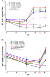
Swine influenza H1N1 and H3N2 ELISA and neutralizing antibody responses in pigs. Pigs (n = 6) were immunized then boosted at day 21 via intradermal routes with 4 × 10⁴ or 8 × 10³ HAU H1N1 either alone or with varying doses (4, 20, 100 or 500 μg) of PCEP adjuvant as indicated. Control groups were injected with either PBS or were unimmunized (naïve). They were challenged with 8 × 10⁵ PFU virulent H3N2 SIV via the intratracheal route on day 35 and killed 6 days after challenge. Serum antibody titres for SIV H1N1 (A) and H3N2 (B) were assayed by ELISA over time. Red font is used for the day of challenge for emphasis. Serum SIV H1N1 (C) and H3N2 (D) neutralizing antibody titres were assessed up to day 41. Data for animal groups are presented with mean values indicated by a horizontal bar and standard error of the mean are shown. p < 0.05 (*), p < 0.01 (**) and p < 0.001 (***).
Figure 3
Figure 3
Swine influenza H1N1 and H3N2 ELISA and neutralizing antibody responses in pigs. Pigs (n = 6) were immunized then boosted at day 21 via intradermal routes with 4 × 10⁴ or 8 × 10³ HAU H1N1 either alone or with varying doses (4, 20, 100 or 500 μg) of PCEP adjuvant as indicated. Control groups were injected with either PBS or were unimmunized (naïve). They were challenged with 8 × 10⁵ PFU virulent H3N2 SIV via the intratracheal route on day 35 and killed 6 days after challenge. Serum antibody titres for SIV H1N1 (A) and H3N2 (B) were assayed by ELISA over time. Red font is used for the day of challenge for emphasis. Serum SIV H1N1 (C) and H3N2 (D) neutralizing antibody titres were assessed up to day 41. Data for animal groups are presented with mean values indicated by a horizontal bar and standard error of the mean are shown. p < 0.05 (*), p < 0.01 (**) and p < 0.001 (***).
Figure 4
Figure 4
Adjuvant PCEP induces immune responses against inactivated swine influenza virus H1N1. Pigs (n = 6) were immunized then boosted at day 21 via intradermal routes with 4 × 10⁴ or 8 × 10³ HAU H1N1 plus 0–500 μg PCEP adjuvant as indicated. They were challenged with 8 × 10⁵ PFU virulent H3N2 SIV via the intratracheal route on day 35 and killed 6-days after challenge. Tracheobronchial and prescapular lymph nodes were collected and cells were harvested at 41 days post immunization. Individual single-cell suspensions of 5 × 10⁵ lymph node cells were re-stimulated with killed SIV H1N1 for 16 h for IFN-γ (A), and 48 h for IL-13 (B) and IL-17A (C) and measured by ELISPOT analysis. Data for animal groups are presented as a bar graph with the mean and standard error of the mean presented. p < 0.05 (*), p < 0.01 (**) and p < 0.001 (***).
Figure 4
Figure 4
Adjuvant PCEP induces immune responses against inactivated swine influenza virus H1N1. Pigs (n = 6) were immunized then boosted at day 21 via intradermal routes with 4 × 10⁴ or 8 × 10³ HAU H1N1 plus 0–500 μg PCEP adjuvant as indicated. They were challenged with 8 × 10⁵ PFU virulent H3N2 SIV via the intratracheal route on day 35 and killed 6-days after challenge. Tracheobronchial and prescapular lymph nodes were collected and cells were harvested at 41 days post immunization. Individual single-cell suspensions of 5 × 10⁵ lymph node cells were re-stimulated with killed SIV H1N1 for 16 h for IFN-γ (A), and 48 h for IL-13 (B) and IL-17A (C) and measured by ELISPOT analysis. Data for animal groups are presented as a bar graph with the mean and standard error of the mean presented. p < 0.05 (*), p < 0.01 (**) and p < 0.001 (***).
Figure 5
Figure 5
Lesion scores in lungs of vaccinated and challenged animals. Pigs (n = 6) were immunized then boosted at day 21 via intradermal routes with 4 × 10⁴ or 8 × 10³ HAU H1N1 plus 0–500 μg PCEP adjuvant or PBS as indicated. Naïve animals were neither immunized, nor challenged. The challenge dose was 8 × 10⁵ PFU virulent H3N2 SIV via the intratracheal route on day 35 and all animals were killed 6 days after challenge. The lung lesion scores were assessed (as indicated in the methods) up to 6-days post challenge. Each data point indicates an individual animal and the horizontal bar represents the median. The colors of each data point are coordinated with the group colors from Figure 1, Figure 2 and Figure 3.

References

    1. Dandagi G.L., Byahatti S.M. An insight into the swine-influenza A (H1N1) virus infection in humans. Lung India Off. Organ Indian Chest Soc. 2011;28:34. doi: 10.4103/0970-2113.76299. - DOI - PMC - PubMed
    1. Vincent A.L., Ma W., Lager K.M., Janke B.H., Richt J.A. Swine influenza viruses: A North American perspective. Adv. Virus Res. 2008;72:127–154. - PubMed
    1. Anderson T.K., Nelson M.I., Kitikoon P., Swenson S.L., Korslund J.A., Vincent A.L. Population dynamics of cocirculating swine influenza A viruses in the United States from 2009 to 2012. Influenza Other Resp. Virus. 2013;7:42–51. doi: 10.1111/irv.12193. - DOI - PMC - PubMed
    1. Brown I.H. History and Epidemiology of Swine Influenza in Europe. In: Richt J., Webby R., editors. Swine Influenza. Current Topics in Microbiology and Immunology. Volume 370 Springer; Berlin/Heidelberg, Germany: 2011. - PubMed
    1. Smith G.J., Vijaykrishna D., Bahl J., Lycett S.J., Worobey M., Pybus O.G., Ma S.K., Cheung C.L., Raghwani J., Bhatt S., et al. Origins and evolutionary genomics of the 2009 swine-origin H1N1 influenza A epidemic. Nature. 2009;459:1122–1125. doi: 10.1038/nature08182. - DOI - PubMed

LinkOut - more resources