. 2020 May 18;21(10):3564.
doi: 10.3390/ijms21103564.

Human Spinal Motor Neurons Are Particularly Vulnerable to Cerebrospinal Fluid of Amyotrophic Lateral Sclerosis Patients

Affiliations

Human Spinal Motor Neurons Are Particularly Vulnerable to Cerebrospinal Fluid of Amyotrophic Lateral Sclerosis Patients

Stefan Bräuer et al. Int J Mol Sci. .

Abstract

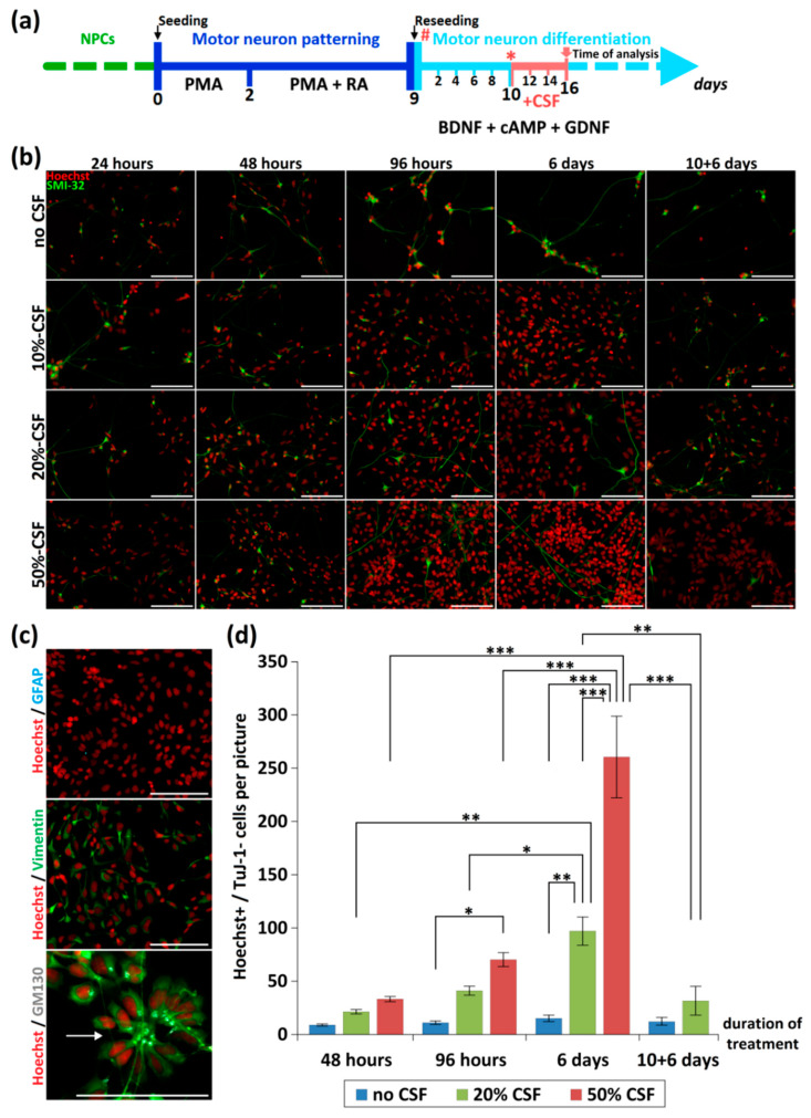
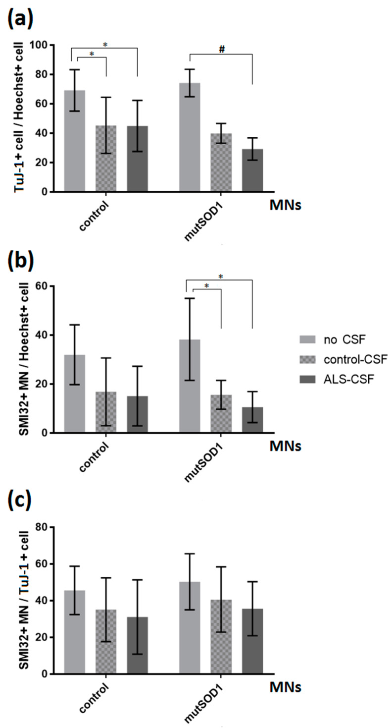
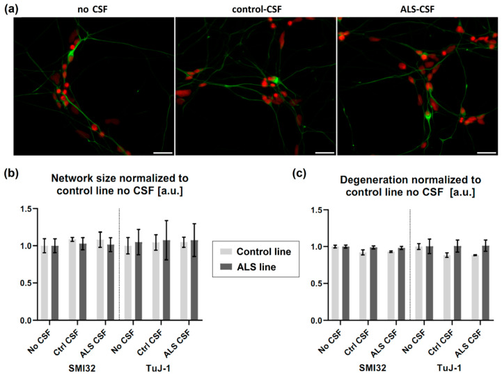
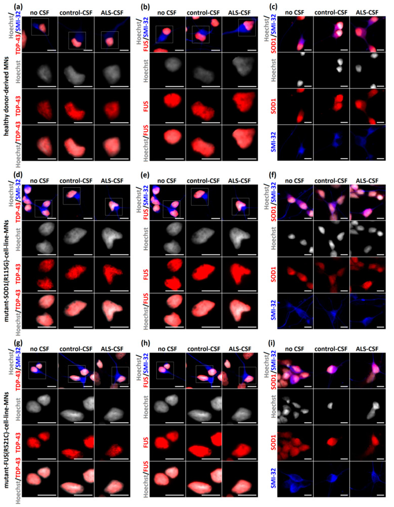
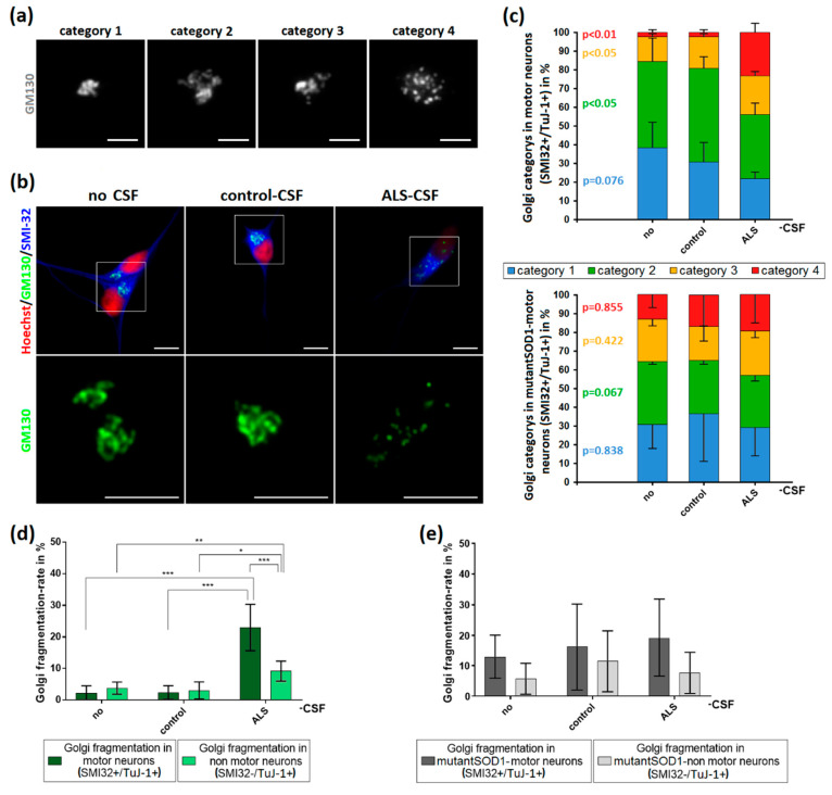
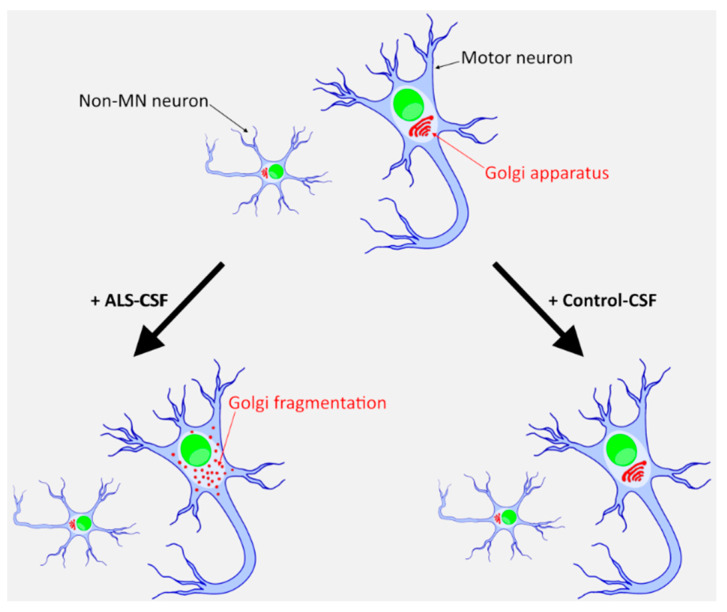
Amyotrophic lateral sclerosis (ALS) is the most common and devastating motor neuron (MN) disease. Its pathophysiological cascade is still enigmatic. More than 90% of ALS patients suffer from sporadic ALS, which makes it specifically demanding to generate appropriate model systems. One interesting aspect considering the seeding, spreading and further disease development of ALS is the cerebrospinal fluid (CSF). We therefore asked whether CSF from sporadic ALS patients is capable of causing disease typical changes in human patient-derived spinal MN cultures and thus could represent a novel model system for sporadic ALS. By using induced pluripotent stem cell (iPSC)-derived MNs from healthy controls and monogenetic forms of ALS we could demonstrate a harmful effect of ALS-CSF on healthy donor-derived human MNs. Golgi fragmentation-a typical finding in lower organism models and human postmortem tissue-was induced solely by addition of ALS-CSF, but not control-CSF. No other neurodegenerative hallmarks-including pathological protein aggregation-were found, underpinning Golgi fragmentation as early event in the neurodegenerative cascade. Of note, these changes occurred predominantly in MNs, the cell type primarily affected in ALS. We thus present a novel way to model early features of sporadic ALS.

Keywords: ALS; Golgi fragmentation; amyotrophic lateral sclerosis; cerebrospinal fluid; fused in sarcoma; superoxide dismutase 1.


Conflict of interest statement

The authors declare no conflict of interest.

Figures

Figure 1
Figure 1
Effect of CSF-application on neurons and NPCs. (a) Depicted is the differentiation and treatment scheme. During the initial dose finding experiments, the time point of CSF-application was varied (# and *). For the final experiments CSF was applied 10 days after reseeding (*) and cells were analyzed after 6 days of CSF treatment (red arrow). (b) Different concentrations of CSF (0%, 10%, 20%, 50%) were applied to human iPSC-derived MN-cell cultures directly after seeding for 24 h, 48 h, 96 h and 6 days. Additionally, we included a scenario in which we initiated treatment after 10 days of differentiation, followed by 6 days CSF treatment (10 + 6 days). Depending on the duration and concentration of CSF-incubation, a proliferation of NPCs could be seen. (c) This effect was independent from the CSF-origin (data not shown) and led to a dramatic overgrowth of GFAP/Vimentin+ NPCs, which partially formed neural rosettes (arrow). (d) Quantification of non-neuronal cells (Hoechst+/Tuj-1) during CSF treatment. */**/*** represents p < 0.05/0.01/0.001 as calculated by two-way ANOVA with Bonferroni post-hoc test). Scale bar = 100 µm.
Figure 2
Figure 2
Effects of ALS-CSF on the relations of neuronal cells in control- and mutant SOD1-MNs. (a) Left: A significant decrease in the relations of TuJ-1+cell/Hoechst when treated with either control- (* p < 0.05 by one-way ANOVA with Tukey post-hoc test) or ALS-CSF (* p < 0.05 by one-way ANOVA with Tukey post-hoc test), but no significant effect comparing respective CSF conditions. Right: The mutant SOD1-cell line exhibited a significant decrease in the relations of TuJ-1+cell/Hoechst when exposed to ALS-CSF compared to the no CSF condition (# = Kruskal-Wallis test H(2) = 7.636, p < 0.05, z = 2.763, p < 0.05 with Dunn-Bonferroni post-hoc test), but again no significant effect comparing control- with ALS-CSF (29.21% vs. 39.91%, z = 1.279, p = 0.602 with Dunn-Bonferroni post-hoc test). (b) Left: The application of CSF had no significant effect in either cell line analyzing the relation of SMI32+MN/Hoechst in the control-cell line. Right: In SOD1 mutant cells, we detected a significant decrease in the amount of total MNs in relation to all cells (MN/Hoechst) comparing either ALS- or control-CSF-treatment condition and non-treated cells (* p < 0.05 by one-way ANOVA with Tukey post-test). (c) The application of CSF had no significant effect in either cell line analyzing the relation SMI32+MN/TuJ-1+cell. Data are depicted as mean ± SEM.
Figure 3
Figure 3
No signs of structural degeneration. (a) There was no differing degeneration of the neuronal network (green = SMI32, red = Hoechst) detectable in either cell line treated with CSF. (b) There was no significant effect of ALS-CSF on neither the neuronal network size, (c) nor network degeneration. Scale bar = 50 µm.
Figure 4
Figure 4
No aggregation of disease-relevant proteins after exposition to ALS-CSF. (ac) ALS-CSF caused no detectable aggregates of TDP-43 (a), FUS (b), or SOD1 (c) when applied to healthy donor-derived MNs. Furthermore, there were no differences of cytoplasmic or nuclear localization of any disease relevant protein and—importantly—no cytoplasmic mislocalization of TDP-43 and FUS, respectively. (df) Similar results were detected after applying CSF to the mutant-SOD1-(p.R115G)- and (gi) mutant-FUS-(p.R521C)-cell-line MNs. Scale bar = 10 µm.
Figure 5
Figure 5
Induction of Golgi fragmentation predominantly in MNs by the application of ALS-CSF. (a) For analysis, Golgi complexes were categorized depending on their structure. Category 1 was defined as small, compact Golgi apparatus. Single cisterns were difficult to differentiate. In contrast, category 2 included bigger “loosened” Golgi complexes. Cisterns were easier to differentiate. Unlike in the previous ones, category 3 Golgi complexes were not closed compartments anymore. They still formed a cisternal structure, but it was possible to differentiate small cisterns/vesicles not being in contact with the main part of the Golgi. The Golgi apparatus in category 4 was fragmented and only this category accounted for the fragmented ones in our statistics in (d,e). It lost its cohesion and cisternal structure thus became vesicular and the small compartments were dispersed across the cell. (b) The structural Golgi changes in control-human iPSC-induced MNs after ALS-CSF application comprised the loss of cisternal configuration and overall Golgi integrity. (c) The diagrams depict the distribution of the respective Golgi categories in control- (upper diagram) and mutant-SOD1-MNs (lower diagram) ± CSF treatment. (d) The exposition to ALS-CSF caused a significantly higher rate of Golgi fragmentation in WT-human iPSC-induced MNs compared to MNs treated with control-CSF or without CSF. Similar results could be observed comparing SMI32−/TuJ-1+-non MNs treated with ALS-CSF to control-CSF or no CSF-. Of note, SMI32+-MNs exposed to ALS-CSF had a significantly higher Golgi fragmentation rate compared to SMI32−/TuJ-1+-non MNs. (e) The application of ALS-CSF to mutant-SOD1 cells caused no significant rate of Golgi fragmentation neither in SMI32+-MNs nor SMI32-/TuJ-1+-non MNs. Data are depicted as mean ± SEM. */**/** p < 0.05/0.01/0.001 by one-way ANOVA with Tukey post-hoc test. Scale bar = 5 µm.
Figure 6
Figure 6
An iPSC-derived neuronal system to model sporadic ALS. Only ALS-CSF, and not healthy donor CSF, induces a degenerative phenotype in healthy donor derived iPSCs. This effect is mainly seen in MNs and only to a much lesser extent in other neuronal subtypes.

Similar articles

Cited by

References

    1. Brauer S., Zimyanin V., Hermann A. Prion-like properties of disease-relevant proteins in amyotrophic lateral sclerosis. J. Neural Transm. 2018;125:591–613. doi: 10.1007/s00702-018-1851-y. - DOI - PubMed
    1. Smith R., Myers K., Ravits J., Bowser R. Amyotrophic lateral sclerosis: Is the spinal fluid pathway involved in seeding and spread? Med. Hypotheses. 2015;85:576–583. doi: 10.1016/j.mehy.2015.07.014. - DOI - PubMed
    1. Couratier P., Hugon J., Sindou P., Vallat J.M., Dumas M. Cell culture evidence for neuronal degeneration in amyotrophic lateral sclerosis being linked to glutamate AMPA/kainate receptors. Lancet. 1993;341:265–268. doi: 10.1016/0140-6736(93)92615-Z. - DOI - PubMed
    1. Gomez-Pinedo U., Galan L., Yanez M., Matias-Guiu J., Valencia C., Guerrero-Sola A., Lopez-Sosa F., Brin J.R., Benito-Martin M.S., Leon-Espinosa G., et al. Histological changes in the rat brain and spinal cord following prolonged intracerebroventricular infusion of cerebrospinal fluid from amyotrophic lateral sclerosis patients are similar to those caused by the disease. Neurologia. 2018;33:211–223. doi: 10.1016/j.nrleng.2016.07.002. - DOI - PubMed
    1. Gomez-Pinedo U., Yanez M., Matias-Guiu J., Galan L., Guerrero-Sola A., Benito-Martin M.S., Vela A., Arranz-Tagarro J.A., Garcia A.G. Cellular changes in motor neuron cell culture produced by cytotoxic cerebrospinal fluid from patients with amyotrophic lateral sclerosis. Neurologia. 2014;29:346–352. doi: 10.1016/j.nrl.2013.08.001. - DOI - PubMed

Supplementary concepts