. 2020 May 20;12(5):565.
doi: 10.3390/v12050565.

Hepatitis B Virus-X Downregulates Expression of Selenium Binding Protein 1

Affiliations

Hepatitis B Virus-X Downregulates Expression of Selenium Binding Protein 1

Young-Man Lee et al. Viruses. .

Abstract

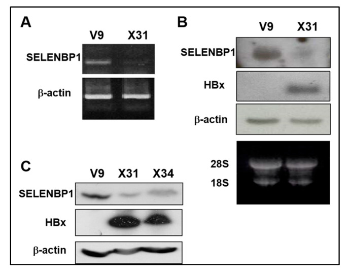
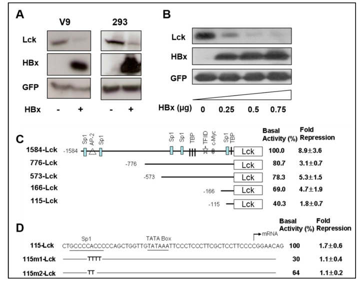
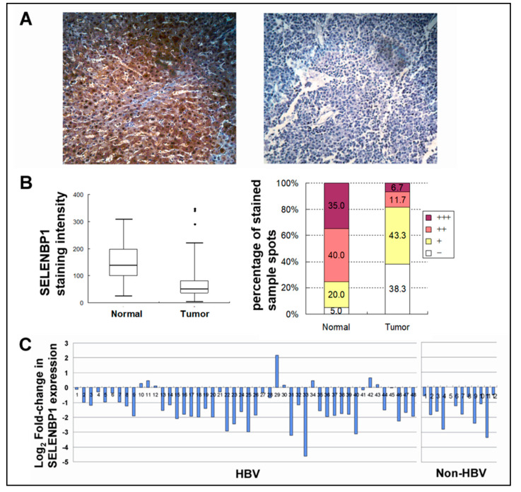
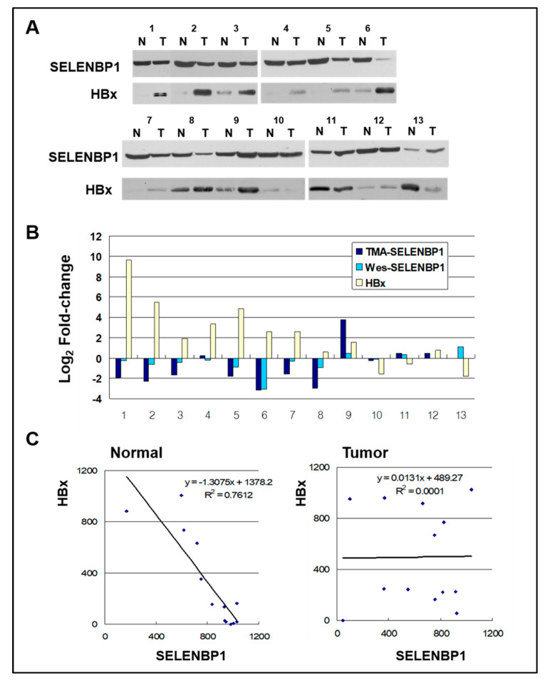
Selenium binding protein 1 (SELENBP1) has been known to be reduced in various types cancer, and epigenetic change is shown to be likely to account for the reduction of SELNEBP1 expression. With cDNA microarray comparative analysis, we found that SELENBP1 is markedly decreased in hepatitis B virus-X (HBx)-expressing cells. To clarify the effect of HBx on SELENBP1 expression, we compared the expression levels of SELENBP1 mRNA and protein by semi-quantitative RT-PCR, Northern blot, and Western blot. As expected, SELENBP1 expression was shown to be reduced in cells expressing HBx, and reporter gene analysis showed that the SELENBP1 promoter is repressed by HBx. In addition, the stepwise deletion of 5' flanking promoter sequences resulted in a gradual decrease in basal promoter activity and inhibition of SELENBP1 expression by HBx. Moreover, immunohistochemistry on tissue microarrays containing 60 pairs of human liver tissue showed decreased intensity of SELENBP1 in tumor tissues as compared with their matched non-tumor liver tissues. Taken together, our findings suggest that inhibition of SELENBP1 expression by HBx might act as one of the causes in the development of hepatocellular carcinoma caused by HBV infection.

Keywords: X protein; hepatitis B virus; hepatocellular carcinoma; selenium binding protein 1.


Conflict of interest statement

The authors declare no conflict of interest.

Figures

Figure 1
Figure 1
Reduced SELENBP1 expression in HBx-expressing cells. (A) Expression of SELENBP1 mRNA was analyzed by semi-quantitative RT-PCR analysis. RT-PCR was carried out on total RNA obtained from Chang V9 and Chang X31 cells using SELENBP1 specific oligonucleotides. (B) The SELENBP1 and HBx mRNA levels in total RNA were assayed by Northern blot analysis. (C) Whole cell lysates prepared from Chang V9, Chang X31, and Chang X34 cells were subjected to SDS-PAGE and immunoblotted using anti-SELENBP1 and anti-HA.
Figure 2
Figure 2
Repression of the SELENBP1 promoter by HBx. (A) HBx expression plasmid or control plasmid was co-transfected into Chang V9 and HEK293 cells with Lck plasmid. Plasmid Myk-eGFP was used for normalizing the transfection efficiency, and pcDNA3.1 was used as a negative control. Two days after transfection, cell lysates were analyzed by Western blotting for investigation of the HBx-mediated down-regulation of SELENBP1 promoter. (B) HEK293 cells were co-transfected with 0, 0.25, 0.5, and 0.75 μg of pRcCMV-HBx and Lck expression reporter plasmid. Two days after transfection, expression levels of Lck, HBx, and GFP in the transfected cells were determined by Western blot analyses. Representative bands are shown after 3 independent experiments. (C) The 1584-bp length SELENBP1 promoter and its truncated constructs are schematically shown, and positions of putative transcriptional binding factor sites in the SELENBP1 promoter are presented. Promoter activity was determined by measuring the level of Lck protein expression using NIH Image J software. The basal activity of 1584-Lck is designated to be 100%, and each promoter activity from truncated forms of the promoter, in the absence of HBx, is shown as a value relative to this basal activity. Fold repression was calculated by comparing the relative promoter activity of HBx-expressing cells with the basal activity of the control. Results as described fold repression are means ± SEM of at least three independent experiments. (D) Mutant constructs are identical to the wild-type 115-Lck sequence with the exception of the sequences shown in Sp1 site of each mutant construct. These constructs were cotransfected with an effector plasmid into 293 cells, and relative promoter activity and fold repression were determined.
Figure 3
Figure 3
SELENBP1 expression in normal and hepatocellular carcinoma (HCCs). (A) Representative images of SELENBP1 immunohistochemistry in non-tumor normal liver tissue (left panel) and matched liver tumor tissue (right panel) (Zeiss, x200). (B) Box plot showing the staining intensity of SELENBP1 in normal and tumor tissues (left panel). Percentages of strong (+++), moderate (++), weak (+), and negative (-) staining are depicted (right panel). Expression level of SELENBP1 is significantly reduced in tumor tissues as compared with counterpart normal liver tissues. (C) The graph represents the levels of fold-change measured against matched non-tumor liver tissue. The level of expression is shown in log2–fold-changes. Each bar represents each individual patient tissue array of 60 pairs of HCC and their matched non-tumor liver tissues on microarray.
Figure 4
Figure 4
SELENBP1 and HBx in normal and HCCs. (A) Sixty micrograms of total protein from normal (N) and tumor (T) liver tissues of HCC patients with hepatitis B virus (HBV) were separated by SDS-PAGE and transferred to a membrane. The membranes were probed using anti-SELENBP1 and anti-HBx. (B) Comparison of changes in SELENBP1 protein expression level as measured by TMA and Western blot. Fold changes in HBx protein expression measured by Western blots are also shown. Values are expressed in fold change (Log2) compared to normal tissues. (C) Relationship between SELENBP1 and HBx protein levels as determined by linear regression analysis in normal and tumor tissues. A significant strong correlation was observed between SELENBP1 and HBx expression in normal tissues, and no correlation in tumor tissues. Regression equations and lines are shown in the graph.

References

    1. Bansal M.F., Mukhopadhyay T., Scott J., Cook R.G., Mukhopadhyay R., Medina D. DNA sequencing of a mouse liver protein that binds selenium: Implications for selenium’s mechanism of action in cancer prevention. Carcinogenesis. 1990;11:2071–2073. doi: 10.1093/carcin/11.11.2071. - DOI - PubMed
    1. Chang P.W., Tsui S.K., Liew C.C., Lee C.Y., Waye M.M., Fung K.P. Isolation, characterization, and chromosomal mapping of a novel cDNA clone encoding human selenium binding protein. J. Cell. Biochem. 1997;64:217–224. doi: 10.1002/(SICI)1097-4644(199702)64:2<217::AID-JCB5>3.0.CO;2-#. - DOI - PubMed
    1. Porat A., Sagiv Y., Elazar Z. A 56-kDa selenium-binding protein participates in intra-Golgi protein transport. J. Biol. Chem. 2000;275:14457–14465. doi: 10.1074/jbc.275.19.14457. - DOI - PubMed
    1. Torrealba J.R., Colburn M., Golner S., Chang Z., Scheunemann T., Fechner J.H., Roenneburg D., Hu H., Alam T., Kim H.T. Selenium-binding protein1 in smooth muscle cells is downregulated in a Rhesus monkey model of chronic allograft nephropathy. Am. J. Transplant. 2005;5:58–67. doi: 10.1111/j.1600-6143.2004.00651.x. - DOI - PubMed
    1. Song L., Zou H., Chang Y., Xu W., Wu L. The cDNA cloning and mRNA expression of a potential selenium-binding protein gene in the scallop Chlamys farreri. Dev. Comp. Immunol. 2006;30:265–273. doi: 10.1016/j.dci.2005.04.001. - DOI - PubMed

Publication types

MeSH terms