. 2020 May 20;11(5):572.
doi: 10.3390/genes11050572.

MicroRNA-1253 Regulation of WASF2 (WAVE2) and its Relevance to Racial Health Disparities

Affiliations

MicroRNA-1253 Regulation of WASF2 (WAVE2) and its Relevance to Racial Health Disparities

Mercy A Arkorful et al. Genes (Basel). .

Abstract

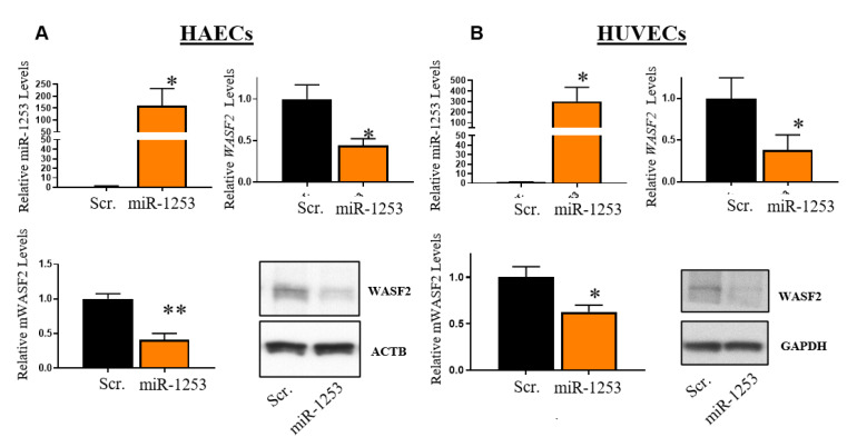
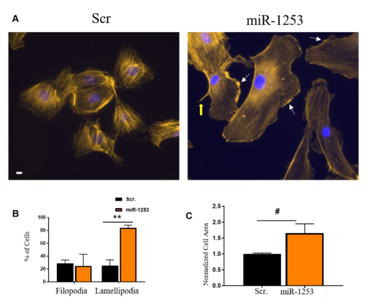
The prevalence of hypertension among African Americans (AAs) in the US is among the highest of any demographic and affects over two-thirds of AA women. Previous data from our laboratory suggest substantial differential gene expression (DGE) of mRNAs and microRNAs (miRNAs) exists within peripheral blood mononuclear cells (PBMCs) isolated from AA and white women with or without hypertension. We hypothesized that DGE by race may contribute to racial differences in hypertension. In a reanalysis of our previous dataset, we found that the Wiskott-Aldrich syndrome protein Verprolin-homologous protein 2 (WASF2 (also known as WAVE2)) is differentially expressed in AA women with hypertension, along with several other members of the actin cytoskeleton signaling pathway that plays a role in cell shape and branching of actin filaments. We performed an in silico miRNA target prediction analysis that suggested miRNA miR-1253 regulates WASF2. Transfection of miR-1253 mimics into human umbilical vein endothelial cells (HUVECs) and human aortic endothelial cells (HAECs) significantly repressed WASF2 mRNA and protein levels (p < 0.05), and a luciferase reporter assay confirmed that miR-1253 regulates the WASF2 3' UTR (p < 0.01). miR-1253 overexpression in HUVECs significantly increased HUVEC lamellipodia formation (p < 0.01), suggesting the miR-1253-WASF2 interaction may play a role in cell shape and actin cytoskeleton function. Together, we have identified novel roles for miR-1253 and WASF2 in a hypertension-related disparities context. This may ultimately lead to the discovery of additional actin-related genes which are important in the vascular-related complications of hypertension and influence the disproportionate susceptibility to hypertension among AAs in general and AA women in particular.

Keywords: African American; actin cytoskeletal regulators; differential gene expression; endothelial cell; health disparities; hypertension; microRNA; race; women.


Conflict of interest statement

The authors declare no conflict of interest.

Figures

Figure 1
Figure 1
Target prediction analysis for miR-1253. (A) Venn diagram of the predicted miR-1253 targets overlapping between the DIANA-microT and TargetScan algorithms. (B) Venn diagram of overlapping, predicted miR-1253 targets that are within a previously curated [9] list of hypertension-related genes. (C) Venn diagram of the predicted hypertension-related miR-1253 targets that are significantly, differentially expressed in peripheral blood mononuclear cells (PBMCs) in hypertensive women. (D) List of significant top diseases and disorders (top) and molecular and cellular functions (bottom) in human aortic endothelial cells (HAECs) transfected with 50 nM miR-1253 mimic. These pathways were identified by Ingenuity Pathway Analysis.
Figure 2
Figure 2
Gene expression analysis of the actin cytoskeleton in hypertensive women.
Figure 3
Figure 3
miR-1253 targeting of the WASF2 3′ UTR. (A) Schematic of the miRTarget WASF2 3′ UTR vectors (plasmid 1 and 2). The predicted binding sites of miR-1253 to the WASF2 3′ UTR are indicated in red with designated base pair positions. (B) Base pair schematic of binding site #3 of miR-1253 to the 3′ UTR region of WASF2, as predicted by TargetScan. (C) The relative expression of luciferase (Luc) reporter in the presence of 50 nM miR-1253 for 48 h and compared with scrambled control. Data were normalized to an internal renilla control and normalized to 1.0. ** p < 0.01; *** p < 0.001, by two-tailed student’s T-test.
Figure 4
Figure 4
Overexpression of miR-1253 in HAECs and HUVECs reduces expression of WASF2. The 50 nM miR-1253 was transfected into HAECs (n = 3) (A) and HUVECs (n = 5) (B) for 48 h, and overexpressed in each cell line compared with a scrambled negative control mimic (scr.; top left). WASF2 mRNA expression was normalized to GAPDH in each cell line and shown relative to a scrambled control (scr.; top right). WASF2 proteins levels were normalized to β actin (HAECs) or GAPDH (HUVECs) and shown relative to a scrambled control (scr.; bottom left). Representative immunoblots are shown for WASF2 and loading controls in each cell line (bottom right); * p < 0.05, ** p < 0.01, by one-tailed T-test (for confirmation of miR-1253 and mRNA expression levels in each cell line) or two-tailed student’s T-test for all protein levels. Columns represent the mean ± S.D.
Figure 5
Figure 5
miR-1253 increased lamellipodia in HUVECs. (A) Representative pictures of HUVECs transfected with either scrambled control mimics (scr., left panel) or miR-1253 (right panel) and stained with rhodamine phalloidin for actin filaments and DAPI for nuclei visualization. White arrows indicate examples of scored lamellipodia and the thick yellow arrow indicates an example of scored filopodia. (B) Percent of cells visualized and counted for filopodia or lamellipodia in cells transfected with the scrambled or miR-1253 mimic versus total number of DAPI-stained cells (n = 3). (C) Quantitation of cell surface area of HUVECs transfected with the scr. control or miR-1253 mimic. ** p < 0.01, # p = 0.09; two-tailed student’s T-test. Scale bar = 10 µm. Columns represent the mean ± S.D. of three independent experiments and between a total of 28–89 (among scrambled conditions) and 25–46 cells counted (among mimic conditions) per experiment.

Similar articles

Cited by

References

    1. Benjamin E.J., Muntner A., Alonso M.S., Bittencourt C.W., Callaway A.P., Carson A.M., Chamberlain A.R., Chang S., Cheng S.R., Das F.N., et al. Virani, Epidemiology American Heart Association Council on, Committee Prevention Statistics, and Subcommittee Stroke Statistics. Heart Disease and Stroke Statistics-2019 Update: A Report from the American Heart Association. Circulation. 2019;139:E56–E528. doi: 10.1161/CIR.0000000000000659. - DOI - PubMed
    1. Patel S.A., Winkel M., Ali M.K., Narayan K.V., Mehta N.K. Cardiovascular mortality associated with 5 leading risk factors: National and state preventable fractions estimated from survey data. Ann. Intern. Med. 2015;163:245. doi: 10.7326/M14-1753. - DOI - PubMed
    1. Nédélec Y., Sanz J., Baharian G., Szpiech Z.A., Pacis A., Dumaine A., Grenier J.-C., Freiman A., Sams A.J., Hebert S., et al. Genetic Ancestry and Natural Selection Drive Population Differences in Immune Responses to Pathogens. Cell. 2016;167:657–669.e21. doi: 10.1016/j.cell.2016.09.025. - DOI - PubMed
    1. Thames A.D., Irwin M.R., Breen E.C., Cole S.W. Experienced discrimination and racial differences in leukocyte gene expression. Psychoneuroendocrinology. 2019;106:277–283. doi: 10.1016/j.psyneuen.2019.04.016. - DOI - PMC - PubMed
    1. Wang B.-D., Ceniccola K., Yang Q., Andrawis R., Patel V., Ji Y., Rhim J., Olender J., Popratiloff A., Latham P., et al. Identification and Functional Validation of Reciprocal microRNA-mRNA Pairings in African American Prostate Cancer Disparities. Clin. Cancer Res. 2015;21:4970–4984. doi: 10.1158/1078-0432.CCR-14-1566. - DOI - PMC - PubMed

Publication types

MeSH terms