. 2020 Aug 6;56(2):2000775.
doi: 10.1183/13993003.00775-2020. Print 2020 Aug.

A fully automatic deep learning system for COVID-19 diagnostic and prognostic analysis

Affiliations

A fully automatic deep learning system for COVID-19 diagnostic and prognostic analysis

Shuo Wang et al. Eur Respir J. .

Abstract

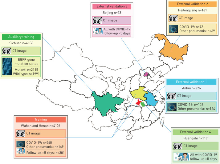
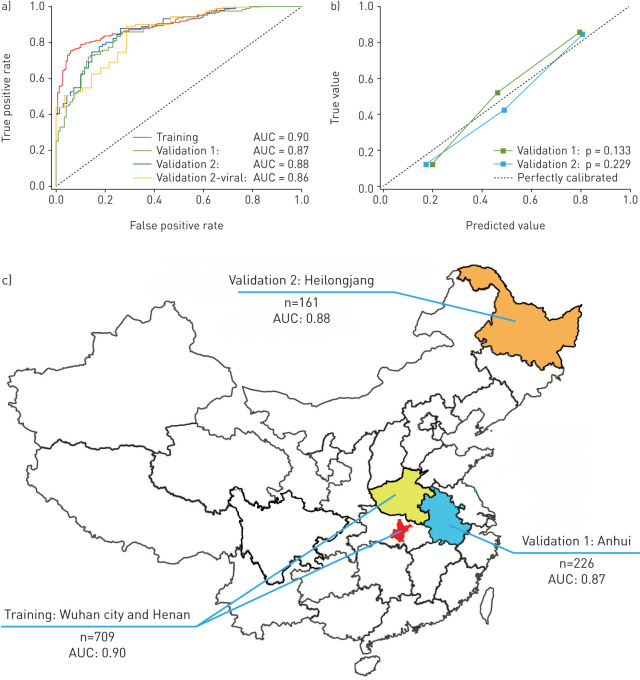
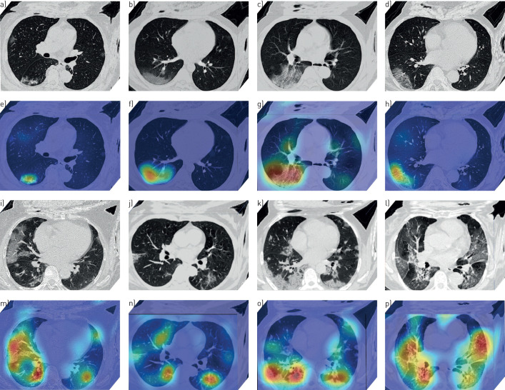
Coronavirus disease 2019 (COVID-19) has spread globally, and medical resources become insufficient in many regions. Fast diagnosis of COVID-19 and finding high-risk patients with worse prognosis for early prevention and medical resource optimisation is important. Here, we proposed a fully automatic deep learning system for COVID-19 diagnostic and prognostic analysis by routinely used computed tomography.We retrospectively collected 5372 patients with computed tomography images from seven cities or provinces. Firstly, 4106 patients with computed tomography images were used to pre-train the deep learning system, making it learn lung features. Following this, 1266 patients (924 with COVID-19 (471 had follow-up for >5 days) and 342 with other pneumonia) from six cities or provinces were enrolled to train and externally validate the performance of the deep learning system.In the four external validation sets, the deep learning system achieved good performance in identifying COVID-19 from other pneumonia (AUC 0.87 and 0.88, respectively) and viral pneumonia (AUC 0.86). Moreover, the deep learning system succeeded to stratify patients into high- and low-risk groups whose hospital-stay time had significant difference (p=0.013 and p=0.014, respectively). Without human assistance, the deep learning system automatically focused on abnormal areas that showed consistent characteristics with reported radiological findings.Deep learning provides a convenient tool for fast screening of COVID-19 and identifying potential high-risk patients, which may be helpful for medical resource optimisation and early prevention before patients show severe symptoms.


Conflict of interest statement

Conflict of interest: S. Wang has nothing to disclose. Conflict of interest: Y. Zha has nothing to disclose. Conflict of interest: W. Li has nothing to disclose. Conflict of interest: Q. Wu has nothing to disclose. Conflict of interest: X. Li has nothing to disclose. Conflict of interest: M. Niu has nothing to disclose. Conflict of interest: M. Wang has nothing to disclose. Conflict of interest: X. Qiu has nothing to disclose. Conflict of interest: H. Li has nothing to disclose. Conflict of interest: H. Yu has nothing to disclose. Conflict of interest: W. Gong has nothing to disclose. Conflict of interest: Y. Bai has nothing to disclose. Conflict of interest: L. Li has nothing to disclose. Conflict of interest: Y. Zhu has nothing to disclose. Conflict of interest: L. Wang has nothing to disclose. Conflict of interest: J. Tian has nothing to disclose.

Figures

FIGURE 1
FIGURE 1
Datasets used in this study. A total of 5372 patients with computed tomography (CT) images from seven cities or provinces were enrolled in this study. The auxiliary training set included 4106 patients with lung cancer and epidermal growth factor receptor (EGFR) gene mutation status information, and is used to pre-train the COVID-19Net to learn lung features from CT images. The training set includes 709 patients from Wuhan city and Henan province. The external validation set 1 (226 patients) from Anhui province, and the external validation set 2 (161 patients) from Heilongjiang province are used to assess the diagnostic performance of the deep learning (DL) system. The external validation set 3 (53 patients with COVID-19) from Beijing, and the external validation set 4 (117 patients with COVID-19) from Huangshi city are used to evaluate the prognostic performance of the DL system.
FIGURE 2
FIGURE 2
Illustration of the proposed deep learning (DL) system. Using the chest computed tomography (CT) scanning of a patient, the DL system predicts the probability the patient has COVID-19 and the prognosis of this patient directly without any human annotation. The DL system includes three parts: automatic lung segmentation (DenseNet121-FPN), non-lung area suppression, and COVID-19 diagnostic and prognostic analysis (COVID-19Net). To let the COVID-19Net learn lung features from the large dataset we used the auxiliary training process for pre-training, which trained the DL network to predict epidermal growth factor receptor (EGFR) gene mutation status using CT images of 4106 patients. The dense connection in this figure means each convolutional layer is connected to all of its previous convolutional layers inside the same dense block.
FIGURE 3
FIGURE 3
Diagnostic performance of the deep learning (DL) system. a) Receiver operating characteristic curves of the DL system in the training set and the two independent external validation sets. Validation 2-viral is a stratified analysis using the patients with coronavirus 2019 and viral pneumonia in the validation set 2. b) Calibration curves of the DL system in the two external validation sets. c) Area under the curve and distribution of the training set and the two external validation sets.
FIGURE 4
FIGURE 4
Deep learning (DL) discovered suspicious lung area. a–p) Computed tomography (CT) images of eight patients with coronavirus 2019. a–d and i–l) CT images of the patients (these CT images are processed by the DL system). e–h and m–p) Heat maps of the DL discovered suspicious lung area. In the heat map, areas with bright red colour are more important than dark blue areas.
FIGURE 5
FIGURE 5
Deep learning (DL) feature visualisation. a–d) Four 3-dimensional (3D) convolutional filters from different convolutional layers. e) Distribution of patients in the 64-dimensional DL feature space. For display convenience, the 64-dimensional DL feature space is reduced to 2-dimensional by a principle component analysis algorithm.

Similar articles

Cited by

References

    1. Wang D, Hu B, Hu C, et al. . Clinical characteristics of 138 hospitalized patients with 2019 novel coronavirus–infected pneumonia in Wuhan, China. JAMA 2020; 323: 1061–1069. - PMC - PubMed
    1. Li Q, Guan X, Wu P, et al. . Early transmission dynamics in Wuhan, China, of novel coronavirus–infected pneumonia. N Engl J Med 2020; 382: 1199–1207. - PMC - PubMed
    1. Yang X, Yu Y, Xu J, et al. . Clinical course and outcomes of critically ill patients with SARS-CoV-2 pneumonia in Wuhan, China: a single-centered, retrospective, observational study. Lancet Respir Med 2020; 8: 475–481. doi:10.1016/S2213-2600(20)30079-5 - DOI - PMC - PubMed
    1. Ji Y, Ma Z, Peppelenbosch MP, et al. . Potential association between COVID-19 mortality and health-care resource availability. Lancet Glob Health 2020; 8: e480. doi:10.1016/S2214-109X(20)30068-1 - DOI - PMC - PubMed
    1. Xie X, Zhong Z, Zhao W, et al. . Chest CT for typical 2019-nCoV pneumonia: relationship to negative RT-PCR testing. Radiology 2020; 296; E41–E45. - PMC - PubMed

MeSH terms

LinkOut - more resources