. 2020 Jun 9;117(23):12806-12816.
doi: 10.1073/pnas.1917196117. Epub 2020 May 22.

The 6-4 photoproduct is the trigger of UV-induced replication blockage and ATR activation

Affiliations

The 6-4 photoproduct is the trigger of UV-induced replication blockage and ATR activation

Kai-Feng Hung et al. Proc Natl Acad Sci U S A. .

Abstract

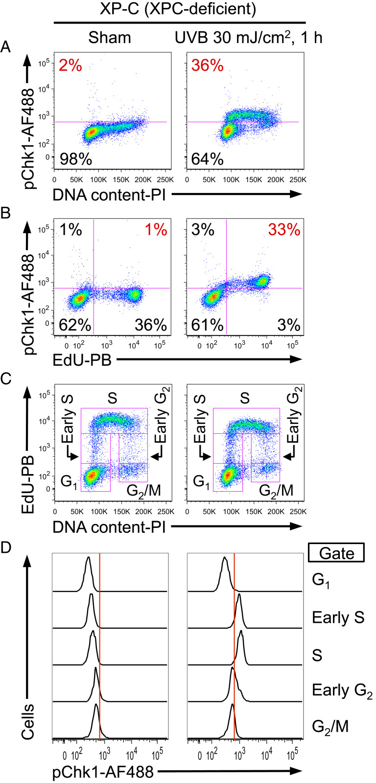
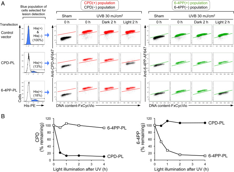
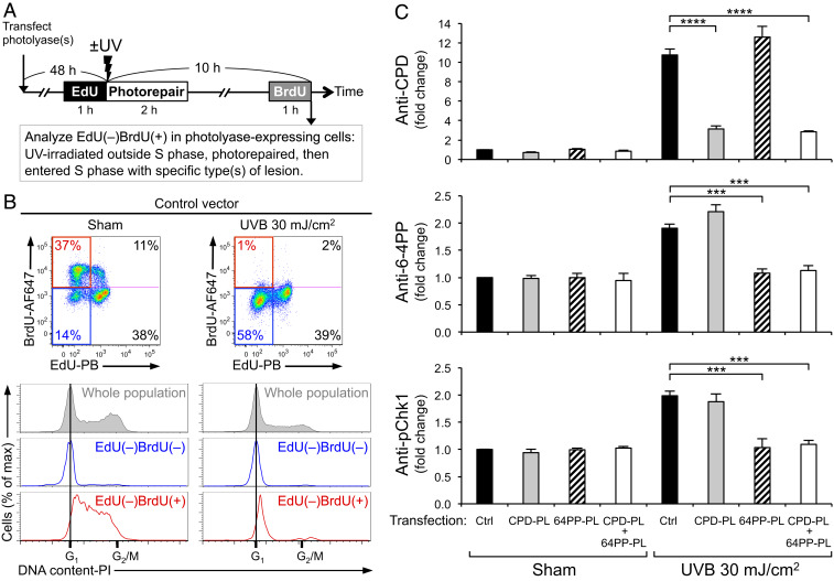
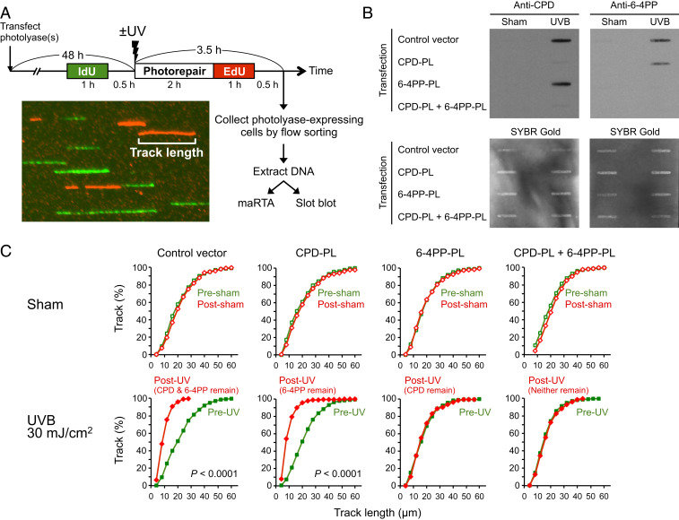
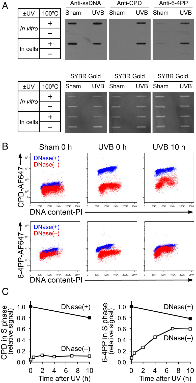
The most prevalent human carcinogen is sunlight-associated ultraviolet (UV), a physiologic dose of which generates thousands of DNA lesions per cell, mostly of two types: cyclobutane pyrimidine dimers (CPDs) and 6-4 photoproducts (6-4PPs). It has not been possible, in living cells, to precisely characterize the respective contributions of these two lesion types to the signals that regulate cell cycle progression, DNA replication, and cell survival. Here we coupled multiparameter flow cytometry with lesion-specific photolyases that eliminate either CPDs or 6-4PPs and determined their respective contributions to DNA damage responses. Strikingly, only 6-4PP lesions activated the ATR-Chk1 DNA damage response pathway. Mechanistically, 6-4PPs, but not CPDs, impeded DNA replication across the genome as revealed by microfluidic-assisted replication track analysis. Furthermore, single-stranded DNA accumulated preferentially at 6-4PPs during DNA replication, indicating selective and prolonged replication blockage at 6-4PPs. These findings suggest that 6-4PPs, although eightfold fewer in number than CPDs, are the trigger for UV-induced DNA damage responses.

Keywords: 6-4PP; CPD; Chk1; DNA damage response; DNA replication.


Conflict of interest statement

The authors declare no competing interest.

Figures

Fig. 1.
Fig. 1.
UV-induced phosphorylation of Chk1 is strictly limited to S phase as revealed by flow cytometry. (AD) XP-C cells (GM15983) were pulse labeled with EdU for 1 h, followed by sham (Left) or UVB irradiation (Right), and harvested 1 h after irradiation. (A) Phosphorylation of Chk1 at Ser345 (pChk1) was evaluated as a function of DNA content. The percentage of pChk1(+) cells is shown in red. (B) UV-induced pChk1 strongly correlates with EdU incorporation. The percentage of EdU(+)pChk1(+) cells is shown in red. (C) EdU incorporation and DNA content were used to identify five cell cycle subpopulations (G1, early S, S, early G2, and G2/M phases; pink boxes identify gates used). (D) pChk1 was evaluated for the cell cycle subpopulations defined in C. Chk1 phosphorylation was essentially restricted to UV-irradiated cells that were in early S or S phase. Data from one representative experiment of three independent experiments are shown.
Fig. 2.
Fig. 2.
Selection of cells with a single type of UV lesion using photolyase and flow cytometry. (A) Flow cytometry identifies photolyase-expressing cells that exhibit light-dependent, lesion-specific repair (photorepair) in all cell cycle phases. XP-C cells (GM15983) transfected with control vector, polyhistidine (His)-tagged CPD-photolyase (CPD-PL), or His-tagged 6-4PP-photolyase (6-4PP-PL) were sham or UVB irradiated and were harvested immediately (0 h) or after 2 h with (light) or without (dark) visible light illumination. Control vector-transfected cells [whole population including His(+) and His(–)] or photolyase-expressing cells [His(+)] were selected for lesion detection (as indicated in blue in histogram with percentage). Middle and Right show the levels of CPD or 6-4PP as a function of DNA content (FxCycle Violet). CPD(+), 6-4PP(+), and lesion-negative populations are indicated in red, green, and black, respectively. (B) Time course of CPD and 6-4PP photorepair by lesion-specific photolyase. XP-C cells were transfected with CPD-PL (closed circles) or 6-4PP-PL (open squares). Cells were sham or UVB irradiated (30 mJ/cm2) and subsequently illuminated with visible light until harvest at the indicated time points. Remaining lesions in photolyase-expressing cells (His tag-positive) were assessed by flow cytometry. Background signal (based on sham-treated cells) was subtracted, and lesion signal at 0 h (immediately after UV) was set to 100%. Data from one representative experiment of three independent experiments are shown.
Fig. 3.
Fig. 3.
The 6-4PP, but not CPD, potently induces phosphorylation of Chk1. (A) Experimental design for analyzing cells that enter S phase with a specific type of lesion. XP-C cells (GM15983) transfected with control vector (Ctrl), CPD-photolyase (CPD-PL), and/or 6-4PP-photolyase (64PP-PL) were labeled with EdU prior to UV. Following UVB 30 mJ/cm2 irradiation, cells were illuminated with visible light for photorepair. Cells were labeled with BrdU prior to harvest. Photolyase-expressing cells (polyhistidine tag-positive) were analyzed for levels of CPD, 6-4PP, and Chk1 phosphorylation at Ser345 (pChk1) within the EdU(–)BrdU(+) population. (B) The EdU(–)BrdU(+) population of UV-irradiated cells is in early S phase. Cells were stained with propidium iodide (PI) for DNA content. Cell cycle profiles of whole population (gray) and its subpopulations of EdU(–)BrdU(–) (blue) and EdU(–)BrdU(+) (red) are shown. Data from one representative experiment of four independent experiments are shown. (C) Phosphorylation of Chk1 is potently induced in cells with 6-4PP, but not in cells with CPD, upon S-phase entry. The levels of CPD, 6-4PP, and pChk1 (Ser345) in cells that newly entered S phase [EdU(–)BrdU(+) population] were evaluated using antibody-based flow cytometry assay. The mean ± SEM (four independent experiments) of fluorescence signals (fold change compared with sham-irradiated control vector cells) are shown. Statistical significance was determined by one-way ANOVA and Dunnett’s test, comparing each UV-irradiated, photolyase-transfected group with a single control (UV-irradiated control group). ***P < 0.001; ****P < 0.0001.
Fig. 4.
Fig. 4.
DNA replication is impeded by 6-4PP, but not CPD lesions. (A) Experimental design to quantitate DNA replication progression in cells with specific type(s) of lesion(s). XP-C cells (GM15983) transfected with the indicated photolyase (PL) were sham or UVB irradiated and pulse labeled with IdU and EdU separately for 1 h each. Photolyase-expressing cells (polyhistidine tag-positive) were collected by flow sorting. Genomic DNA extracted from sorted (photolyase-expressing cells) or unsorted (for control vector) cells was subjected to immuno-slot blot and microfluidic-assisted replication track analysis (maRTA; a representative image of segments of labeled DNA “tracks” is shown). (B) Slot-blot validation of lesion-specific photorepair of DNA used for maRTA. Extracted genomic DNA was heat denatured and then spotted onto membranes. Membranes were probed with anti-CPD or anti-6-4PP antibodies and subsequently stained with SYBR Gold for total DNA detection. Data from one representative experiment of two independent experiments are shown. (C) DNA replication is impeded in the presence of 6-4PP, but not CPD. Genomic DNA was aligned through microchannels and stained for IdU and EdU (maRTA). Replication track lengths of first (IdU; presham/UV) and second (EdU; postsham/UV) labels were measured in two independent experiments, and combined data are shown as cumulative distributions (n = 500 tracks for each label). The vertical axis indicates the cumulative fraction of tracks that are equal to or shorter than the corresponding track length indicated on the horizontal axis. The remaining lesion type(s) (validated in B) is indicated in parentheses above each UV graph. Statistical significance was determined by Mann–Whitney U test. Two conditions showed P < 0.0001 as indicated.
Fig. 5.
Fig. 5.
The 6-4PP lesion preferentially becomes surrounded by ssDNA. (A) Lesion-specific antibodies recognize CPD or 6-4PP on ssDNA, but not on dsDNA. For “in vitro” UV irradiation, genomic DNA was extracted from unirradiated cells and subsequently irradiated with UVB 30 mJ/cm2 in vitro. For “in cells” irradiation, genomic DNA was extracted from cells that were irradiated with UVB 30 mJ/cm2. DNA was left untreated or heat denatured at 100 °C for 10 min and spotted onto membranes for immuno-slot blot using antibodies specific for ssDNA, CPD, or 6-4PP. The same membranes were stained with SYBR Gold for total DNA detection. (B) UV lesions on ssDNA under non-DNase condition become detectable in S phase long after UV, but not immediately after UV. XP-C cells (GM15983) were harvested immediately (0 h) or 10 h after UVB 30 mJ/cm2. Fixed cells were left untreated or treated with DNase. CPD and 6-4PP were detected using lesion-specific antibodies validated in A to recognize the lesions only when on ssDNA. DNA content was assessed using propidium iodide (PI). Flow cytometry data from two samples [DNase(+) and DNase(–)] were overlaid on each plot. (C) In S-phase cells, 6-4PP lesions are increasingly surrounded by ssDNA. Signal intensities of CPD and 6-4PP in S phase at various time points following UV were measured using the same flow cytometry assay as in B. Relative signal intensities of CPD or 6-4PP in S phase were calculated relative to 0 (sham irradiation at 0 h) and 1 (UVB 30 mJ/cm2 at 0 h with DNase treatment). Data from one representative experiment of three independent experiments are shown.

Similar articles

Cited by

References

    1. Rogers H. W., Weinstock M. A., Feldman S. R., Coldiron B. M., Incidence estimate of nonmelanoma skin cancer (keratinocyte carcinomas) in the U.S. population, 2012. JAMA Dermatol. 151, 1081–1086 (2015). - PubMed
    1. Mouret S. et al. ., Cyclobutane pyrimidine dimers are predominant DNA lesions in whole human skin exposed to UVA radiation. Proc. Natl. Acad. Sci. U.S.A. 103, 13765–13770 (2006). - PMC - PubMed
    1. Ciccia A., Elledge S. J., The DNA damage response: Making it safe to play with knives. Mol. Cell 40, 179–204 (2010). - PMC - PubMed
    1. Jans J. et al. ., Powerful skin cancer protection by a CPD-photolyase transgene. Curr. Biol. 15, 105–115 (2005). - PubMed
    1. Sancar A., DNA excision repair. Annu. Rev. Biochem. 65, 43–81 (1996). - PubMed

Publication types