Review
. 2020 Aug;106(16):1202-1210.
doi: 10.1136/heartjnl-2020-316540. Epub 2020 May 22.

Comprehensive review of evaluation and management of cardiac paragangliomas

Affiliations
Review

Comprehensive review of evaluation and management of cardiac paragangliomas

Sri Harsha Tella et al. Heart. 2020 Aug.

Abstract

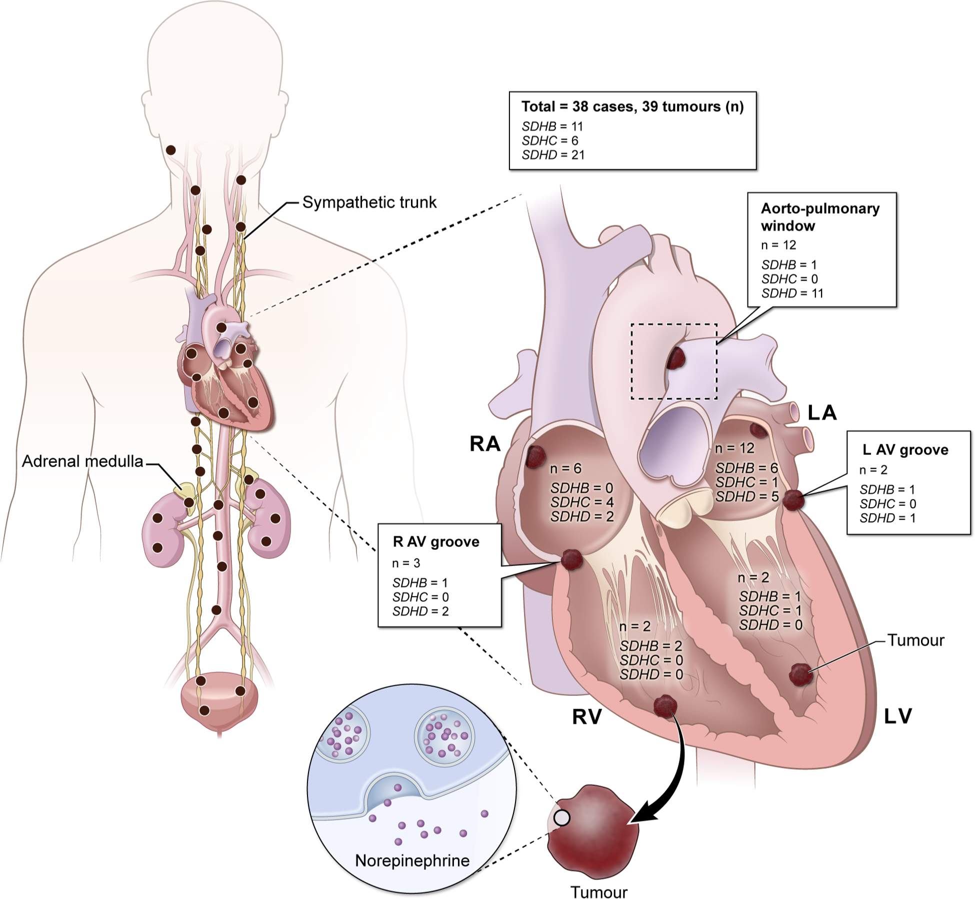
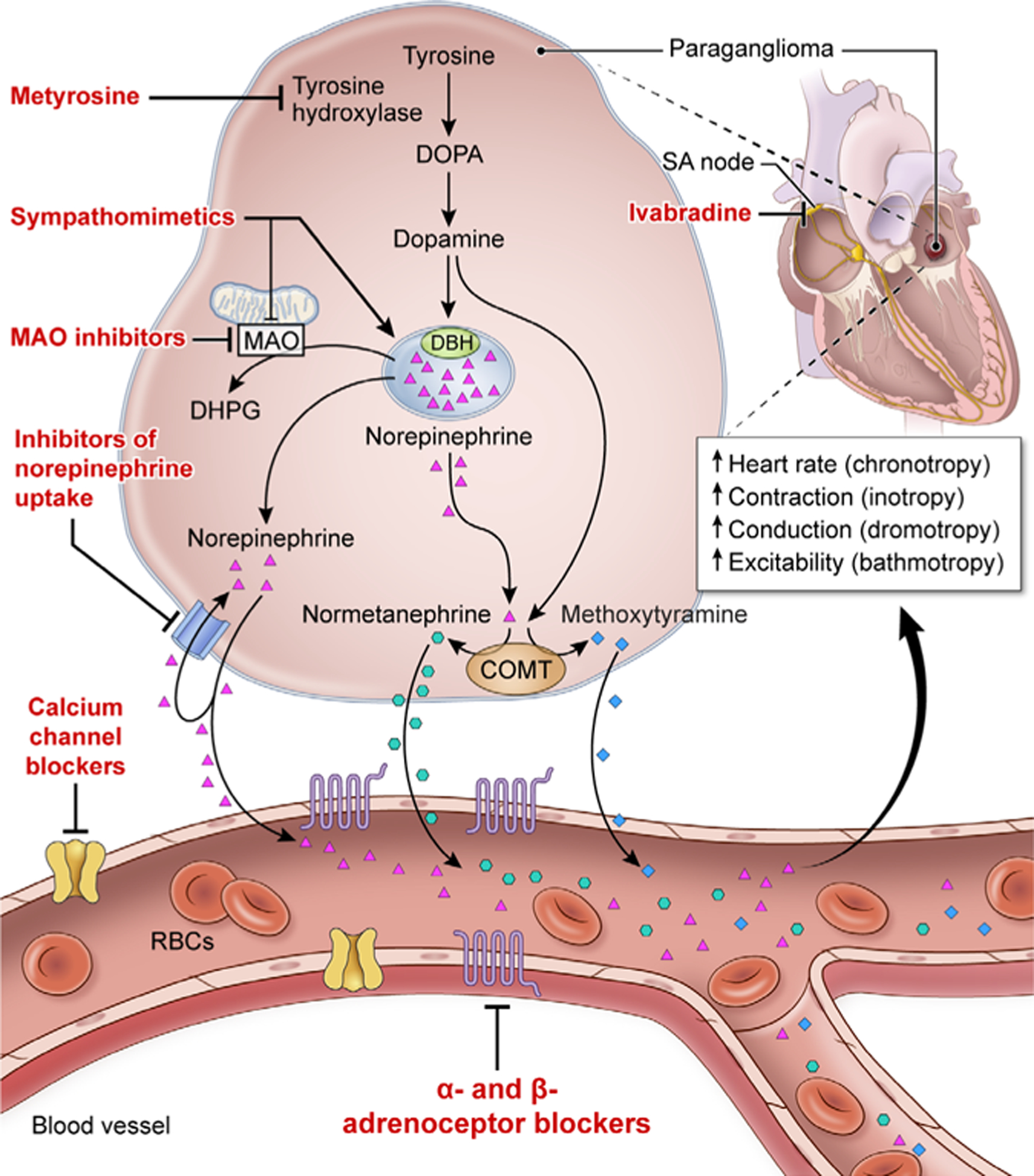
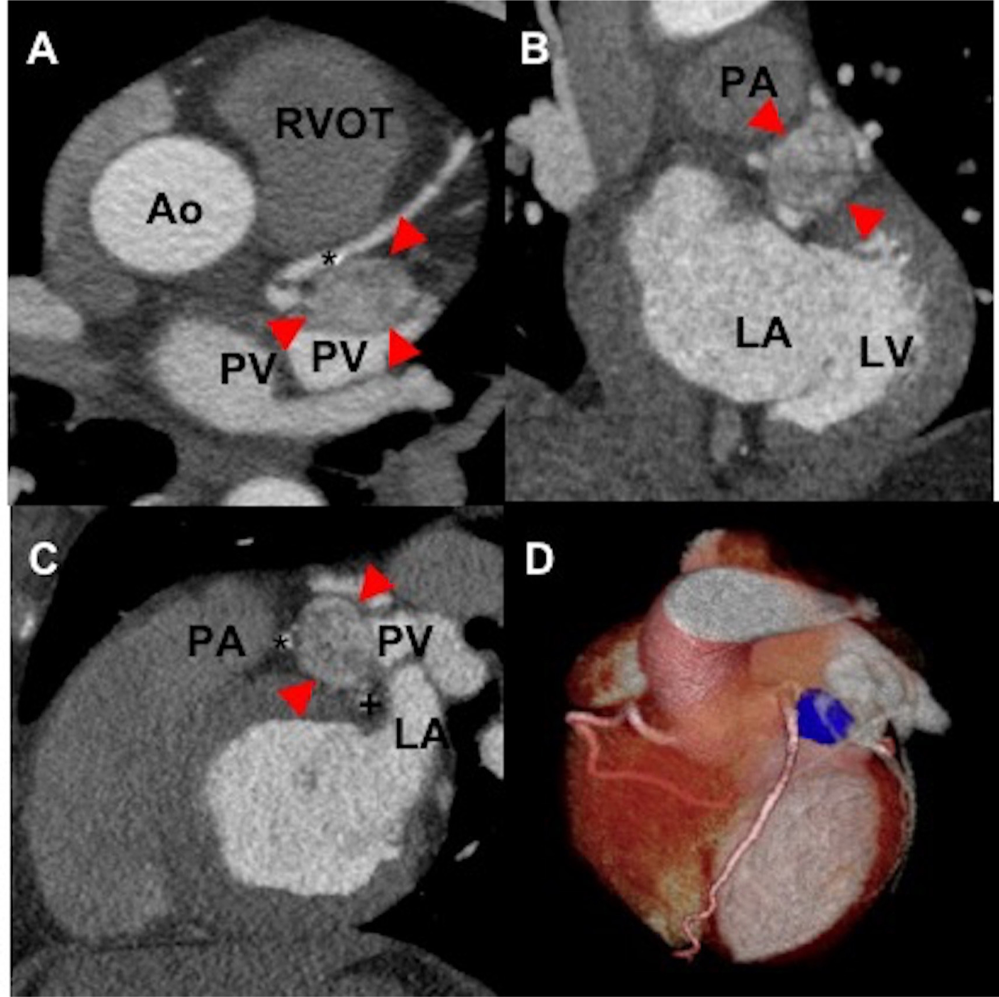
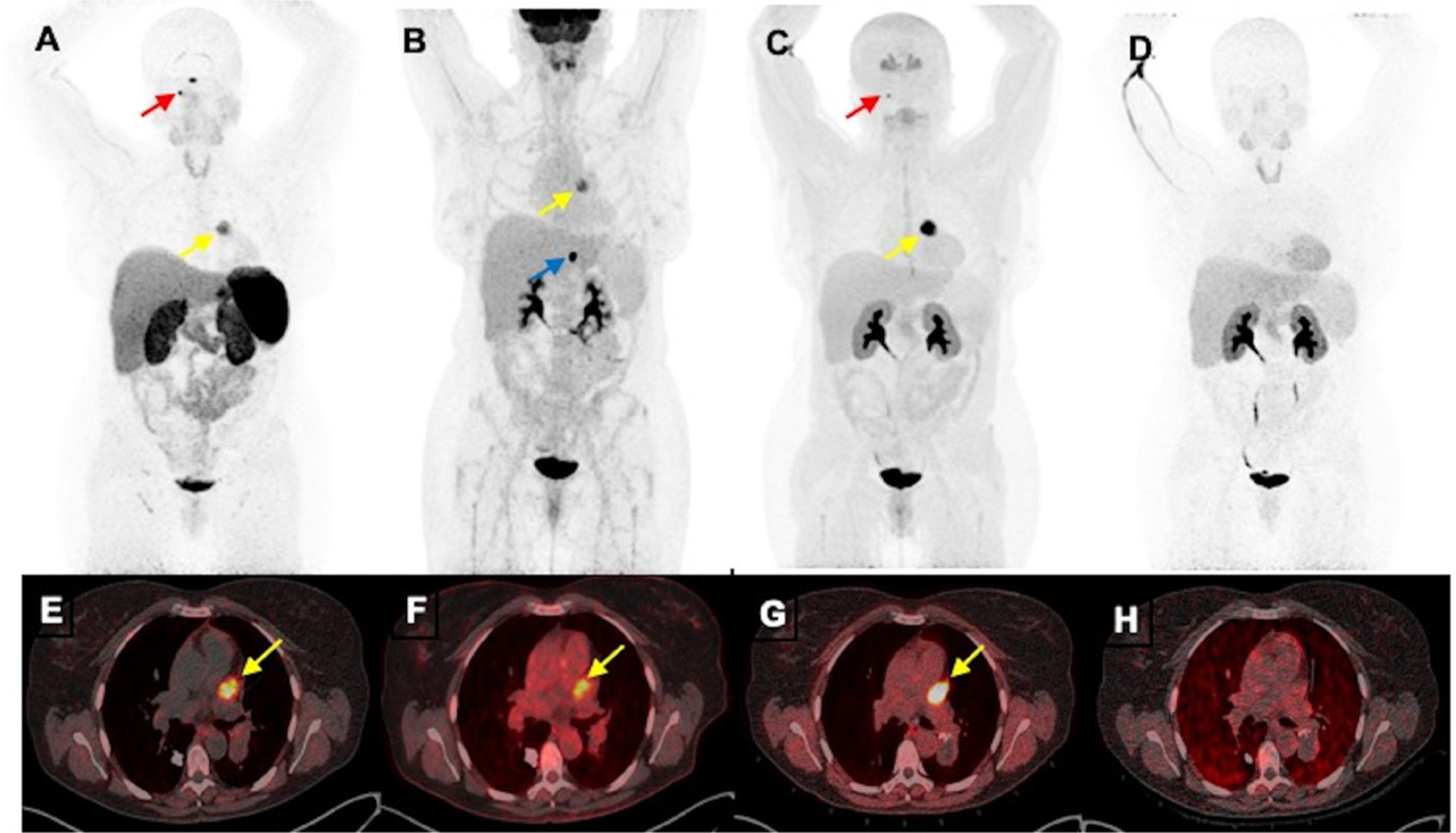
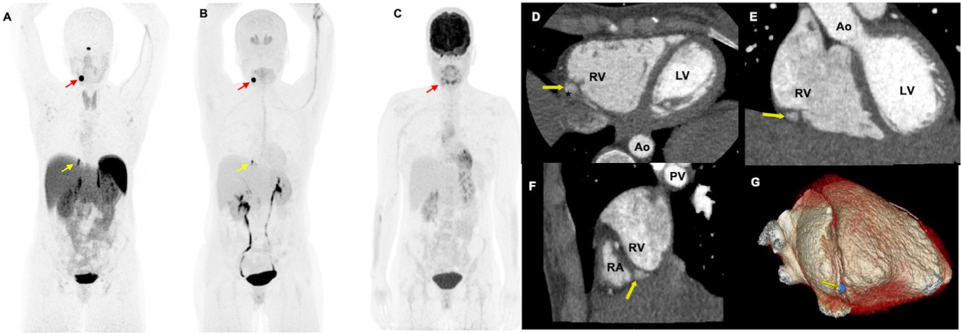
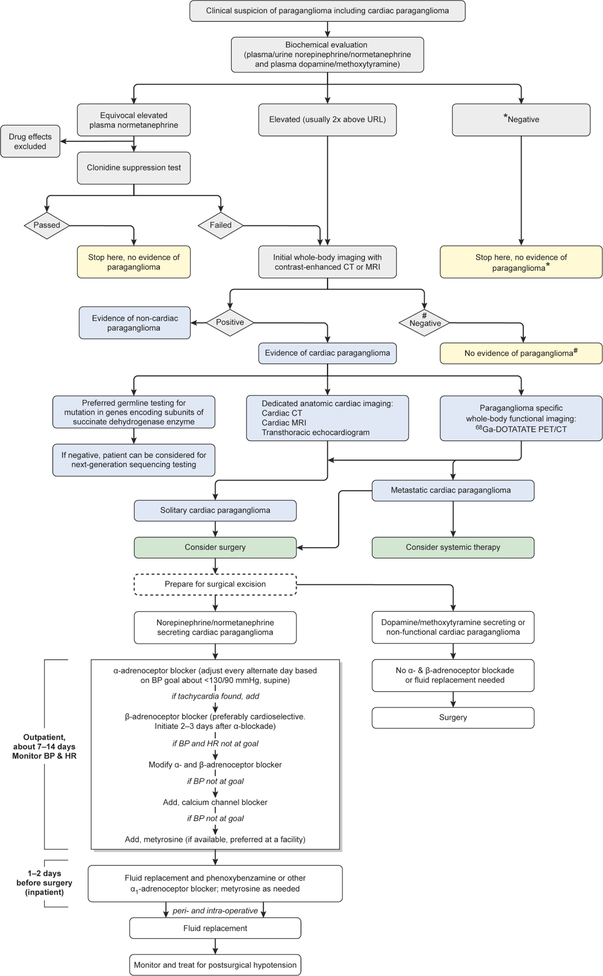
Cardiac paraganglioma (PGL) is a rare neuroendocrine tumour causing significant morbidity primarily due to norepinephrine secretion potentially causing severe hypertension, palpitations, lethal tachyarrhythmias, stroke and syncope. Cardiologists are faced with two clinical scenarios. The first is the elevated norepinephrine, whose actions must be properly counteracted by adrenoceptor blockade to avoid catastrophic consequences. The second is to evaluate the precise location of a cardiac PGL and its spread since compression of cardiovascular structures may result in ischaemia, angina, non-noradrenergic-induced arrhythmia, cardiac dysfunction or failure. Thus, appropriate assessment of elevated norepinephrine by its metabolite normetanephrine is a gold biochemical standard at present. Furthermore, dedicated cardiac CT, MRI and transthoracic echocardiogram are necessary for the precise anatomic information of cardiac PGL. Moreover, a cardiologist needs to be aware of advanced functional imaging using 68Ga-DOTA(0)-Tyr(3)-octreotide positron emission tomography/CT, which offers the best cardiac PGL-specific diagnostic accuracy and helps to stage and rule out metastasis, determining the next therapeutic strategies. Patients should also undergo genetic testing, especially for mutations in genes encoding succinate dehydrogenase enzyme subunits that are most commonly present as a genetic cause of these tumours. Curative surgical resection after appropriate α-adrenoceptor and β-adrenoceptor blockade in norepinephrine-secreting tumours is the primary therapeutic strategy. Therefore, appropriate and up-to-date knowledge about early diagnosis and management of cardiac PGLs is paramount for optimal outcomes in patients where a cardiologist is an essential team member of a multidisciplinary team in its management.

Keywords: cardiac CT imaging; cardiac imaging and diagnostics; genetics; positron emission tomographic (PET) imaging; systemic review.


Conflict of interest statement

Competing interests: None declared.

Figures

Figure 1
Figure 1
Schematic representation of mutation-wise distribution of cardiac paragangliomas in genes encoding subunits of succinate dehydrogenase enzyme that were reported to PubMed between the years 2000 and 2019.
Figure 2
Figure 2
Schematic representation of synthesis, release and action of catecholamines and the respective metabolites released from cardiac paraganglioma and drugs that act on the catecholamine pathway. The α-adrenoceptor and β-adrenoceptor blockers reducethe effects of catecholamines on end organs such as blood vessels, heart and others that harbour adrenoceptors. Metyrosine blocks the rate-limiting step in catecholamine synthesis by inhibiting tyrosine hydroxylase. Calcium channel blockers cause smooth muscle relaxation in the blood vessel. Ivabradine, an If current inhibitor, acts on the sinoatrial node (SA node), thereby targeting the chronotropic effect of catecholamines at SA node. Agents such as sympathomimetic drugs (ephedrine, caffeine, amphetamine and nicotine) cause displacement of norepinephrine from the stores (vesicular sequestration, dominant mechanism indicated by bold arrow) and partly by inhibiting monoamine oxidase (MAO). MAO inhibitors block the conversion of catecholamines to dihydroxyphenylglycol (DHPG), thereby increasing the concentrations of norepinephrine. Antidepressants such as selective-norepinephrine re-uptake inhibitors, and tricyclic antidepressants that inhibit norepinephrine re-uptake leading to increased concentration of norepinephrine. RBC, red blood cell.
Figure 3
Figure 3
The axial (A), coronal (B), sagittal (C) and reformatted three-dimensional images (D) of a contrast-enhanced cardiac-gated CT of a woman aged 50 years without mutation in genes encoding subunits of succinate dehydrogenase enzyme demonstrates a well-circumscribed cardiac mass (arrows) measuring 3×2×2 cm. This mass is located superior to left atrium (B, C) with the inferior border extending to the left atrioventricular groove. The mass is bordered laterally by the left atrial appendage (C) and medially by the main pulmonary artery (superiorly, (B, C)) and proximal left anterior descending artery (*, (C)). The proximal left circumflex courses just inferior to the mass (+, (D)). There is no evidence of luminal compression in the left anterior descending or left circumflex arteries by the mass. Ao, aortic root; LA, left atrium; LV, left ventricle; PA, pulmonary artery; PV, pulmonary vein; RVOT, right ventricular outflow tract; asterisk (*), left anterior descending artery; plus (+), left circumflex.
Figure 4
Figure 4
Functional positron emission tomography/CT (PET/CT) imaging of a cardiac paraganglioma of a woman aged 50 years without mutation in genes encoding subunits of succinate dehydrogenase enzyme. The anterior maximum intensity projection images (A–D) and fused axial PET/CT images (E–H) of 68Ga-DOTA(0)-Tyr(3)-octreotide PET/CT (A, E), 18F-fluorodeoxyglucose (B, F) and 18F-dihydroxyphenylalanine (C, G) PET/CT scans demonstrate a cardiac paraganglioma (yellow arrows) located in between pulmonary trunk and left atrium and superior aspect of left ventricle. However, this lesion lacks avidity on 18F-fluorodopamine (D, H). Furthermore, 68Ga-DOTA(0)-Tyr(3)-octreotide and 18F-dihydroxyphenylalanine PET/CT demonstrate a small right glomus jugulare paraganglioma (red arrows; A, B) and 18F-fluorodeoxyglucose PET/CT alone demonstrates a gastrohepatic nodule (blue arrow, (B)).
Figure 5
Figure 5
Functional positron emission tomography/CT (PET/CT) and contrast-enhanced cardiac-gated CT imaging of a woman aged 34 years with mutation in succinate dehydrogenase D gene. The anterior maximum intensity projection images (A–C) of 68Ga-DOTA(0)-Tyr(3)-octreotide PET/CT (A) and 18F-dihydroxyphenylalanine demonstrates a pericardiac paraganglioma (yellow arrows; A, B) as well as a left carotid body paraganglioma (red arrows; A, B). However, 18F-fluorodeoxyglucose (C) PET/CT scan demonstrates faint uptake in the left carotid body paraganglioma (red arrow, (C)) but lacks avidity in the pericardiac paraganglioma. This pericardiac paraganglioma was not localised on contrast-enhanced chest CT. The patient further underwent a contrast-enhanced cardiac-gated CT imaging to further characterise this pericardiac mass. The axial (D), coronal (E), sagittal (F) and reformatted three-dimensional images (G) demonstrate a well-circumscribed pericardiac mass (yellow arrows; D–G measuring 0.6×0.7×1.1 cm. This mass is located adjacent to mid-distal right carotid artery (*, (D)) without impingement of vessel lumen. The mass is outside the right ventricular free wall (yellow arrows, (D–F)) in the basal portion of the right atrial-ventricular groove (F) and within the pericardium. Ao, aortic root; LV, left ventricle; PV, pulmonary vein; RA, right atrium; RV, right ventricle; asterisk (*), right carotid artery.
Figure 6
Figure 6
Flow chart enumerating the diagnosis and management of cardiac paraganglioma. *Results are to be interpreted with caution that biochemical evaluation may be negative in non-functioning paraganglioma. #Results are to be interpreted with caution that anatomical imaging may miss small paragangliomas and functional imaging can act as complimentary diagnostic tool. BP, blood pressure; HR, heart rate, 68Ga-DOTATATE, 68Ga-DOTA(0)-Tyr(3)-octreotide; PET/CT, positron emission tomography/CT.

References

    1. Lam AK-Y, Lam AK. Update on adrenal tumours in 2017 World Health organization (who) of endocrine tumours. Endocr Pathol 2017;28:213–27. - PubMed
    1. Martucci VL, Emaminia A, del Rivero J, et al. Succinate dehydrogenase gene mutations in cardiac paragangliomas. Am J Cardiol 2015;115:1753–9. - PMC - PubMed
    1. Patel J, Sheppard MN. Pathological study of primary cardiac and pericardial tumours in a specialist UK centre: surgical and autopsy series. Cardiovasc Pathol 2010;19:343–52. - PubMed
    1. Fishbein L, Leshchiner I, Walter V, et al. Comprehensive molecular characterization of pheochromocytoma and paraganglioma. Cancer Cell 2017;31:181–93. - PMC - PubMed
    1. Ghayee HK, Havekes B, Corssmit EPM, et al. Mediastinal paragangliomas: association with mutations in the succinate dehydrogenase genes and aggressive behavior. Endocr Relat Cancer 2009;16:291–9. - PMC - PubMed

Publication types

MeSH terms

Substances