. 2020 May 22;11(1):2578.
doi: 10.1038/s41467-020-16306-5.

N6-methyladenosine regulates glycolysis of cancer cells through PDK4

Affiliations

N6-methyladenosine regulates glycolysis of cancer cells through PDK4

Zihan Li et al. Nat Commun. .

Abstract

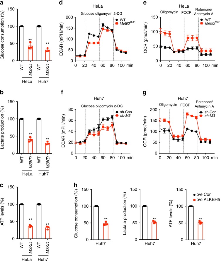
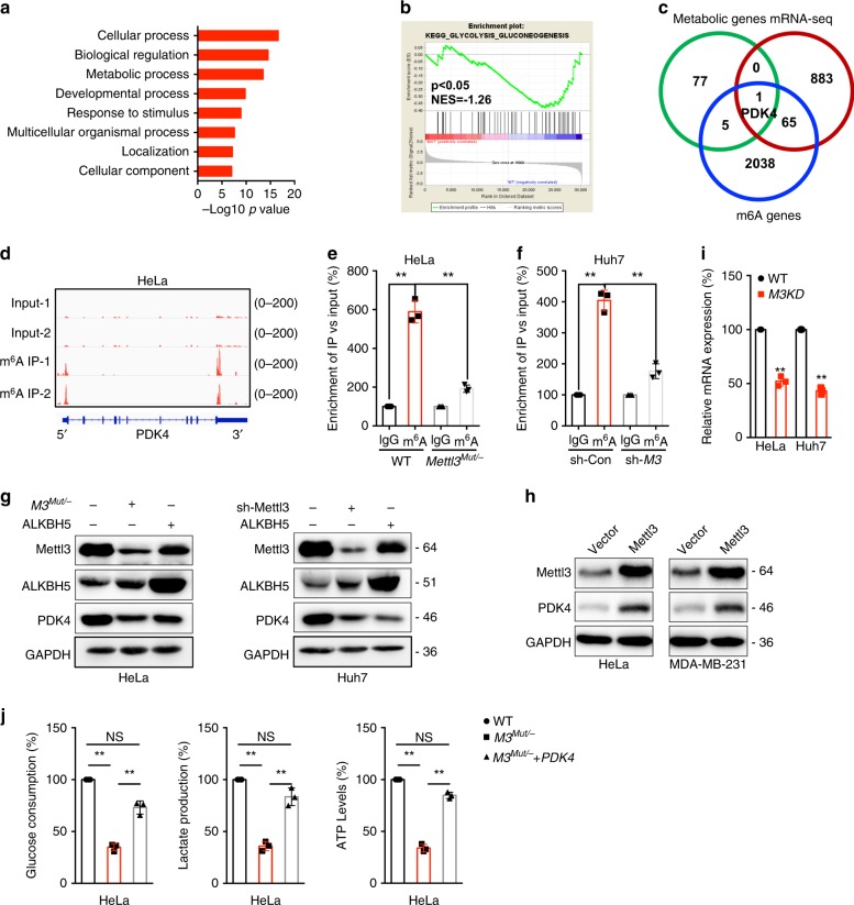
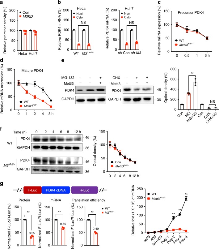
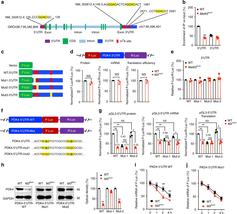
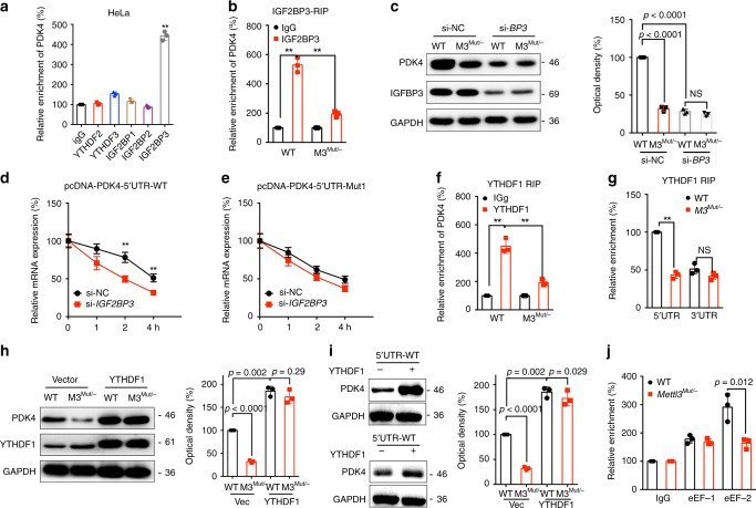
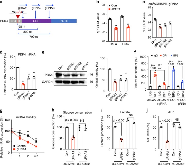
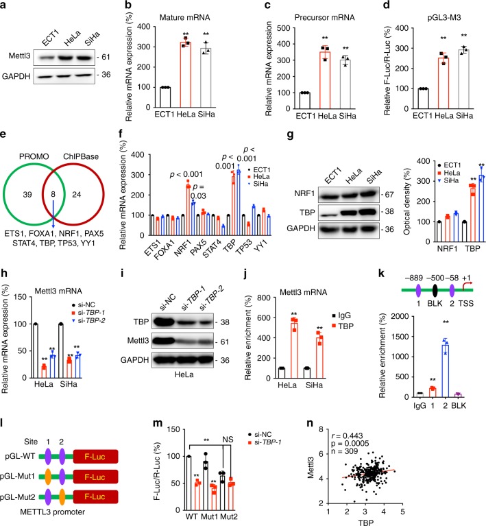
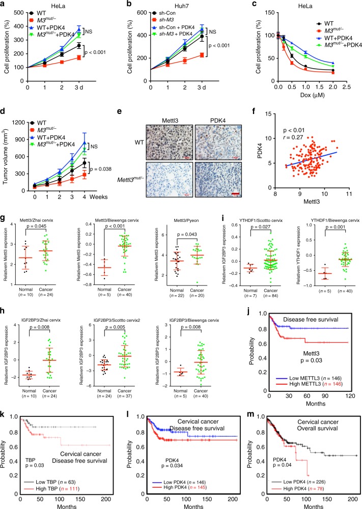
Studies on biological functions of N6-methyladenosine (m6A) modification in mRNA have sprung up in recent years. We find m6A can positively regulate the glycolysis of cancer cells. Specifically, m6A-sequencing and functional studies confirm that pyruvate dehydrogenase kinase 4 (PDK4) is involved in m6A regulated glycolysis and ATP generation. The m6A modified 5'UTR of PDK4 positively regulates its translation elongation and mRNA stability via binding with YTHDF1/eEF-2 complex and IGF2BP3, respectively. Targeted specific demethylation of PDK4 m6A by dm6ACRISPR system can significantly decrease the expression of PDK4 and glycolysis of cancer cells. Further, TATA-binding protein (TBP) can transcriptionally increase the expression of Mettl3 in cervical cancer cells via binding to its promoter. In vivo and clinical data confirm the positive roles of m6A/PDK4 in tumor growth and progression of cervical and liver cancer. Our study reveals that m6A regulates glycolysis of cancer cells through PDK4.


Conflict of interest statement

The authors declare no competing interests.

Figures

Fig. 1
Fig. 1. m6A regulates glycolysis and ATP generation of cancer cells.
ac The glucose consumption (a), lactate production (b), and ATP levels (c) in Mettl3Mut/- HeLa, sh-Mettl3 Huh7 and their corresponding control cells. d, e The cellular ECAR (d) and OCR (e) were measured in wild type and Mettl3Mut/- HeLa cells. f, g The cellular ECAR (f) and OCR (g) were measured in sh-control and sh-Mettl3 Huh7 cells. b The glucose consumption, lactate production, and ATP levels in Huh7 cells transfected with vector control or ALKBH5 constructs. Data are presented as the mean ± SD from three independent experiments. **p < 0.01 by two tailed t test for (a) (p = 0.005 and p < 0.0001), (b) (p < 0.0001), (c) (p < 0.0001), and (h) (p < 0.0001).
Fig. 2
Fig. 2. PDK4 mediates m6A regulated glycolysis and ATP generation of cancer cells.
a Gene ontology analysis was performed on a subset of downregulated genes in Mettl3Mut/- HeLa cells. Log2 fold change, (KD: WT) < −0.5, was applied as the threshold cutoff. Several biological processes involved in metabolic processes were enriched and highlighted in bold; Significance shown as –Log10 Bonferroni p-value after multiple hypothesis correction. b GSEA reveals negative enrichment of genes in glycolysis gluconeogenesis sets of Mettl3Mut/- HeLa cells. c Venn diagram shows substantial and significant overlap among metabolic genes, variated genes in Mettl3Mut/- HeLa cells (>2 folds), and m6A enriched genes in wild type HeLa cells (>3 folds than input). d m6A peaks were enriched in 5′UTR and 3′UTRs of PDK4 genes from m6A RIP-seq data; e m6A RIP-qPCR analysis of PDK4 mRNA in wild type and Mettl3Mut/- HeLa cells. f m6A RIP-qPCR analysis of PDK4 mRNA in sh-Con and sh-Mettl3 Huh7 cells. g The expression of PDK4 in Mettl3Mut/- HeLa, sh-Mettl3 Huh7, or over expression of ALKBH5 and their corresponding control cells were measured by western blot analysis. h Cells were transfected with vector control or Mettl3 construct for 24 h, the expression of PDK4 was measured. i The mRNA of PDK4 in Mettl3Mut/- HeLa, sh-Mettl3 Huh7 and their corresponding control cells were measured by qRT-PCR. j The glucose consumption, lactate production, and ATP levels in wild type or Mettl3Mut/- HeLa cells transfected with PDK4 constructs for 24 h. Data are presented as the mean ± SD from three independent experiments. A representative from a total of two to three independent experiments is shown for (g) and (h). **p < 0.01, NS, no significant, by random permutation test for (b), by two-way ANOVA for (e) (p = 0.0001 and p = 0.0004, respectively) and (f) (p < 0.0001 and p = 0.0007, respectively), two-tailed unpaired Student’s t test for (i) (p < 0.0001), and one-way ANOVA for (j) (p < 0.0001 and p = 0.0008 for glucose consumption, p < 0.0001 and p = 0.0011 for lactate production, and p < 0.0001 for ATP levels, respectively).
Fig. 3
Fig. 3. m6A regulates the mRNA stability and translation of PDK4 in cancer cells.
a Cells were transfected with pGL3-Basic-PDK4-luc reporter and pRL-TK plasmid for 24 h. Results were presented as the ratios between the activity of the reporter plasmid and pRL-TK. b The relative levels of nuclear versus cytoplasmic PDK4 mRNA in wild-type and Mettl3Mut/- cells, or sh-control and sh-Mettl3 Huh7 cells. c After treatment with Act-D to inhibit transcription, the precursor mRNA levels of PDK4 were checked in wild type and Mettl3Mut/- cells. d After treatment with Act-D for the indicated times, the mature mRNA levels of PDK4 were checked in wild type and Mettl3Mut/- cells. e HeLa cells were pre-transfected with vector control or Mettl3 construct for 24 h and then further treated with CHX (10 μg/ml) or MG-132 (5 μM) for 6 h, the expression of PDK4 was detected by western blot analysis (left) and quantitatively analyzed (right). f Cells were treated with 10 μg/ml CHX for the indicated time periods, the expression of PDK4 was detected by western blot analysis (left) and quantitatively analyzed (right). g Wild-type or Mettl3Mut/- HeLa cells were transfected with pmirGLO-PDK4 reporter for 24 h. The translation outcome was determined as a relative signal of F-luc divided by R-luc, the mRNA abundance was determined by qRT-PCR of F-luc and R-luc, and the translation efficiency of PDK4 is defined as the quotient of reporter protein production (F-luc/R-luc) divided by mRNA abundance. h qRT-PCR checked the mRNA levels of PDK4 in non-ribosome portion (<40S), 40S, 60S, 80S, and polysome fractions in wide type and Mettl3Mut/- HeLa cells. Data are presented as the mean ± SD from three independent experiments. *p < 0.05, **p < 0.01, NS, no significant, by two-tailed unpaired Student’s t test for (a) and (c) (p < 0.0001 for all comparisons), and two-way ANOVA for (b) (p > 0.05), and by one-way ANOVA for (e) (p = 0.005) and (h) (p < 0.0001 for all comparisons).
Fig. 4
Fig. 4. Methylation sites of PDK4 involved in m6A regulated expression of PDK4.
a Schematic representation of positions of m6A motifs within PDK4 mRNA. b The m6A in 5′UTR or 3′UTR of PDK4 in wild type or Mettl3Mut/- HeLa cells were analyzed by m6A-RIP-qPCR using fragmented RNA. c Schematic representation of mutated (GGAC to GGCC) 3′UTR of pmirGLO vector to investigate the roles of m6A in 3′UTR in PDK4 expression. d Wild-type or Mettl3Mut/- HeLa cells were transfected with pmirGLO-PDK4-3′UTR-WT reporter for 24 h. The protein, mRNA and translation efficiency were determined. e The relative luciferase activity of F-Luc/R-Luc of pmirGLO-3′UTR-WT, or pmirGLO-3′UTR-Mut-1/-2/-3 in wild type and Mettl3Mut/- HeLa cells; f Schematic representation of mutation in 5′UTR to investigate the m6A roles on PDK4 expression. g Wild-type or Mettl3Mut/- HeLa cells were transfected with pGL3-PDK4-5′UTR-WT or pGL3-PDK4-5′UTR-Mut1/2 reporter for 24 h. The protein, mRNA and translation efficiency were determined. h pcDNA-PDK4-5′UTR-WT or pcDNA-PDK4-5′UTR-Mut1/2 was transfected into wild type or Mettl3Mut/- HeLa cells for 24 h. Protein expression was measured by western blot analysis (left) and quantitatively analyzed (right). i, j pcDNA-PDK4-5′UTR-WT (i) or pcDNA-PDK4-5′UTR-Mut1 (j) was transfected into wild type or Mettl3Mut/- HeLa cells for 24 h and then further treated with Act-D for the indicated times. The mRNA of PDK4 was checked by qRT-PCR. Data are presented as the mean ± SD from three independent experiments. *p < 0.05, **p < 0.01, NS, no significant, by two-tailed unpaired Student’s t test for (b) (p < 0.0001), (d) (p > 0.05), and (h) (p < 0.0001 for all), and two-way ANOVA for (c) and (i) (p < 0.001 for all).
Fig. 5
Fig. 5. Factors involved in m6A regulated expression of PDK4.
a RIP-qPCR analysis of PDK4 mRNA in wild type HeLa cells by use of antibody of YTHDF2, YTHDF3, and IGF2BP1~3. b IGF2BP3 RIP-qPCR analysis of PDK4 mRNA in wild type or Mettl3Mut/- HeLa cells. c Wild type or Mettl3Mut/- HeLa cells were transfected with si-NC or si-IGF2BP3 for 24 h, the expression of PDK4 was checked by western blot analysis (left) and quantitatively analyzed (right). d, e HeLa cells were transfected with si-NC, si-IGF2BP3, pcDNA-PDK4-5′UTR-WT (d) or pcDNA-PDK4-5′UTR-Mut1 (e) for 24 h and then further treated with Act-D for the indicated times. The mRNA of PDK4 was checked by qRT-PCR. f YTHDF1 RIP-qPCR analysis of PDK4 mRNA in wild type or Mettl3Mut/- HeLa cells. g Binding of YTHDF1 with the 5′UTR or 3′UTR in wild type or Mettl3Mut/- HeLa cells were analyzed by YTHDF1 RIP-qPCR using fragmented RNA. h Wild type or Mettl3Mut/- HeLa cells were transfected with vector or YTHDF1 construct for 24 h, the expression of PDK4 was checked by western blot analysis (left) and quantitatively analyzed (right). i HeLa cells were transfected with vector, YTHDF1 construct, pcDNA-PDK4-5′UTR-WT, and pcDNA-PDK4-5′UTR-Mut1 for 24 h, the expression of PDK4 was checked by western blot analysis (left) and quantitatively analyzed (right). j eEF-1 and eEF-2 RIP-qPCR analysis of PDK4 mRNA in wild type or Mettl3Mut/- HeLa cells. Data are presented as the mean ± SD from three independent experiments. *p < 0.05, **p < 0.01, NS, no significant, by two-tailed unpaired Student’s t test for (a) (p < 0.0001) and (g) (p < 0.0001 and p = 0.11, respectively), and two-way ANOVA for (b) (p < 0.0001 and p = 0.004, respectively), (c, d) (p < 0.0001 for all), (f) (p = 0.0003 and p = 0.001, respectively), (hj).
Fig. 6
Fig. 6. Targeting m6A of PDK4 by dm6ACRISPR re-programs metabolic of cancer cells.
a Schematic representation of positions of m6A site within PDK4 mRNA and the regions targeted by three gRNAs, respectively. b The threshold cycle (Ct) of qPCR showing SELECT results for detecting m6A site in the potential m6A site of PDK4 5′UTR in Mettl3Mut/- HeLa, sh-Mettl3 Huh7 and their corresponding control cells. c The threshold cycle (Ct) of qPCR showing SELECT results for detecting m6A site in 5′UTR of PDK4 in HeLa cells transfected with dCas13b-ALKBH5 combined with gRNA control or gRNA1/2/3, respectively, for 24 h. d, e The mRNA (d) or protein (e) expression of PDK4 in HeLa cells transfected with dCas13b-ALKBH5 combined with gRNA control or gRNA1/2/3, respectively, for 24 h. f RIP-qPCR analysis of PDK4 mRNA in HeLa cells transfected with dCas13b-ALKBH5 combined with gRNA control (dC-A5) or gRNA for PDK4 (dC-A5 + gRNA) for 24 h by use of antibodies against YTHDF1 and IGF2BP3, respectively. g HeLa cells were transfected with gRNA control, gRNA1 for PDK4, and dCas13b-ALKBH5 for 24 h and then further treated with Act-D for the indicated times. The mRNA of PDK4 was checked by qRT-PCR. hj The glucose consumption (h), lactate production (i), and ATP levels (j) in HeLa cells transfected with gRNA control, gRNA1 for PDK4, and dCas13b-ALKBH5 or dCas13b-ALKBH5-Mut for 24 h. Data are presented as means ± SD from three independent experiments. **p < 0.01, NS, no significant, by two-tailed unpaired Student’s t test for (b) (p = 0.001 and p = 0.002, respectively), (hj) one-way ANOVA for (c) (p < 0.001), (d) (p < 0.001), and (e) (p < 0.001), and two-way ANOVA for (f) and (j) (p < 0.001).
Fig. 7
Fig. 7. TBP is responsible for the upregulation of Mettl3 in cervical cancer cells.
ac The protein (a), mature mRNA (b), or precursor mRNA (c) of Mettl3 in ECT1/E6E7, HeLa, and SiHa cells were checked. d The promoter activities of Mettl3 in ECT1/E6E7, HeLa, and SiHa cells were checked by dual-luciferase assay. e Venn diagram shows the overlap of transcription factors of Mettl3 predicted by PROMO and ChIPBase, respectively. f The mRNA expression of potential transcription factors of Mettl3 in ECT1/E6E7, HeLa, and SiHa cells were checked by qRT-PCR analysis. g The protein expression of NRF1 and TBP was checked by western blot analysis (left) and quantitatively analyzed (right). h, i Cells were transfected by siRNA negative control (si-NC) or siRNAs of TBP for 24 h, the mRNA (h) and protein (i) levels of Mettl3 were checked. j The binding between TBP and promoter of Mettl3 was checked by ChIP-PCR using IgG or TBP antibody. k Binding between TBP transcriptional factor and the promoter of Mettl3 at the potential binding site 1 and 2 or negative site BLK was checked by ChIP-PCR. l Schematic representation of the mutated promoter in pGL3-Basic-Mettl3-luc reporter to investigate the role of TBP in Mettl3 expression. m HeLa cells were co-transfected with pGL3-Mettl3-WT-Luc, pGL3-Mettl3-Mut1-Luc, pGL3-Mettl3-Mut2-Luc, pRL-TK plasmid and si-NC or si-TBP-1 for 24 h. Results were presented as the ratio between the activity of the reporter plasmid and pRL-TK. n Correlation between Mettl3 and TBP in cervical cancer patients (n = 309) from ChIPBase database. Data are presented as the mean ± SD from three independent experiments. **p < 0.01. NS, no significant, by one-way ANOVA for (b) (p < 0.001 for all), (c) (p < 0.001 for all), (d) (p < 0.001 for all), (f, g) (p < 0.001 for all), and (h) (p < 0.001 for all), two-tailed unpaired Student’s t test for (j) (p < 0.001 for all), (k) (p < 0.001 for all), and (n) (p = 0.0005), and by two-way ANOVA for (m) (p < 0.001 for all).
Fig. 8
Fig. 8. PDK4 is involved in m6A regulated cancer progression.
The relative cell proliferation of wild type and Mettl3Mut/- HeLa cells stably transfected with vector control or PDK4 constructs; The relative cell proliferation of sh-control and sh-Mettl3 Huh7 cells stably transfected with vector control or PDK4 constructs; Wild type and Mettl3Mut/- HeLa cells stably transfected with vector control or PDK4 constructs were treated with increasing concentrations of Dox for 24 h, and the cell proliferation was tested. a The tumor growth curves of wild type and Mettl3Mut/- HeLa cells stably transfected with vector control or PDK4 constructs. b IHC (Mettl3 and PDK4)-stained paraffin-embedded sections obtained from wild type and Mettl3Mut/- HeLa cells. The scale bar is 50 μM. c Correlation between Mettl3 and PDK4 in cervical cancer patients (n = 169) from TCGA database. gi The relative mRNA expression of Mettl3 (G), IGF2BP3 (H), or YTHDF1 in Oncomine datasets. j, k The Kaplan–Meier survival curves of DFS based on Mettl3 (j) or TBP (k) expression in cervical cancer patients from TCGA data base. l, m The Kaplan–Meier survival curves of DFS (l) or OS (m) based on PDK4 expression in cervical cancer patients from TCGA data base. Data are presented as means ± SD from three independent experiments. A representative from a total of three independent experiments is shown for (e). NS, no significant, by one-way ANOVA for (a), (b), and (d), by two-tailed unpaired Student’s t test for (fi), and by two-sided log-ranjk test for (jm).

References

    1. Perry JK, Kelley DE. Existence of methylated messenger RNA in mouse L cells. Cell. 1974;1:37–42.
    1. Roundtree IA, Evans ME, Pan T, He C. Dynamic RNA modifications in gene expression regulation. Cell. 2017;169:1187–1200. - PMC - PubMed
    1. Liu J, et al. A METTL3-METTL14 complex mediates mammalian nuclear RNA N6-adenosine methylation. Nat. Chem. Biol. 2014;10:93–95. - PMC - PubMed
    1. Ping XL, et al. Mammalian WTAP is a regulatory subunit of the RNA N6-methyladenosine methyltransferase. Cell Res. 2014;24:177–189. - PMC - PubMed
    1. Jia GF, et al. N6-Methyladenosine in nuclear RNA is a major substrate of the obesity-associated FTO. Nat. Chem. Biol. 2011;7:885–887. - PMC - PubMed

Publication types

MeSH terms