. 2020 May 22;11(1):2582.
doi: 10.1038/s41467-020-16318-1.

Truncation of mutant huntingtin in knock-in mice demonstrates exon1 huntingtin is a key pathogenic form

Affiliations

Truncation of mutant huntingtin in knock-in mice demonstrates exon1 huntingtin is a key pathogenic form

Huiming Yang et al. Nat Commun. .

Erratum in

Abstract

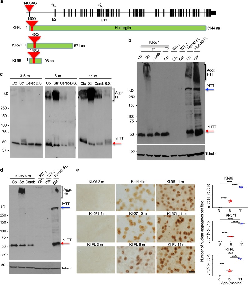
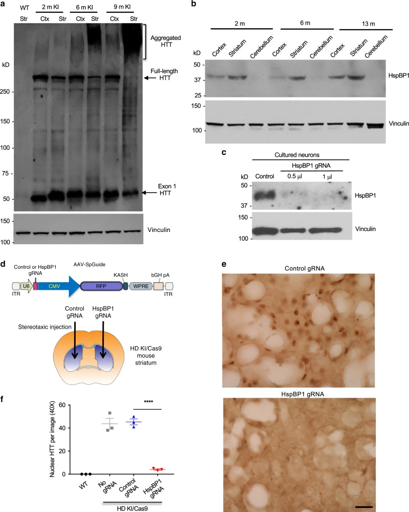
Polyglutamine expansion in proteins can cause selective neurodegeneration, although the mechanisms are not fully understood. In Huntington's disease (HD), proteolytic processing generates toxic N-terminal huntingtin (HTT) fragments that preferentially kill striatal neurons. Here, using CRISPR/Cas9 to truncate full-length mutant HTT in HD140Q knock-in (KI) mice, we show that exon 1 HTT is stably present in the brain, regardless of truncation sites in full-length HTT. This N-terminal HTT leads to similar HD-like phenotypes and age-dependent HTT accumulation in the striatum in different KI mice. We find that exon 1 HTT is constantly generated but its selective accumulation in the striatum is associated with the age-dependent expression of striatum-enriched HspBP1, a chaperone inhibitory protein. Our findings suggest that tissue-specific chaperone function contributes to the selective neuropathology in HD, and highlight the therapeutic potential in blocking generation of exon 1 HTT.


Conflict of interest statement

The authors declare no competing interests.

Figures

Fig. 1
Fig. 1. CRISPR/Cas9 targeting endogenous HTT at different sites in mice.
a Different HTT exons were targeted by CRISPR/Cas9 to generate truncated HTT. b The summary showing the survival rates of homozygous mice with exon 1 deletion (d177) or HTT truncations at different exons (Exon 2-T, 13-T, and 31-T). WT wild type, Het heterozygous, Hom homozygous. c Sequencing analysis confirmed that d177 mutant mice have 177 nucleotides deletion in exon 1, resulting in deletion of N-terminal 59 amino acids without changing other amino acid sequences in the mouse HTT. d Western blotting analysis of d177 mouse brains showing that the d177 HTT (-exon 1) is smaller than full-length mouse HTT (WT HTT). The blots were probed by the antibody MAB2166. More than three times of experiments were performed. e The growth and motor functions of heterozygous (Het) or homozygous (Hom) d177 and wild-type (WT) littermate mice. Data are presented as minimum to maximum showing all points. A p-value < 0.05 is considered as significance, and two-way ANOVA followed by Tukey’s multiple comparison tests was used for analysis (mice numbers: n = 8 for WT, n = 10 for Hom d177, n = 8 for Het d177. Rotarod, F = 0.07562, p = 0.9272; balance beam, F = 0.1509, p = 0.8602; body weight, F = 0.106, p = 0.8996). Data are presented as mean values ± SEM. Source data are provided as a Source Data file.
Fig. 2
Fig. 2. Generation of HD KI mice expressing different N-terminal HTT fragments.
a Generation of new HD KI mouse models by truncating exon 2 and exon 13 of the mouse HTT gene in HD140Q KI (KI-FL) mice via CRISPR/Cas9, resulting in the expression of truncated mutant HTT containing the first 96 and 571 amino acids in KI-96 and KI-571 mice, respectively, with an additional 140Q repeat. b mEM48 western blotting showing the specific expression of mutant HTT in KI-571 mouse brains. This N-terminal mutant HTT (nHTT) is at the same size as the smallest mutant HTT in KI-FL mouse brain and forms aggregated HTT in the striatum. c mEM48 western blotting revealing an age-dependent aggregation of mutant HTT in KI-571 mouse brain. d mEM48 western blotting showing the specific expression of mutant HTT in KI-96 mice, which also has the same size as the smallest mutant HTT in KI-FL mouse brain and preferentially forms aggregated HTT in the striatum. More than three times of experiments were performed independently for bd. In ad, Ctx cortex, Str striatum, Cereb cerebellum. F1 F1 generation, F2 F2 generation. Aggr. HTT aggregated HTT, nHTT n-terminal HTT, fHTT full-length HTT. e mEM48 immunostaining showing an age-dependent accumulation of mutant HTT in the striatum in KI-96, KI-571, and KI-FL mice. Scale bar: 10 μm. Quantification of immunostaining results in Fig. 2e were obtained by counting six images per brain region per mouse (n = 3 mice per genotype, ***p < 0.001, ****p < 0.0001, one-way ANOVA followed by Tukey’s multiple comparison tests, KI-FL, F = 408.1, p < 0.0001; KI-571, F = 1195, p < 0.0001; KI-96, F = 720.8, p < 0.0001). Data are presented as mean values ± SEM. Source data are provided as a Source Data file.
Fig. 3
Fig. 3. Preferential accumulation and aggregation of mutant HTT in the striatum of different KI mice.
a Comparison of mEM48 staining of various brain regions in R6/2 and KI (KI-96, KI-571, KI-FL) mice showing the preferential accumulation of mutant HTT in the striatum of KI mice. The micrographs (5×) from the striatum (Str), cortex (Ctx), hippocampus, cerebellum, and brain stem are presented. Scale bar: 100 μm. b Quantitative analysis of density of mEM48 labeled nuclei in different brain regions in KI mice. The data were obtained by counting six images per brain region per mouse (n = 3 mice per genotype, ****p < 0.0001, one-way ANOVA followed by Tukey’s multiple comparison tests, KI-FL, F = 405.5, p < 0.0001; KI-571, F = 150.3, p < 0.0001; KI-96, F = 375.9, p < 0.0001). Data are presented as mean values ± SEM. Source data are provided as a Source Data file.
Fig. 4
Fig. 4. Comparison of the expression levels of mutant HTT in HD KI mouse striatum.
a Immunostaining of the mouse striatum with mEM48 showing that R6/2 mice have much more abundant nuclear HTT staining than KI-96, KI-571, and KI-FL mice. Scale bar: 20 μm. b Quantitative analysis of the density of nuclear HTT aggregates in the striatum of R6/2 and different KI mice. The data were generated by mean value of counting six images each mouse (n = 3 mice per genotype, ****p < 0.0001, one-way ANOVA followed by Tukey’s multiple comparison tests, F = 69.11, p < 0.0001). Data are presented as mean values ± SEM. c PCR to amplify the CAG repeat showing R6/2 and all KI mice carry the similar CAG repeat numbers. WT wild type. d Western blotting using mEM48 revealing that R6/2 mouse striatum expresses more abundant soluble mutant HTT and aggregated HTT than KI mouse striatum. R6/2 mouse at 8 weeks and KI mice at 3.5 months of age were examined. More than three independent experiments were performed. e PCR primers used to detect mutant HTT transcripts in R6/2 and HD KI mouse brains. f RT-PCR revealing a much higher level of exon 1 HTT mRNAs in R6/2 mice than other KI mice. The negative controls are PCR results from mRNA samples without reverse transcriptase to rule out genomic DNA amplification products. g The ratios of HTT cDNA bands to the control gapdh band in f. Data are presented as mean values ± SEM and were obtained from three independent experiments each mouse (n = 3 mice per genotype, ****p < 0.0001, one-way ANOVA followed by Tukey’s multiple comparison tests, F = 114.9, adjusted p < 0.0001). h Quantitative RT-PCR using primers S1 and A1 also confirmed the significantly higher level of exon 1 HTT mRNA in the R6/2 mouse striatum than KI mouse striatum. Data are presented as mean values ± SEM and were obtained from three independent experiments each mouse (n = 3 mice per genotype, **p < 0.01, ***p < 0.001, one-way ANOVA followed by Tukey’s multiple comparison tests, F = 17.44, p = 0.0002). Source data are provided as a Source Data file.
Fig. 5
Fig. 5. Truncation of mutant HTT in the striatum of adult HD140Q KI mice.
a mEM48 western blotting showing age-dependent decrease of soluble mutant exon 1 HTT and corresponding increase of aggregated HTT in the HD KI striatum. Quantification of western blotting results is also shown. Data are presented as mean values ± SEM and were obtained from three independent experiments each mouse (n = 3 mice per genotype, **p < 0.01, ***p < 0.001, one-way ANOVA followed by Tukey’s multiple comparison tests, aggregated HTT/vinculin analysis, F = 19.79, p = 0.0023; mutant exon 1 HTT/vinculin analysis, F = 49.81, p = 0.0002). b HD140Q KI mice were crossed with Cas9 mice to yield KI/Cas9 mice. AAV HTT-gRNA was then injected into the striatum of KI/Cas9 mice at 2 months of age. Eight weeks after injection, the striatum of the injected mice was isolated for analyzing truncated HTT. c A fluorescent image of the injected mouse striatum verifying the viral transduction of AAV HTT-gRNA that also expresses red fluorescent protein (RFP). Scale bar: 20 μm. d T7E1 analysis of the HTT DNAs from the injected region confirming that the only HTT-gRNA, but not control gRNA, could target the HTT gene to generate fragmented DNA products. e Western blotting analysis of striatum lysates of AAV-injected mice showing that truncation of mutant HTT at exon 2 and exon 13 by HTT-gRNAs (HTT-91 gRNA and HTT-571 gRNA) could reduce full-length mutant HTT and increase exon 1 HTT but did not alter the amount of aggregated HTT. f mEM48 immunostaining of the AAV-injected striatum of HD140Q KI mice showing that nuclear HTT accumulation after AAV HTT-gRNA injection is not different from that with the AAV control gRNA injection. Scale bar: 20 μm. More than three times of experiments were performed independently for cf. Source data are provided as a Source Data file.
Fig. 6
Fig. 6. Phenotype analysis of different KI mice.
a Comparison of motor function (rotarod performance, balance beam, grip strength) and body weight of wild type and different types of KI (KI-96, KI-571, and KI-FL) mice. Each group consists of 14 mice. Data are presented as minimum to maximum showing all points. *p < 0.05, **p < 0.01, ***p < 0.001, ****p < 0.0001, two-way ANOVA followed by Tukey’s multiple comparison tests, rotarod analysis, F = 13.73, p < 0.0001; grip strength analysis, F = 9.187, p < 0.0001; balance beam analysis, F = 20.45, p < 0.001; body weight analysis, F = 4.777, p = 0.0029. Data are presented as mean values ± SEM. b Gfap immunostaining of wild type (WT) and KI-571, KI-FL mouse striatum at 6 and 11 months of age. Scale bar: 20 μm. c Quantification of relative Gfap immunostaining signals in wild type (WT) and KI-571, KI-FL mouse striatum. The data were obtained from six images in the striatum per mouse (n = 3 mice per genotype, ***p < 0.001, one-way ANOVA followed by Tukey’s multiple comparison tests, F = 65.67, p < 0.0001). Data are presented as mean values ± SEM. Source data are provided as a Source Data file.
Fig. 7
Fig. 7. HspBP1 expression accounts for the nuclear accumulation of mutant HTT in KI mouse striatum.
a mEM48 western blot analysis of the cortex and striatum of HD140Q KI mice at different ages revealing that mutant exon 1 HTT is constantly expressed but selectively forms aggregates in the aged striatum. More than three times of experiments were performed independently. b Western blotting of wild-type mouse brains showing that HspBP1 is more abundant in the striatum and its expression increases with age. c Western blotting of cultured mouse striatal neurons showing that targeting HspBP1 by AAV Cas9/HspBP1-gRNA could effectively reduce HspBP1 expression. d Injection of AAV control gRNA and AAV HspBP1-gRNA into the striatum of KI/Cas9 mice at 25 weeks of age. The mouse brain image was remixed from the clipart https://commons.wikimedia.org/wiki/File:Mouse_Dorsal_Striatum.pdf, under the Creative Commons Attribution-Share Alike International license. e Eight weeks after injection, mEM48 immunostaining of the injected striatum showing that deletion of HspBP1 could markedly reduce the nuclear accumulation of mutant HTT. Scale bar: 40 μm. f Quantitative analysis of nuclear HTT staining in the AAV-injected mouse striatum. The data were obtained from five images in the striatum per mice (n = 3 mice per group, ****P < 0.0001, one-way ANOVA followed by Tukey’s multiple comparison tests, F = 82.84, p < 0.0001). Data are presented as mean values ± SEM. Source data are provided as a Source Data file.

References

    1. Orr HT, Zoghbi HY. Trinucleotide repeat disorders. Annu Rev. Neurosci. 2007;30:575–621. - PubMed
    1. Bates GP, et al. Huntington disease. Nat. Rev. Dis. Prim. 2015;1:15005. - PubMed
    1. Ross CA, Tabrizi SJ. Huntington’s disease: from molecular pathogenesis to clinical treatment. Lancet Neurol. 2011;10:83–98. - PubMed
    1. Vonsattel JP, et al. Neuropathological classification of Huntington’s disease. J. Neuropathol. Exp. Neurol. 1985;44:559–577. - PubMed
    1. Lee CY, Cantle JP, Yang XW. Genetic manipulations of mutant huntingtin in mice: new insights into Huntington’s disease pathogenesis. FEBS J. 2013;280:4382–4394. - PMC - PubMed

Publication types

Substances